

 PDF
PDF ePub
ePub Citation
 Print
Print


INTRODUCTION
Methotrexate (MTX), a folic acid antagonist, has been widely used in dermatology to treat various diseases, such as moderate to severe psoriasis, psoriatic arthritis, and collagen vascular disease1,2. This drug may cause various mucocutaneous adverse reactions, including mucositis, bone marrow depression, hepatitis, and renal insufficiency3,4. Although development of skin ulceration in a small number of patients has been reported5-7, multifocal mucocutaneous involvement, which shows similar clinical features with Behçet's disease has not been reported. Herein we describe a 63-year-old woman with oral, genital ulcer and superficial thrombophlebitis or erythema multiforme-like lesions, which developed after administration of MTX.
CASE REPORT
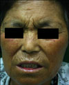
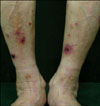
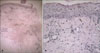
A 63-year-old woman presented with erythematous painful ulcers in the oral cavity and vulva, and multiple erythematous to purpuric crusted patches and papules on both legs. Five weeks ago, she presented with chief complaints of loss of vision in the right eye. Her fundus examination was within normal limits. On anterior segment fluorescein and indocyanine green, the patient was diagnosed as having diffuse necrotizing scleritis. She started treatment with MTX (10 mg daily). Three weeks after starting the treatment with MTX, erythematous ulcers appeared in her oral cavity and vulva. Concomitantly, erosive erythematous crusted patches and papules appeared on both legs. The patient was referred to the department of dermatology for evaluation of Behçet's disease. A physical examination showed erythematous ulcers in her oral cavity (Fig. 1), vagina, and erosive erythematous patches and papules on both lower legs (Fig. 2). Differential diagnosis included Behçet's disease and cutaneous side effects of MTX. Laboratory tests showed hemoglobin of 8.3 g/dl, white blood cell count 1.99×10³ cells/mm3 and platelet count 72×103/mm3. Liver and renal function tests were normal. Antinuclear antibodies and HLA-B51 were positive and anti-ENA antibody group was negative. Pathergy test was also negative. Histopathological findings showed flattening of rete ridges and dykeratotic keratinocytes, vacuolar degeneration, and hyperpigmentation in the epidermis. In the dermis dense perivascular lympho-histiocytic infiltration was seen, without evidence of vasculitis (Fig. 3). Although systemic steroid (prednisolone 50 mg/day) was given in combination with colchicines for 1 week, there was no clinical improvement. After reducing the dose of MTX (7.5 mg, weekly twice) for a month and subsequently stopping the MTX and starting treatment with pyridoxine, the mucocutaneous lesions were healed completely. After 12 months, there had been no recurrence of the lesions. Although the patient presented with oral and genital ulcers mimicking Behçet's disease and HLA-B51 was positive, the skin lesions showed significant correlation with the MTX treatment. Therefore we diagnosed the condition as acute cutaneous side effects of MTX.
DISCUSSION
MTX (4-amino-4deoxy-10-methyl-pteroyl glutamic acid) is a folic acid analogue, and it blocks intracellular DNA synthesis1-3. It is has been used successfully for over 50 years for a wide variety of cutaneous diseases4. This drug is approved for treatment of moderate to severe psoriasis, and cutaneous T cell lymphoma, and it has been known to be effective in many other dermatoses, including atopic dermatitis, vascular disease, and blistering disease5-7. Rapidly proliferating cells are susceptible to MTX because most cells are in the S-phase, where this drug exerts its effects1,4. Gastrointestinal mucosa, hair, and bone marrow are very susceptible to MTX because of the high rate of cellular turnover3. Although cutaneous adverse reaction to MTX is not common, skin ulcerations, necrosis, erosions of the psoriatic plaques and bullous acral erythema, have been reported3-6.
In our case, the patient presented with oral, genital ulcers, and erythematous crusted patches. On initial physical examination, we suspected this case as Behçet's disease. However, the evidence of neutrophilic vasculitis was not seen on histologic examination and pathergy test showed negative result. By further history taking, we learned that her mucocutaneous lesions had occurred after the treament with high-dose oral MTX. Laboratory findings showed pancytopenia, which suggested bone marrow suppression due to MTX. After stopping of MTX (7.5 mg, weekly twice), the mucocutaneous lesions were healed, and there has been no recurrence of the lesions for 12 months. Although the patient complained of ocular symptoms, scleritis is rarely associated with Behçet's disease8. Combining histologic findings and clinical course, we diagnosed the condition as acute cutaneous side effects of MTX.
MTX may cause direct cell toxicity, especially in tissues where cell turnover is increased, due to trauma or underlying dermatosis. Therefore in the literature, most cases of cutaneous ulcers showed some form of pre-existing cutaneous pathology, i.e. either physical insults or underlying dermatosis. However, in our case, the patient did not remember any remarkable history of trauma or other dermatosis in the area of the mucocutaneous lesion.
Our case is particular in that the patient showed similar clinical features to Behçet's disease, such as oral, ocular and genital lesions and superficial thrombophlebitis or erythema multiforme-like lesions. Hence, we report a case of cutaneous side effects of MTX that mimicked Behçet's disease.
 XML Download