

 PDF
PDF ePub
ePub Citation
 Print
Print


INTRODUCTION
Eczema herpeticum is also known as Kaposi's varicelliform eruption, and this refers to a herpetic superinfection of a pre-existing skin disease. There have been reports of eczema herpeticum occurring in atopic dermatitis, dyskeratosis follicularis, Darier's disease1,2, pemphigus foliaceus3, mycosis fungoides, ichthyosis vulgaris, Hailey-Hailey disease4-7, Sézary syndrome8 and in patients with burns9. When eczema herpeticum is recognized early, it is easily and effectively treated with antiviral agents.
Hailey-Hailey disease is a blistering dermatosis that is inherited as an autosomal dominant trait and it usually presents around the third and fourth decades. This disease is characterized by recurrent eruptions, usually on the intertriginous areas, i.e., the axillae, the groin and/or on the neck. Vesicles, erosions and crusts may be present in these erythematous lesions. A decreased numbers of desmosomes have been implicated in the pathogenesis of Hailey-Hailey disease. The therapeutic options for this disease are limited.
The occurrence of eczema herpeticum together with Hailey-Hailey disease is rare. There is little information linking herpes simplex virus with the exacerbation of Hailey-Hailey disease. Only four such cases have been reported in the English and German medical literature. To the best our knowledge, this is first such case report in Korea. This report describes eczema herpeticum in a patient with Hailey-Hailey disease, and this was confirmed by skin biopsy and the clinical features.
CASE REPORT
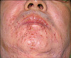
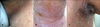
A 47 years old man visited our Department of Dermatology with a presentation of painful vesicles on the chin and he'd had these lesions for the previous 10 days (Fig. 1). The multiple umbilicated vesicles on the chin were suspected to be eczema herpeticum according to the clinical features. This man had also presented with a painful, erythematous, fissured plaque involving the flexural areas for the previous year (Fig. 2). He complained of an unpleasant smell from his lesions. Physical examination revealed recurrent painful erosions, vesicopustules and scaly erythematous plaques in the intertriginous areas, i.e., the axilla, inguinal folds and the neck. The lesions were intermittently aggravated and especially during the summer, which restricted his mobility. The bullous and erosive lesions epithelized slowly without leaving scars. He has a medical history of diabetes and hypertension.
The KOH mount was negative. The abnormal laboratory test results included an elevated total white blood cell count of 19,740 cells/mm3. Other investigations, including blood sugar, urinalysis, liver function tests and serological tests for syphilis, were within the normal limits. Serology for IgM and IgG antibodies to herpes simplex was not performed.
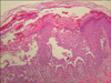
A biopsy specimen from an active skin lesion on the chin showed marked infiltration of the chronic inflammatory cells in the dermis. Inclusion bodies were not seen. The skin biopsy from the right groin confirmed the diagnosis of Hailey-Hailey disease. It showed focal hyperkeratosis and dyskeratotic cells similar to corps ronds within the granular layer. A cleft was present within the epidermis, and the epithelium beneath the cleft showed partial acantholysis. Suprabasal bulla and the appearance of a dilapidated brick wall in the epidermis were seen (Fig. 3). So, we diagnosed this case as eczema herpeticum with Hailey-Hailey disease.
He was treated by a combination of systemic antibiotics and oral famciclovir, topical pimecrolimus and wet dressing. The vesicles on the chin were rapidly controlled by antiviral therapy within a week. But during the usage of topical pimecrolimus, the patient complained of local irritation, and then this medication was changed to topical steroid cream. After these treatments, slight improvement of his Hailey-Hailey disease lesions was noted.
DISCUSSION
Eczema herpeticum is a potentially life-threatening viral infection that arises in pre-existing skin conditions. In some cases, it may progress to a fulminating, life-threatening infection and it can have severe sequelae, including herpes keratitis, disseminated infection with visceral involvement and death. It begins as clusters of umbilicated vesiculopustules in the areas where the skin has been affected by a preexisting dermatitis.
Disruption of the stratum corneum secondary to skin disease is the most common predisposing factor10. There have been reports of eczema herpeticum occurring in atopic dermatitis, Darier's disease1,2, pemphigus foliaceous3, pemphigus vulgaris, pityriasis rubra pilaris, Hailey-Hailey disease4-7, irritant contact dermatitis, cutaneous T cell lymphoma, seborrheic dermatitis8, Wiskott-Aldrich syndrome8, congenital icthyosiform erythroderma and Sézary syndrome8. Eczema herpeticum may also occur when there has been recent injury to the skin such as second-degree burns9, autografted skin and post dermabrasion. A commonality among all these diseases is the disruption of the integrity of the epidermis.
Viral cultures of fresh vesicular fluid and the direct observation of the infected cells scraped from the ulcerative lesions by direct fluorescent antibody (DFA) staining are the most useful and reliable diagnostic tests available10,11. A Tzanck smear of an opened vesicle or erosion can provide a rapid diagnosis when it shows the characteristic epithelial multinucleated giant cells and acantholysis. If the lesions are atypical, equivocal or old, then biopsy or the polymerase chain reaction (PCR) should be considered.
Therapy should be instituted without delay when there is a high suspicion or a positive Tzanck preparation. The early use of both antiviral drugs and antibiotics is extremely important; their use should not be delayed pending laboratory tests. The most commonly used antiviral drugs are the nucleoside analogs, which inhibit viral DNA polymerase. The initial treatment is generally with high-dose intravenous acyclovir, which is the most widely studied and used drug for treating eczema herpeticum11. Valacyclovir and famciclovir are also very effective with better oral bioavailability and a more convenient dosing schedule for patients. The antibiotic therapy is tailored to the organism found on culture, and this is most commonly the Staphylococcus species. When a bacterial infection is not present, the patients should be given a topical antibiotic cream like silver sulfadiazine for prevention11. Patients with recurrent HSV infections and a chronic skin disease that predisposes them to eczema herpeticum should be offered prophylaxis with either valacyclovir or acyclovir.
Hailey-Hailey disease was first described by the Hailey brothers in 193912. It usually appears in the third or fourth decade, although it can occur at any age. It is a chronic autosomal dominant disorder with incomplete penetrance. Approximately two thirds of patients have a family history of this disorder. A history of multiple relapses and remissions is characteristic. It typically begins as a painful erosive skin rash in a flexural area. It usually involves the genital area, neck, axillae and popliteal fossae and it recurs in the same locations. Patients typically have recurrent vesicles on an erythematous background. The vesicles rupture and leave an eroded base, and then they generally become crusted. Secondary bacterial infection, which is not uncommon, can give rise to an unpleasant smell. White bands on the fingernails and pits in the palms can also occur13,14. Heat, sweating and friction often exacerbate the disease, and most patients have worse symptoms during the summer months14.
The responsible defect has been identified in the gene named ATP2C1 on the 57 chromosome 3q21-24, and the gene encodes the human secretory pathway Ca(2+)-ATPase (hSPCA1) localized in the Golgi apparatus15. It controls the Ca2+ stored in the Golgi bodies. More than 82 different ATP2C1 mutations have currently been described16. The defect that is responsible has been identified on a gene called ATP2C1 and the gene is found on chromosome 3q21-24. This gene codes for the protein SPCA1 (Secretory Pathway Calcium/manganese-ATPase), which is a calcium and manganese pump. Ca2+ is needed for the assembly of functional adherens junctions and desmosomes. The keratinocytes stick together via desmosomes and it seems that the desmosomes do not assemble properly if there is insufficient calcium. The epidermal Ca2+ gradient in the skin is therefore attenuated in the patients with Hailey-Hailey disease. The total Ca2+ concentrations were reported to be substantially decreased in the superficial layers of the epidermis, whereas the basal layer of the epidermis and the dermis had unchanged Ca2+ concentrations16. This results in a mechanical defect in the tonofilament-desmosome complex or intercellular substance, leading to the cleavage of the epidermal cells, that is, epidermal acantholysis16. The disease persists when acantholysis is coupled with repeated minor shearing stress. Viral infections have been implicated in the exacerbation of lesions4,14. It is well known that direct virus transmission by contact with infected persons may evoke eczema herpeticum. These facts suggest that the fragility of the epidermal cells, which results from eczemalike epidermal lesions, makes it easy for herpes simplex virus to infect and proliferate there. Interestingly, the typical lesions of eczema herpeticum in our case occurred in an exposed area like the chin. The location of the eczema herpeticum in our case suggests that the skin involvement was the result of direct inoculation of the herpes simplex virus. Herpes simplex virus infection may be from autoinoculation or from an infected contact11.
The characteristic pathologic finding of Hailey-Hailey disease is suprabasal acantholysis17. Severe acantholysis is seen in some lesions, and this finding is diagnostic in the proper clinical setting. Histologically, Hailey-Hailey disease has a characteristic 'dilapidated brick wall appearance'. The pathology in some biopsies may only consist of a suprabasal slit with minimal acantholysis, and such a biopsy may be indistinguishable from that seen in pemphigus vulgaris. Yet the clinical presentation is distinctive. Some cases of Grover's disease (transient or not-sotransient acantholytic dermatosis) may share pathologic features with Hailey-Hailey disease, although dyskeratosis is more commonly seen in Grover's disease.
A variety of therapeutic modalities have been recommended for the treatment of Hailey-Hailey disease. Unfortunately there is no cure for Hailey-Hailey disease. The current therapeutic strategies attempt to suppress Hailey-Hailey outbreaks and allow the patient to live comfortably with this condition. The therapeutic options include antibiotics, corticosteroids14, systemic and topical cyclosporine18, oral retinoid and the topical vitamin D analogue19 and surgical methods such as excision and grafting20 and CO2 lasers.
To the best of our knowledge, this is the first case report from Korea in which we have described the coexistence of eczema herpeticum and Hailey-Hailey disease.
 XML Download