

 PDF
PDF ePub
ePub Citation
 Print
Print


INTRODUCTION
Lipomas, whose principle components are mature adipocytes, are the most common neoplasms of mesenchyme. A number of different subtypes of lipoma have been described which vary by location and by the presence of other tissue elements1. Angiomyxolipoma (AML) is a rare variant of lipoma first described by Mai et al2 in 1996. It presents as a well-circumscribed tumor characterized by an admixture of mature adipose tissue, paucicellular myxoid stroma, and an abundance of thin- and thick-walled blood vessels. Until now only eight cases had been reported in literatures2-8. Here we report another case of angiomyxolipoma arising in subcutaneous tissue.
CASE REPORT
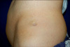
A 69-year old Korean man presented with a 3-year history of a slowly growing painless subcutaneous mass on the left iliac crest. It was accompanied by a mild tenderness that resulted from an accidental trauma. On physical examination, there was found to be a solitary, movable, relatively well-demarcated subcutaneous mass that measured a maximum of 2 cm across (Fig. 1).
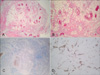
Histopathology demonstrated a multilobular well-circumscribed mass surrounded by a thin fibrous capsule (Fig. 2A, B). The adipose tissue and myxoid tissue alternated in small nests in a ratio of approximately 4:6. The myxoid areas were stained with alcian blue (pH 2.5) and found to contain spindled cells (Fig. 2C). Both components contained numerous dilated thin- and thick-walled blood vessels without vascular thrombus. Very little perivascular lymphocyte infiltration was evident, and there was no evidence of tumor necrosis, cellular atypia, mitotic figures, or lipoblasts.
An immunohistochemical study found vimentin expression in both the adipose and the myxoid tissue. The spindle cells in the myxoid areas co-expressed CD34 (Fig. 2D) but not S-100 protein, desmin, or smooth muscle actin (SMA). The mature adipocytes were focally positive for S-100 protein. The vascular endothelium expressed CD34 and SMA. The tumor was negative for desmin and HMB 45.
A surgical excision was performed, and a yellowish-white, gelatinous rubbery tumor was removed. Currently, 2 months after the excision, there is no evidence of recurrence.
DISCUSSION
An adipose tissue tumor is the most common mesenchymal neoplasm, but its variants are rare. A number of different subtypes of lipoma have been described that vary according to their location and the presence of other tissue elements, including synovial, parosteal, intraosseous, lumbosacral and thymolipoma; and spindle cell/pleomorphic, chondroid lipoma, and angio-, fibro-, myxo-, chondro-, osteo-, and myolipoma1.
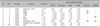
Eight cases of angiomyxolipoma, or vascular myxolipoma, have been described previously (Table 1)2-8. The patients' ages ranged from 32 to 69 years, averaging 51.8, and most were male. The location has generally been in subcutaneous tissue, although the first and last cases were located on the spermatic cord and in the subungual region, respectively.
The histopathologic features of angiomyxolipoma are characteristic and include an admixture of a myxoid area with relatively few cells, mature adipose tissue without lipoblasts, and numerous thin- and thick-walled vascular structures. Immunohistochemical studies show that the spindle cells of the myxoid areas express vimentin and CD34 but do not express SMA, desmin, or S-100 protein. The mature adipocytes are immunoreactive for S-100 protein, and the blood vessels are positive for vimentin and SMA. Together, the features point toward a differential diagnosis that includes variants of lipomas, such as myxolipoma, angiolipoma angiomyolipoma, superficial angiomyxoma, myxoid spindle cell lipoma, myxoid liposarcoma, low-grade myxofibrosarcoma, and superficial angiomyxoma.
Myxolipoma and angiolipoma each lack one of the three major components of angiomyxolipoma, and angiomyolipoma displays bundles of smooth-muscle cells with HMB-45 immunoreactivity. Superficial angiomyxoma is a poorly circumscribed mass of adipose tissue accompanied by a mixed inflammatory infiltration of neutrophils and, frequently, epithelial structures9. Myxoid spindle-cell lipoma and angiomyxolipoma share the unique histologic features of mature adipose tissue plus spindle cells in myxoid background. However, its lack of ropy collagen and its poor vascularity differentiate it from angiomyxolipoma. Furthermore, the vascular variant of spindle-cell lipoma lacks prominent myxoid components, a major feature of angiomyxolipoma10. Pseudoangiomatous spindle-cell lipoma contains spindle cells in the myxoid area and branched or cleft-like pseudovascular spaces. Malignant tumors of myxoid liposarcoma and low-grade myxofibrosarcoma should also be differentiated from angiomyxolipoma. A myxoid liposarcoma is composed of lipoblasts, which express S-100 protein, and vascular structures in a fine branching pattern11. Low-grade myxofibrosarcoma frequently exhibits numerous elongated curvilinear vessels or a plexiform vascular pattern in a myxoid matrix with increased pleomorphism12. In our case the histopathologic findings showed only thin- and thick-walled blood vessels without specific vascular patterns, such as branching patterns, curvilinear vessels, or plexiform vascular patterns.
With an electron microscope examination, Mai et al2 identified spindle cells with fat vacuoles (so-called 'preadipocytes') in the transitional areas between the myxoid and lipomatous components, similar to findings for spindle-cell lipoma. A cytogenetic study of angiomyxolipoma revealed chromosomal aberrations involving translocations t(7;13)(p15;q13) and t(8:12)(q12;13), which are similar to those found in ordinary lipoma, spindle-cell/pleomorphic lipoma, and myxoma4, but the primary origin of the angiomyxolipoma is still to be determined.
Anxiomyxolipoma is a very rare benign neoplasm with characteristic histopathologic and immunohistochemical features. Additional immunohistochemical studies can help to differentiate angiomyxolipoma from other lipomatous neoplasms, but histopathologic findings that show a mixture of adipose tissue, myxoid component, and abundant blood vessels without immature or atypical cells should suggest to dermatologists and pathologists a diagnosis of angiomyxolipoma with its benign nature.
 XML Download