

 PDF
PDF Citation
 Print
Print


Introduction
Sleep is an essential factor for healthy life.[1,2] However, many critically ill patients are known to experience sleep deprivation.[3,4] They have poor sleep quality characterized by suppressed slow wave sleep (SWS) and rapid eye movement (REM) sleep because their sleep is interrupted frequently, regardless of normal total sleep time (TST).[5] The cause of sleep deprivation is not clearly understood, but multiple factors including noise,[6] bright light, inflammatory mediators,[4] tests such as blood sampling, taking vital signs[7] and mechanical ventilation,[8] appear to cause sleep disruption in intensive care unit (ICU) patients. Sleep deprivation can lead to the deterioration in neurocognitive performance,[9] immune function[10] and respiratory control,[11] posing a serious barrier to effective intensive care.
ICU patients are commonly treated with sedatives to reduce pain, relieve anxiety and induce sleep.[12] Midazolam is one of the most commonly used sedatives in ICU settings.[13] As a gamma-aminobutyric acid (GABA) receptor-specific drug, midazolam is metabolized in the liver and excreted through the kidney with the elimination half life of 3 hours.[14] Even many researches were explored sedative effects of midazolam on ICU patients,[15] the evidences of midazolam’s effect on sleep, which definitely differs from sedation, are scarce.[16,17] Nonetheless, many intensivists mistakenly believe that sedation and sleep refer to the same thing. In addition, there have been few reports on the association between the appropriate dose of midazolam and proper sleep in ICU patients.
This study aimed to examine the association of intravenous midazolam infusion at different doses with sleep quantity and quality in ICU patients using 24-hour polysomnography (PSG) and to determine an appropriate dose of midazolam for Korean ICU patients.
Materials and Methods
1) Study population
This study was conducted with patients in the medical ICU of tertiary teaching hospital and approved by the institutional board review committee (IRB No. B-1307/212-010). This study was conducted as a part of an ongoing prospective ICU sleep study carried out from 1. October. 2013. The inclusion criteria were patients aged 18 or above undergoing mechanical ventilation and sedation with continuous intravenous infusion of midazolam. Informed consent was obtained from patients or their family members. Patients were excluded if they had a medical history of Parkinson’s disease, severe dementia, cerebral infarction/cerebral hemorrhage, if they were addicted to alcohol or taking psychiatric medication due to depression or anxiety disorder, etc. Patients who had an infectious condition of the central nervous system, renal insufficiency or liver cirrhosis at the time of admission were also excluded. After selecting qualified patients, we recored their age, sex, body mass index, Acute Physiology and Chronic Health Evaluation II (APACHE II) and Sequential Organ Failure Assessment (SOFA) scores on admission. ICU midazolam was administered at the loading dose of 0.05 mg/kg/h and Richmond Agitation Sedation Scale (RASS) score was maintained in range of 0 to −2 by measuring sedation level every 4 hours. The midazolam dose was seldom changed during 24-hour PSG. In addition to midazolam, remifentanyl was administered for pain control. The dose of remifentanyl was determined by physicians on the basis of pain level of patients. The ICU length of stay, duration of mechanical ventilation after discharge from the ICU and mortality were also identified.
2) Polysomnography (PSG)
The quantity and quality of sleep of patients were measured for 24 hours using Somnowatch™, plus (SOMNOmedics, Rander-sacker, Germany). This polygraphic screening device was comprised of two pairs of electroencephalographys and one pair of electromyography, electrooculography and electrocardiograph recorders. We performed electroencephalogram after washing patients’ scalp and placing electrodes in the scalp locations (A1 or A2/C3 and C4) according to the International 10–20 system.[18] Electromyographic electrodes were placed on the right and left sides of the chin. Electrooculographic electrodes were placed 1 cm above the right outer canthus and 1 cm below the left outer canthus. Electrocardiographic electrodes were directly placed on both sides of the chest to measure Lead II. Each electrode was checked every 4 hours and replaced if its impedance value increased. PSG recordings were interpreted by two experts using Standard Rechtschaffen and Kales criteria.[19] If a significant difference was discovered in interpretation provided by the two individuals, the respective PSG data was reinterpreted through discussion.
3) Statistical analysis
Medians and interquartile range (IQR) were used to describe continuous variables, and percentages (%) were used to express baseline characteristics, such as sex, diagnosis, and survival rate. Sleep efficiency (%) was calculated as the ratio of TST to total time of lying down.[20] Wake index (n/h) was defined as the number of awakenings per hour. Spearman’s correlation was used to detect the association between midazolam dose and TST and the association between midazolam dose and sleep stages (REM, non-REM Level 1, 2, 3) and identify a specific sleep stage that is most affected by the sedative. p values less than 0.05 were considered statistically significant. The software package SPSS version 21.0 (IBM Corporation, Armonk, NY, USA) was used to analyze the data.
Results
Of 20 patients who underwent PSG for 24 hours during the study period, nine of them received midazolam as sedative by continuous infusion for 24 hours. Of those, three patient’s PSG data could not be interpreted because of severe artifacts, and one patient who received 0.08 mg/kg/h of intravenous midazolam was found to be coma in PSG. Therefore, PSG recordings of five patients were analyzed in this study. Baseline patient characteristics are summarized in Table 1. The median patient age, APACHE II and SOFA score was 67, 20 and 6, respectively. The median ICU length of stay was 3.0 days at the time of PSG test. The median (IQR) dose of midazolam and remifentanyl was 0.02 (0.015–0.04) mg/kg/h and 0.02 (0.00–0.09) μg/kg/min, respectively, for 24 hours. RASS score was −1 (−1∼−2). In arterial blood gas analysis (ABGA), the level of carbon dioxide was 38.3 (33.9–41.2) mmHg.
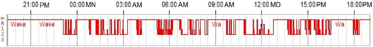
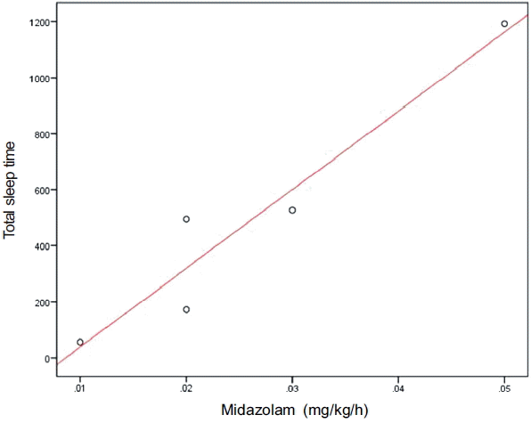
PSG data of the five patients were summarized in Table 2. Median TST was 494 min, and the median amount of REM was 10 min. However, no patients experienced stage 3 sleep. The median of wake index was 16.1, meaning that the patients awakened approximately 16 times per hour. The hypnogram shows sleep characteristics of a representative patient (Fig. 1). The patient repeatedly awakened, making his sleep regularly fragmented and circadian rhythm disrupted. A significantly postive correlation was found between midazolam dose and TST (r = 0.975, p = 0.005, Fig. 2). By individual, the patient who received 0.01 mg/kg/h of midazolam showed TST of 55 min during a 24-hour bed time, whereas the patients who received a higher dose of midazolam in range of 0.02 to 0.03 mg/kg/h showed TST of 172 to 526 min. The patient who received 0.05 mg/kg/h of midazolam showed TST of 1,192 min (Table 3). The effects of midazolam dose on sleep stages revealed a statistically significant correlation with stage 2 sleep (r = 0.975, p = 0.005), whereas effects on REM (r = 0.103, p = 0.87) and stage 1 sleep (r = 0.564, p = 0.322) were not correlated with midazolam dose.
Discussion
This prospective pilot study explored the association between midazolam dose and sleep quantity and quality in ICU patients using PSG. As a result, a positive correlation was observed between midazolam dose and TST. In particular, an adequate amount of sleep was found at the dose of 0.02 to 0.03 mg/kg/h of midazolam. However, the patients spent their most of TST in stage 1 and 2 sleep with little REM sleep, and they did not experience stage 3 sleep at all. In addition, their sleep fragmentation was also severe because they awakened 16 times per hour.
Midazolam is the only benzodiazepine available for continuous intravenous infusion in ICU patients in many countries.[21–23] It is the most popular sedative in the ICU because of its short elimination half life and inexpensive price.[13] Currently, the Korean Ministry of Food and Drug Safety recommends continuous intravenous infusion of midazolam at maintenance dose of 0.03 to 0.2 mg/kg/h for sedation in ICU patients. This dose level and range are higher and wider than 0.02–0.1 mg/kg/h allowed in North America.[24] However, there have been no evidence-based data suggesting an adequate midazolam dose for ICU patients. Thus, the results of this study was meaningful because this is the first study to evaluate sleep quantity and quality in ICU by an objective manner using PSG.
The importance of sleep is increasing for ICU patients. [3,4,6,9,10,25] ICU environmental factors such as noise, light, tests and treatment are combined with external stimuli to disrupt circadian rhythm and eventually sleep.[8,26] Lack of sleep is one of the most frequent complaints among ICU patients,[27] whose cell mediated and humoral immunity is suppressed and cytokine release is affected as a result. Suppressed immune function can in turn cause or worsen infection and slow down wound healing.[28] Sleep deprivation also affects muscles of respiration, which not only lowers tidal volume[29] but also makes respiratory response to carbon dioxide slow.[30] In this case, weaning from mechanical ventilation can be difficult.[4] In this study, the low-dose of midazolam (0.01 mg/kg/h) led to TST of 55 min for 24 hours even though RASS score was −1. On the contrary, the patient who received 0.08 mg/kg/h of midazolam showed suppressed EEG presenting coma. The dose of 0.08 mg/kg/h is allowed in both Korea and North America, but the result of this study indicates that the dose level is too high to use for ICU patients. A patient showed TST of nearly 20 hours during 24-hour bed time at a dose of 0.05 mg/kg/h of midazolam. This finding also suggests that 0.05 mg/kg/h of midazolam is also too high for ICU patients. Hypersomnia is well known to disrupt circadian rhythm and contribute to the increasing mortality rate.[1]
In this study, the patients showed an adequate amount of TST at the dose midazolam of 0.02 to 0.03 mg/kg/h. However, the patients also had very bad sleep quality in which REM sleep was drastically reduced and stage 3 sleep was not observed. The poor sleep quality is consistent with the findings of previous studies that investigated sleep characteristics of ICU patients.[25] Stage 3 sleep tends to decrease with age, but humans spend 20–25% of their TST in REM sleep, regardless of age.[27] REM sleep boosts memory consolidation, and reduced REM sleep can increase the risk of delirium.[25] However, the cause of a reduced amount of REM sleep in ICU patients is not clearly understood. Some researchers claim that ICU patients cannot experience REM sleep because of frequent sleep fragmentation.[27] This study also found frequent awakenings of 16 times per hour, i.e. once every 3–4 min, confirming very bad sleep quality in ICU patients.
Noteworthily, this study used PSG to analyze the effects of midazolam on sleep of ICU patients by an objective manner. It is already well understood that the use of midazolam increases TST but reduces REM sleep in insomnia patients.[31] However, the effects of midazolam on sleep of ICU patients have scarcely been known. In 1996, Treggiari-Venzi et al.[32] investigated the relationship between midazolam and sleep quality in ICU patients using a questionnaire. However, questionnaire-based investigation is less effective in producing objective results than PSG.
This pilot study had a several limitations. It could not provide further analysis of the relationship between sleep and clinical indicators such as RASS and carbon dioxide concentration due to small sample size. We also used remifentanyl, which can inhibit REM sleep,[33] to manage pain of the patients because they were undergoing tracheal intubation and mechanical ventilation. Remifentanyl may be partly responsible for their decreasing REM sleep.
In conclusion, we found continuous intravenous infusion of midazolam at dose of 0.02–0.03 mg/kg/h resulted in an adequate amount of sleep in ICU patients. Although medical efforts to improve sleep quality of ICU patients by adjusting environmental factors continue, there have been no significant change in sleep pattern of ICU patients.[34] Further attention and investigations of intensivists seem important to achieve desired results.
 XML Download