

 PDF
PDF Citation
 Print
Print


Foreign body aspiration can cause a variety of complications, including atelectasis, obstructive pneumonitis, bronchial stenosis and infections such as endobronchial actinomycosis in adults.[1] Timely recognition and treatment of airway foreign body is therefore crucial to reduce potential complications. Since Ikeda developed the first flexible bronchoscope in 1968,[2] flexible bronchoscopy has been widely performed under local anesthesia, and also preferred for removal of endobronchial foreign body.[3] Despite potential risk of hypoxemia or arrhythmia, flexible bronchoscopy can even be used safely for patients in intensive care unit (ICU).[4] In addition, flexible bronchoscopy is a safe procedure for patients receiving mechanical ventilation, if a larger diameter endotracheal tube can be used and positive end-expiratory pressure (PEEP) can be discontinued.[5]
Among endobronchial foreign bodies, a tooth is not easy to remove due to its round shape and smooth surface. The removal of aspirated tooth would become challenging in ICU patients with artificial airways because there is no appropriate tool that can be used to grasp an aspirated tooth. At the same time, the lumen of endotracheal tube or tracheostomy tube is too narrow to allow an aspirated tooth to pass. Currently, grasping forceps are commonly used for the removal of aspirated foreign objects, but not ideal to grasp an round-shaped object like a tooth.[2] However, fishnet basket’s net is more useful to enclose an object.[1] I report six trials of endobronchial tooth removal using flexible bronchoscopy and fishnet basket.
CASE REPORT
A total of six ICU patients were diagnosed with tooth aspiration at Catholic university of Daegu hospital from March 2005 through February 2010. In all patients, an aspirated tooth in the airway was extracted through flexible bronchoscopy using fishnet basket. Two experienced bronchoscopists had performed the procedure for more than five years. The bronchoscope (Olympus BF-P240, Olympus, Japan) they used had a 5.2 mm insertion tube of outer diameter and a 2.0 mm working channel. In addition, in case the internal diameter of inserted endotracheal or tracheostomy tube was smaller than 7.5 mm, the tube was replaced with an 8.0 mm internal diameter tube. For mechanically ventilated patients, a swivel connector (Portex 15 mm fiberoptic bronchoscope swivel connector, Smiths medical, UK) was connected between artificial airway and ventilator circuit. During the procedure, each patient’s electrocardiogram, heart rate and oxygen saturation level measured by a pulse oximeter (SpO2) were monitored. Whenever an alarm beeped from monitors or ventilator during the procedure, the bronchoscope was removed promptly from the patient. And the procedure was resumed when the patient’s condition became stable.
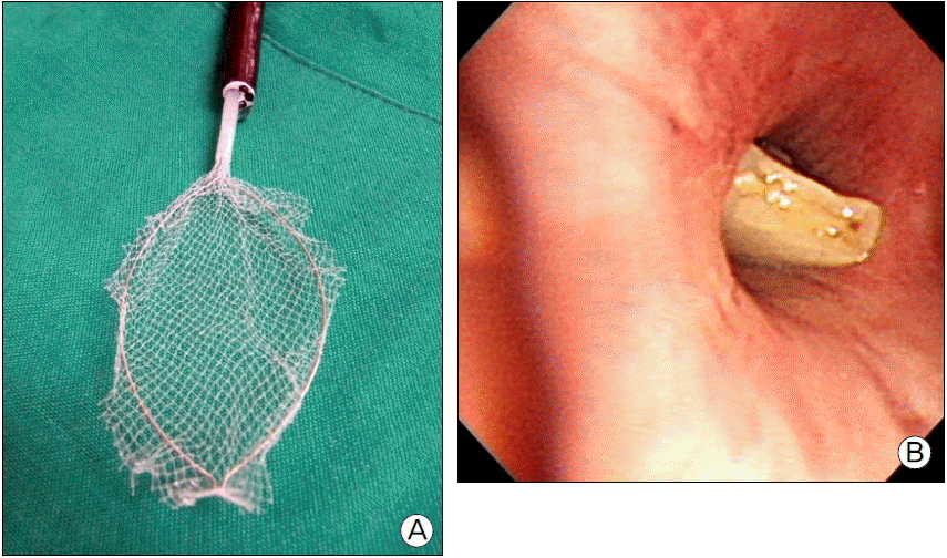
After a bronchoscope was placed into the airway through the lumen of artificial airway, the trachea and bilateral bronchi were examined. When an aspirated tooth was identified in the bronchus, a balloon catheter was inserted distal to the aspirated tooth, the balloon was then inflated. And the inflated balloon was pulled forward, moving the tooth to the right or left main bronchus or trachea. And then fishnet basket (Roth Net® foreign body retriever, US Endoscopy Inc., USA) was inserted through the working channel of bronchoscope (Fig. 1a). The fishnet basket was sheath diameter 2.5 mm, working length 230 mm and net size 3 × 6 cm. Because the sheath was larger than the 2.0 mm diameter working channel, it could not pass through the bronchoscope. It was therefore removed from the basket. The inserted basket covered the tooth and rotated clockwise to enclose the tooth. When the tooth is caught tightly under net, the basket was withdrawn through the working channel. When the enclosed tooth reached at the tip of bronchoscope, it was pulled together with the scope and basket as a unit to the tip of artificial airway. If the unit was small to pass the lumen of artificial airway, it was removed without further difficulty. If the unit was large, the cuff of artificial airway was deflated, and the unit and the artificial airway were pulled together out of the mouth. In this case, the artificial airway was quickly reinserted. The extraction procedure was completed after patient’s condition was assessed.
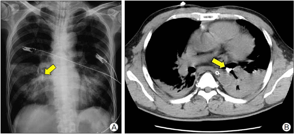
In Case 1, a 63-year-old male patient presented with dyspnea and diagnosed with acute respiratory failure induced by bacterial pneumonia. After undergoing endotracheal intubation, he was transferred to the ICU. The patient was taking medicine for Parkinson’s disease. His chest X-ray displayed tooth aspiration in the airway. Before the procedure, ventilator settings included volume control mode, FiO2 0.5, tidal volume 350 ml, respiratory rate 20/min, PEEP 5 cmH2O and then SpO2 was 100%. No complications identified during the procedure. Respiratory acidosis (pH 7.267) occurred 2 hours after the procedure but disappeared soon. No further complications such as arrhythmia, hypoxemia and pneumothorax occurred within 24 hours after the procedure. In Case 2, a 53-year-old man was transferred from another hospital after developing traumatic aortic dissection following a car accident. He already underwent tracheostomy after endotracheal intubation at the previous hospital. His chest computed tomography (CT) scan revealed a tooth (Fig. 2b). Before the procedure, ventilator settings included volume control mode, FiO2 0.5, inspiratory pressure 15 cmH2O, respiratory rate 20/min, PEEP 10 cmH2O and then SpO2 showed 94%. In the presence of patient-ventilator dyssynchrony during procedure, midazolam 5 mg and vecuronium 4 mg was injected. Hypoxemia occurred 4 hours after the procedure and increased FiO2 level to 1.0 for a while, but FiO2 dropped to 0.6 after tracheal suction. The patient remained stable until 24 hours after the procedure.
In Case 3, a 52-year-old man admitted with traumatic subdural hematoma, accompanied by fracture of the skull and dental injuries, after a fall. After endotracheal intubation, neurosurgery was performed and followed by tracheostomy in the ICU. His chest x-ray showed a tooth. Before the procedure, ventilator settings were volume control mode, FiO2 0.5, inspiratory pressure 12 cmH2O, respiratory rate 12/min, PEEP 5 cmH2O and then SpO2 was 100%. No complications were identified during the procedure and for 24 hours after the procedure. In Case 4, a 51-year-old man presented with traumatic epidural hematoma after a car crash. After endotracheal intubation and surgery, the patient was transferred to the ICU. Mechanical ventilation was not used before the procedure. The SpO2 was 98% with oxygen of 5 L/min. The aspirated tooth located on the left main bronchus was removed in 5 minutes after the procedure began. And no complications were identified.
In Case 5, a 54-year-old woman presented with traumatic epidural hematoma developed after she fell from the bed during hospitalization due to general weakness. After being admitted into the neurosurgery department, the patient received conservative treatment without surgery. A chest X-ray performed following endotracheal intubation showed a tooth (Fig. 2a). Mechanical ventilation was not used before the procedure. The SpO2 was 98% with oxygen of 5 L/min. A tooth impacted in the right lower lobe bronchus was identified during the flexible bronchoscopy (Fig. 1b). Several attempts failed to extract the tooth although the tooth was successfully withdrawn to the right main bronchus. Fishnet basket handling was not effective, causing the tooth to fall deep down into the right upper lobe bronchus. Endotracheal extubation was performed 3 days after the procedure. And the tooth was excreted in the feces. After endotracheal extubation, the tooth was likely coughed out of the segmental bronchus and traveled up the trachea before flowing through the esophagus and in the gastrointestinal tract. In Case 6, a 44-year-old woman admitted to the emergency room with seizure. She has alcoholism and continued loss of consciousness was accompanied by aspiration pneumonia. The patient was therefore moved to the ICU after endotracheal intubation. Before the procedure, ventilator settings included volume control mode, FiO2 of 0.4, tidal volume of 440 ml, respiratory rate of 18/min, PEEP of 5 cmH2O and then SpO2 showed 100%. Attempts to remove an aspirated tooth from the left lower lobe bronchus bronchoscopically were not successful, pushing the tooth further down into the distal portion (Table 1).
DISCUSSION
Foreign body aspiration commonly occurs in older adults. Its incidence is particularly high in residents of mental health facilities.[6] Risk factors for aspiration of foreign objects include the use of sedative and hypnotic drug, general anesthesia, trauma accompanied by consciousness loss, alcohol intoxication, mental retardation, Parkinson’s disease, and neurologic disorder accompanied by swallowing difficulty and seizure.[7,8] At least one of these risk factors was found in all patients in this article. Tracheobronchial aspiration of tooth can happen following the displacement of the tooth from its alveolar socket. And endotracheal intubation appears to affect the displacement directly or indirectly. That is, the tooth can be displaced by the laryngoscope blade during intubation, and the displaced tooth can be aspirated into the airway. A tooth can be also displaced by trauma and aspirated into the airway during intubation. All patients had a history of at least one endotracheal intubation.
Since the first rigid bronchoscope was developed by Killian in 1897, it has been used to remove endobronchial foreign bodies.[1] However it can be performed by only experienced pulmonologists.[1,9] Rigid bronchoscope is not widely used in Korea due to the lack of physician who can use it. Applications of flexible bronchoscope for removal of aspired foreign object began in 1970s. Cunanan used flexible bronchoscope for foreign body removal in 300 cases and reported 89% success.[10] Cunanan cited the following reasons for choosing flexible bronchoscope over rigid bronchoscope: First, the flexible bronchoscope is easier to introduce in special condition such as severely deformed cervical spine. Second, topical anesthesia was possible. Third, the flexible scope allows greater range of visualization, which makes manipulation of the instrument easy. These advantages of flexible bronchoscope support its use for foreign body removal in ICU patients with artificial airway.
Three steps are required to remove an aspirated tooth by flexible bronchoscopy. Once an aspirated tooth is dislodged, the tooth should be grasped securely to remove it.[1] When the aspirated tooth is removed by flexible bronchoscopy, it should be withdrawn out of the mouth not the nose.[11] That is because a tooth may not pass through the nasal cavity as it is narrower than the oral cavity. Once an aspirated tooth is identified, its shape and surrounding structure should be examined carefully. Once the tooth is captured using an instrument, all three (the tooth, instrument and bronchoscope) should be withdrawn together as a unit. If the unit is too large to pass through an artificial airway, the airway device should be withdrawn together as part of the unit.
Instruments used for endobronchial foreign body extraction include grasping forceps, fishnet baskets, Dormia baskets, prong snares, balloon catheters, magnet extractors and cryoprobes.[1,3] Grasping forceps are available in various shapes that look like the character W, rat-tooth or alligator jaw. Thus grasping forceps are suitable to remove an object like coin, pin or clip, [12,13] but not useful to remove a tooth. A prong snare has three to five prongs, and they are not effective in capturing a tooth. The magnet extractor is also useless for tooth removal. Cryoprobes are applicable but they require expensive equipment controlling coolant nitrous oxide. A balloon catheter can be used effectively to bring an aspirated tooth up to the desired position after being inflated distal to the tooth. In this article, a balloon catheter was used to withdraw an aspirated tooth to the main bronchus or trachea.
A fishnet basket can enclose an aspirated tooth with its net, facilitating the removal without dropping the tooth. However it is not easy to manipulate within in small airways because it’s net takes up a large space when being unfolded. An aspirated tooth should be therefore moved to a larger proximal airway near the trachea to use a fishnet basket. Easy manipulation of the basket would increase the chances of success as a result. Lodging the tooth on the right main bronchus however requires extra caution because a mishandled basket may push the tooth farther down into segmental bronchus of the right upper lobe. That is why the removal of aspirated tooth failed in Case 5. An aspirated tooth located in distal portion of the main bronchus is more prone to being pushed deep down into segmental bronchus because a small lumen of the bronchus makes manipulation of the basket very hard. That is why the removal of aspirated tooth failed in Case 6. When an aspirated tooth is impacted in segmental bronchus, a balloon catheter is carefully inserted between the tooth and bronchus. It should be placed distal portion of the tooth in the bronchus and inflated to dislodge the tooth. If the tooth was impacted tightly by pushing force of the fishnet basket, it would not be dislodged by a balloon catheter. Attempts to withdraw the aspirated tooth from the segmental bronchus were not successful in two cases. Moving the aspirated tooth into a more favorable position near the trachea is therefore a crucial factor when removing the tooth with a fishnet basket.
Dormia basket would be also useful for removing the aspirated tooth. Dormia baskets are widely used in the fields of gastroenterology and urology to remove bile duct stone or ureter stone.[3,14] Since tooth and stone have similar structures, a Dormia basket can be used to enclose a tooth. Also, its wing would be easier to manipulate than fishnet basket in small airways. However Dormia baskets also have a limitation because their wing is incapable of grasping an object securely. The baskets may drop an object when bumping onto surrounding structure, including vocal cord, during removal process.
I report that endobronchial tooth was removed successfully in ICU patients undergoing airway intervention with a flexible bronchoscope using fishnet basket. However, mishandled basket resulted in migration of aspirated tooth farther down the segmental bronchus, making it impossible to remove. Careful manipulation is therefore needed to use the fishnet basket.
 XML Download