Journal List > Korean J Physiol Pharmacol > v.18(2) > 1025932

Zhou, Sun, Yang, and Zhang: The Hepatotoxicity and Testicular Toxicity Induced by Arecoline in Mice and Protective Effects of Vitamins C and E

Abstract

Arecoline is a major alkaloid of areca nuts which are widely chewed by southeast Asian and it manifests various toxic effects in different organs of human and animals. In this work, mature mice were treated by vitamins C plus E, arecoline, or both daily for four weeks. The results showed that arecoline significantly increased the levels of serum alkaline phosphatase (ALP), glutamate oxaloacetate transaminase (GOT), glutamate pyruvate transaminase (GPT) and significantly decreased the levels of reduced glutathione (GSH), glutathione-S-transferase (GST), superoxide dismutase (SOD) and catalase (CAT) in the liver tissues. Additionally, the body weight, testis weight, sperm counts, motility and normal sperms also were significantly decreased. The supplement of vitamins C and E can bring the activities of ALP and GPT to normal levels and partially restore the sperm counts compared to the arecoline-treated group but have no other positive effects. In conclusion, the vitamins C and E partially attenuated the arecoline-induced hepatotoxiciy but basically had on protective effects against the arecoline-induced testicular toxicity.

ABBREVIATIONS

ALP

alkaline phosphatase

GOT

glutamate oxaloacetate transaminase

GPT

glutamate pyruvate transaminase

GSH

reduced glutathione

GST

glutathione-S-transferase

SOD

superoxide dismutase

CAT

catalase

ROS

reactive oxygen species

H&E staining

hematoxylin and eosin staining

TEM

transmission electron microscope

INTRODUCTION

Areca nuts (Areca catechu), the fourth most widely used addictive substance after tobacco, alcohol and caffeine [1], are mainly chewed by the people located in the southeast Asia [2]. Although some of therapeutic values of areca nuts chewing are reported [3,4], the toxicity induced by areca nuts chewing recently get more and more attention. Arecoline is a major alkaloid of areca nut and has been reported to be the main etilologic factor of oral, oropharyngeal cancers and oral submucosal fibrosis induced by areca nuts chewing [5,6,7]. Besides, arecoline induced cytotoxic, genotoxic and immunotoxic effects are also well documented [8,9,10]. Interestingly, arecoline is demonstrated to induce hepatotoxicity and testicular toxicity both by generating reactive oxygen species (ROS) [11,12]. Several reports have implicated that excessive generation of these free radicals could result in oxidant stress and disturb antioxidant defense [13,14].
Vitamins C and E are known to be antioxidants. Vitamin C is a water-soluble chain-breaking antioxidant and can scavenge superoxide and hydrogen peroxide [15,16]. Vitamin E (α-tocopherol) is a lipid-soluble vitamin with powerful biological antioxidant [17,18] and can effectively inhibit free radical formation and minimize lipid peroxidation in biological systems [19,20,21]. Moreover, vitamin C is found to have the ability to restore the antioxidant abilities of vitamin E [22]. Therefore, the combined use of vitamin C and vitamin E is believed to have interactive effects [23] and the co-treatment of vitamins C and E is also approved to be able to attenuate methyl parathion- and nickel-induced hepatotoxicity to some extent [24,25].
However, to the best knowledge we have, until now there are few papers covering the effects of vitamin C and vitamin E, alone or combined, on the toxicity induced by arecoline, especially on the hepatotoxicity and testicular toxicity. Therefore, the main aim of this paper is to confirm hepatotoxicity and testicular toxicity of arecoline, most importantly, assess the protective effects of co-treatment by vitamins C and E against hepatotoxicity and testicular toxicity induced by arecoline in mice.

METHODS

Animals and chemicals

Arecoline hydrobromide (98%) was purchased from Acros Organics (USA). Vitamins C, E and corn oil were obtained from the Sigma. Mature male ICR mice weighing 28~32 g were provided by Laboratory Animal Center of Sichuan University (Chengdu, China). All animal experiments were carried out according to the Guidelines for the Care and Use of Laboratory Animals published by the National Institutes of Health and approved by the Ethics Committee of Sichuan University.

Animal treatments

All the experimental mice were housed in plastic cages at a temperature- and humidity-controlled room with a well-regulated light:dark (12 h:12 h) schedule. Free access to a standard laboratory diet and water was offered to mice. The animals were divided into four groups (each group with 10 mice). Group I: mice were intraperitoneally injected with physiological saline as control; Group II: mice were orally administrated with vitamin C (10 mg/kg body weight) dissolved in physiological saline and vitamin E (100 mg/kg body weight) dissolved in the corn oil per day according to the previous study [26]; Group III: mice were intraperitoneally injected arecoline hydrobromide (10 mg/kg body weight) dissolved in physiological saline and each does was divided equally into half and was injected at 9 AM and 6 PM, respectively, according to pervious reports [27,28]; Group IV: mice were first orally administrated with the same does of vitamin C and vitamin E as Group II, 1 hour later, intraperitoneally injected with the same does of arecoline hydrobromide as Group III.

Collection of serum and measurement of body and organ weights

Blood samples were drawn from the supraorbital sinus of the etherized mice and serum was collected for subsequent biochemical analysis at the end of the 4th week. Body and testis from the four groups were weighted at the end of the 4th week.

Biochemical evaluation

Hepatic toxicity marker enzymes in the serum, alkaline phosphatase (ALP), glutamate oxaloacetate transaminase (GOT) and glutamate pyruvate transaminase (GPT), were assessed by commercial test kits (NanJing JianCheng Reserch Institute of Biology and Engineering, China). The activities of reduced glutathione (GSH), catalase (CAT), glutathione-S-transferase (GST) and superoxide dismutase (SOD) in the liver tissues were also assayed by commercial test kits (NanJing JianCheng Reserch Institute of Biology and Engineering, China).

Sperm analysis

Spermatozoa were collected by mincing the epididymides in 37℃ normal saline. Epididymal sperm counts and evaluation of the motility of epididymal sperms were performed using the methods of Zhang et al. [29] and the WHO manual for semen analysis [30]. The examination of sperm morphology was carried out by dropping a drop of sperm suspension on a slide. After air dried, stained with 1% eosin Y, washed and air dried again, the smears on the slides were observed under a light microscope (Olympus DP73, Japan).

Hematoxylin and eosin (H&E) staining

After the dissected liver and testis were fixed in 4% paraformaldehyde, the fixed samples were processed in a series of graded ethanol solutions and embedded in paraffin wax. Paraffin sections were cut at 5 µm height, deparaffinized, rehydrated and stained with hematoxylin and eosin for histological examination under a light microscope (Olympus DP73, Japan).

Transmission electron microscopy (TEM)

After the testicular tissues were fixed in 2.5% glutaraldehyde and 1% paraformaldehyde in a sodium phosphate buffer for 3 hours at 4℃. The fixed tissues were washed and post fixed in 1% osmiumtetroxide in the same buffer for 1 hour at 4℃. Tissues were subsequently dehydrated in a graded ethanol series and were embedded in araldite. Thin sections (1 µm) were cut and stained with 2% uranyl acetate and lead citrate. Finally, the sections were photographed on a Tecnai G2 F20S-TWIN transmission electron microscope (FEI, USA).

Statistical analysis

The data were analyzed using SPSS 17.0 and expressed as mean±SEM. The significance of differences was calculated using one-way analysis of variance (ANOVA) followed by Tamhane's T2 for multiple comparison. p<0.05 was considered statistically significant.

RESULTS

Changes of biochemical parameters in serum and liver tisssue

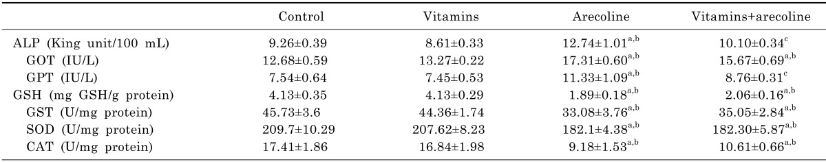
The activities of ALP, GOT, GPT in arecoline group mice showed significant increases compared to the control and vitamins group with all p<0.05, while the vitamins treatment in arecoline-exposed group efficiently brought down the ALP and GPT activities to normal levels and slightly decrease GOT levels but there no was statistically significant difference (Table 1).
Data presented in Table 1 also indicated that treatment with arecoline caused significant reduction in the GSH, GST, SOD, CAT levels of liver tissues compared to the control and vitamins group (p<0.05), and the supplied vitamins in arecoline-treated group had on positive effects on these changes caused by arecoline.

Pathological observation of liver

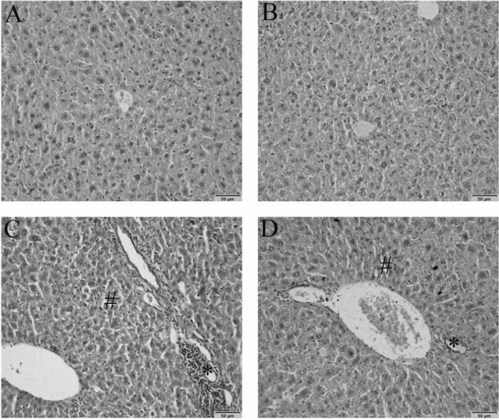
The observation detected by H&E staining showed that the livers of control and vitamins group presented normal features with normal hepatic lobule and normal hepatocytes (Fig. 1A and B). While in the arecoline group, hepatocyte damage was manifested by severe steatosis and inflammatory infiltrates (Fig. 1C). The vitamins treatment had a part prevention against the hepatocyte damage caused by arecoline, but the abnormal features of steatosis and inflammatory infiltrates were also observed in the vitamins plus arecoline group (Fig. 1D).

Evaluation of body and testis weights

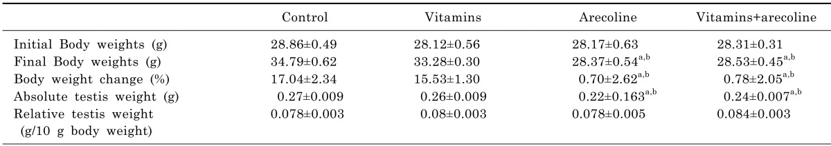
The statistical data in Table 2 showed that body weight and absolute testis weight significantly declined in the arecoline-treated group when compared to the control group and the vitamins group (p<0.05). The addition of vitamins in the arecoline-administrated group can't increase the body weight and absolute testis weight. However, there were no statistically significant differences observed in relative testis weight in the four groups.

Sperm parameter changes

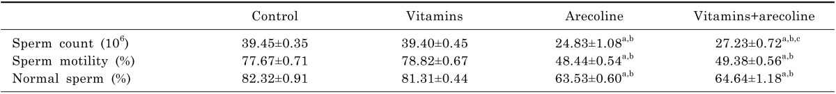
To further explore the protection of vitamins against the testicular toxicity induced by arecoline, the epididymal sperm count, motility and morphology were examined. The epididymal sperm count, sperm motility and normal sperms in the arecoline group were significantly reduced compared to the control and vitamins group (p<0.05) and the supplied vitamins in the arecoline-treated group didn't improve any parameters compared to the control and vitamins groups (Table 3). But the sperm count in the vitamins plus arecoline-treated group was markedly elevated compared to the arecoline-treated group (p<0.05).
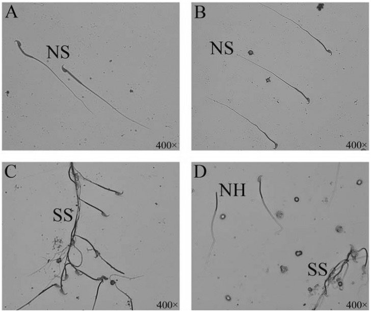
The sperm morphology were shown in Fig. 2, from which no head sperm, sticky sperm and sperms with coiled tail were observed in the arecoline- and vitamins combined with arecoline-treated groups.

Pathological observation of testis

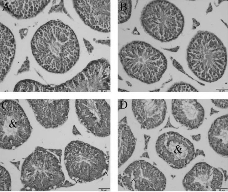
Microscopic observation of the testicular sections by H&E staining revealed that normal morphology and cellular arrangement of seminiferous tubules appeared in the control and vitamins groups (Fig. 3A and B), but in the testicular sections from the arecoline- and vitamins combined with arecoline-treated groups, large vacuoles and no spermatogenesis in the lumen of seminiferous tubules were observed (Fig. 3C and D).
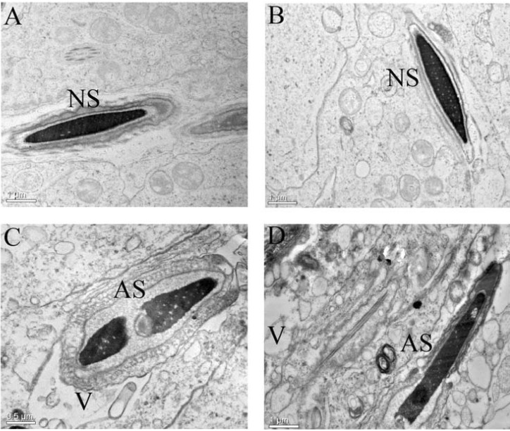
TEM observation further showed that no pathological changes were existed in the spermatogenic cells of mice in the control and vitamins groups (Fig. 4A and B). After arecoline exposure, morphological abnormal spermatozoa and vacuoles were detected in the exposed testis and the supplied vitamins in the arecoline-exposed seemed to have no abilities to alleviate these pathological changes (Fig. 4C and D).

DISCUSSION

Areca nut chewing is a popular oral habit in many parts of Asia for its all kinds of therapeutic effects. But recently diseases caused by the areca nut chewing are gained more and more attention, during which the hepatotoxicity and testicular toxicity induced by arecoline have been widely reported.
Dasgupta et al. [11] find that the arecoline treatment can result in heptatotoxicity by elevating the hepatotoxic marker enzymes (ALP, GPT and GOT) in serum. Consistent with these findings, our results also unravel the same changes. The increase in the serum enzymes is attributed to the damage of hepatocytes [24,31] and our observation of damaged liver tissues by H&E staining in this paper further supported the opinion. To find out the reasons underlying the arecoline-caused hepatocytes damage, we find that arecoline can depress the antioxidants in the liver tissue by decreasing the activities of SOD, CAT, GSH and GST. In line with our findings, arecoline also are proved to produce reactive oxygen species (ROS) in oral cancer, submucosal fibrosis and liver tissues [11,32]. Meanwhile, our test of protective effects of vitamins shows that vitamin C plus vitamin E treatment brings down the ALP and GPT activities and slightly attenuates the other biochemical parameters with no significant difference. Vitamins C and E are biologically required for many organism functions and both of them, when alone or combined use, exhibit reputable hepatoprotective effects in humans and animals due to their antioxidant properties [33,34,35]. So we speculate the partial protective effects against the hepatotoxicity conferred by vitamins also are attributed to their ability to scavenge free radicals produced by arecoline.
When referring to the testicular toxicity induced by arecoline, Wu et al. [12] indicate that the administration of areca nut extracts have no impact on the weights of body and testis in rats but can induce spermatogenic damage and affect sperm counts and sperm motility. In contraction with their indications, we detect dominant changes in the body and testis weights except in the relative testis weight. Along with their indications, the sperm toxicity tests also reveal that sperm count, motility and normal sperms are sharp reduction in the arecoline-treated mice. The decrease in the body weight may be caused by the decline of food intake and the reduction in the testicular weight is attributed to the necrotic changes in the testis [36,37]. Accordingly, we consider that the observed abnormal features in the testis tissues by H&E and TEM analysis are responsible for the decreased testicular weight.
The abnormal features in the testis and the aberrant sperm parameters are reported to result from the reactive oxygen species (ROS) [12]. However, the supplementation of vitamins C and E in this work can't recover the body and testis weights decreased by arecoline treatment and have no efficiently alleviating effects on the sperm toxicity. Although vitamins C and E are revealed to have the ability to ameliorate oxidative stress-related testicular impairments due to their antioxidantive activities [38,39,40], the administration of vitamins with no beneficial effects on the testicular toxicity is also reported [41]. Concerning the situation in the paper, the factors can be complicated. One of them may be that the reactive oxygen species induced by arecoline may be not the sole reason leading to the damage of testis.
In conclusion, the present work finds that the arecoline exposure could cause serious hepatotoxiciy and testicular toxicity and the co-treatment of vitamins C and E could partially improve the arecoline-induced hepatotoxiciy but basically have on protective effects against the arecoline-induced testicular toxicity. However, although the results in this work showed that vitamins had no protection against the testicular toxicity, the final determination about the effects of vitamins on the testicular toxicity induced by arecoline still can't be made, because the route of supplied vitamins, dose of vitamins, even the ratio of vitamin C and E all may involve the final protective effects of vitamins. So more research must be done before a reliable decision is made.

ACKNOWLEDGEMENTS

This study was supported by the Public Project of Chinese Agriculture Ministry (201203041) and the Support Program of Science and Technology of Sichuan in China (2012SZ0045).

ABBREVIATIONS

ALP

alkaline phosphatase

GOT

glutamate oxaloacetate transaminase

GPT

glutamate pyruvate transaminase

GSH

reduced glutathione

GST

glutathione-S-transferase

SOD

superoxide dismutase

CAT

catalase

ROS

reactive oxygen species

H&E staining

hematoxylin and eosin staining

TEM

transmission electron microscope

References

1. Boucher BJ, Mannan N. Metabolic effects of the consumption of Areca catechu. Addict Biol. 2002; 7:103–110. PMID: 11900629.
2. Gupta PC, Warnakulasuriya S. Global epidemiology of areca nut usage. Addict Biol. 2002; 7:77–83. PMID: 11900626.
crossref
3. Mondadori C, Hengerer B, Ducret T, Borkowski J. Delayed emergence of effects of memory-enhancing drugs: implications for the dynamics of long-term memory. Proc Natl Acad Sci U S A. 1994; 91:2041–2045. PMID: 8134347.
crossref
4. Sullivan RJ, Allen JS, Otto C, Tiobech J, Nero K. Effects of chewing betel nut (Areca catechu) on the symptoms of people with schizophrenia in Palau, Micronesia. Br J Psychiatry. 2000; 177:174–178. PMID: 11026959.
5. Stich HF, Stich W, Lam PP. Potentiation of genotoxicity by concurrent application of compounds found in betel quid: arecoline, eugenol, quercetin, chlorogenic acid and Mn2+. Mutat Res. 1981; 90:355–363. PMID: 7335107.
6. Deng YT, Chen HM, Cheng SJ, Chiang CP, Kuo MY. Arecoline-stimulated connective tissue growth factor production in human buccal mucosal fibroblasts: Modulation by curcumin. Oral Oncol. 2009; 45:e99–e105. PMID: 19457704.
crossref
7. Thangjam GS, Agarwal P, Balapure AK, Rao SG, Kondaiah P. Regulation of extracellular matrix genes by arecoline in primary gingival fibroblasts requires epithelial factors. J Periodontal Res. 2009; 44:736–743. PMID: 19438976.
crossref
8. Chou WW, Guh JY, Tsai JF, Hwang CC, Chen HC, Huang JS, Yang YL, Hung WC, Chuang LY. Arecoline-induced growth arrest and p21WAF1 expression are dependent on p53 in rat hepatocytes. Toxicology. 2008; 243:1–10. PMID: 17997002.
crossref
9. Kumpawat K, Deb S, Ray S, Chatterjee A. Genotoxic effect of raw betel-nut extract in relation to endogenous glutathione levels and its mechanism of action in mammalian cells. Mutat Res. 2003; 538:1–12. PMID: 12834749.
crossref
10. Panigrahi GB, Rao AR. Influence of caffeine on arecoline-induced SCE in mouse bone-marrow cells in vivo. Mutat Res. 1983; 122:347–353. PMID: 6656817.
11. Dasgupta R, Saha I, Pal S, Bhattacharyya A, Sa G, Nag TC, Das T, Maiti BR. Immunosuppression, hepatotoxicity and depression of antioxidant status by arecoline in albino mice. Toxicology. 2006; 227:94–104. PMID: 16945459.
crossref
12. Wu PF, Chiang TA, Chen MT, Lee CP, Chen PH, Ko AM, Yang KJ, Chang PY, Ke DS, Ko YC. A characterization of the antioxidant enzyme activity and reproductive toxicity in male rats following sub-chronic exposure to areca nut extracts. J Hazard Mater. 2010; 178:541–546. PMID: 20202746.
crossref
13. Lee SE, Song HJ, Park SY, Nam Y, Min CH, Lee do Y, Jeong JY, Ha HS, Kim HJ, Whang WK, Jeong JH, Kim IK, Kim HR, Min YS, Sohn UD. Effect of ECQ on Iodoacetamide-Induced Chronic Gastritis in Rats. Korean J Physiol Pharmacol. 2013; 17:469–477. PMID: 24227950.
crossref
14. Noorani AA, Kale MK. Pretreatment of albino rats with methanolic fruit extract of Randia Dumetorum (L.) protects against alcohol induced liver damage. Korean J Physiol Pharmacol. 2012; 16:125–130. PMID: 22563258.
15. Mongi S, Mahfoud M, Amel B, Kamel J, Abdelfattah el F. Protective effects of vitamin C against haematological and biochemical toxicity induced by deltamethrin in male Wistar rats. Ecotoxicol Environ Saf. 2011; 74:1765–1769. PMID: 21514672.
crossref
16. Wilson JX. The physiological role of dehydroascorbic acid. FEBS Lett. 2002; 527:5–9. PMID: 12220624.
crossref
17. John S, Kale M, Rathore N, Bhatnagar D. Protective effect of vitamin E in dimethoate and malathion induced oxidative stress in rat erythrocytes. J Nutr Biochem. 2001; 12:500–504. PMID: 11834209.
crossref
18. Cemek M, Buyukben A, Buyukokuroglu ME, Aymelek F, Tur L. Protective roles of vitamin E (a-tocopherol), selenium and vitamin E plus selenium in organophosphate toxicity in vivo: A comparative study. Pestic Biochem Physiol. 2010; 96:113–118.
19. Kalender S, Kalender Y, Ates A, Yel M, Olcay E, Candan S. Protective role of antioxidant vitamin E and catechin on idarubicin-induced cardiotoxicity in rats. Braz J Med Biol Res. 2002; 35:1379–1387. PMID: 12426639.
crossref
20. Kalender S, Kalender Y, Ogutcu A, Uzunhisarcikli M, Durak D, Açikgoz F. Endosulfan-induced cardiotoxicity and free radical metabolism in rats: the protective effect of vitamin E. Toxicology. 2004; 202:227–235. PMID: 15337585.
crossref
21. Kalender S, Ogutcu A, Uzunhisarcikli M, Açikgoz F, Durak D, Ulusoy Y, Kalender Y. Diazinon-induced hepatotoxicity and protective effect of vitamin E on some biochemical indices and ultrastructural changes. Toxicology. 2005; 211:197–206. PMID: 15925023.
crossref
22. Serbecic N, Beutelspacher SC. Anti-oxidative vitamins prevent lipid-peroxidation and apoptosis in corneal endothelial cells. Cell Tissue Res. 2005; 320:465–475. PMID: 15838641.
crossref
23. Stoyanovsky DA, Goldman R, Darrow RM, Organisciak DT, Kagan VE. Endogenous ascorbate regenerates vitamin E in the retina directly and in combination with exogenous dihydrolipoic acid. Curr Eye Res. 1995; 14:181–189. PMID: 7796601.
crossref
24. Uzunhisarcikli M, Kalender Y. Protective effects of vitamins C and E against hepatotoxicity induced by methyl parathion in rats. Ecotoxicol Environ Saf. 2011; 74:2112–2118. PMID: 21782244.
crossref
25. Dahdouh F, Kechrid Z, Djebar MR. Beneficial Effects of Vitamins (C+E) Supplementation against Nickel-induced Hepatotoxicity in Mice. Adv Biores. 2013; 4:67–76.
26. Acharya UR, Mishra M, Patro J, Panda MK. Effect of vitamins C and E on spermatogenesis in mice exposed to cadmium. Reprod Toxicol. 2008; 25:84–88. PMID: 18065194.
crossref
27. Pradhan SN, Maickel RS. Pharmacology in Medicine Principles and Practice. USA: SP Press International Inc.;1986. p. 144.
28. Saha I, Chatterjee A, Mondal A, Maiti BR, Chatterji U. Arecoline augments cellular proliferation in the prostate gland of male Wistar rats. Toxicol Appl Pharmacol. 2011; 255:160–168. PMID: 21741983.
crossref
29. Zhang H, Wang H, Ji YL, Ning H, Yu T, Zhang C, Zhang Y, Zhao XF, Wang Q, Liu P, Meng XH, Xu DX. Lactational fenvalerate exposure permanently impairs testicular development and spermatogenesis in mice. Toxicol Lett. 2009; 191:47–56. PMID: 19683566.
crossref
30. World Health Organization. WHO laboratory manual for the examination of human semen and sperm-cervical mucus interaction. 4th ed. Cambridge: Cambridge University Press;1999. p. 128.
31. Awad ME, Abdel-Rahman MS, Hassan SA. Acrylamide toxicity in isolated rat hepatocytes. Toxicol In Vitro. 1998; 12:699–704. PMID: 20654459.
crossref
32. Chang MC, Ho YS, Lee PH, Chan CP, Lee JJ, Hahn LJ, Wang YJ, Jeng JH. Areca nut extract and arecoline induced the cell cycle arrest but not apoptosis of cultured oral KB epithelial cells: association of glutathione, reactive oxygen species and mitochondrial membrane potential. Carcinogenesis. 2001; 22:1527–1535. PMID: 11532876.
crossref
33. Ebuehi OA, Ogedegbe RA, Ebuehi OM. Oral administration of vitamin C and vitamin E ameliorates lead-induced hepatotoxicity and oxidative stress in the rat brain. Nig Q J Hosp Med. 2012; 22:85–90. PMID: 23175903.
34. Prabu SM, Shagirtha K, Renugadevi J. Naringenin in combination with vitamins C and E potentially protects oxidative stress-mediated hepatic injury in cadmium-intoxicated rats. J Nutr Sci Vitaminol (Tokyo). 2011; 57:177–185. PMID: 21697638.
crossref
35. Adikwu E, Deo O. Hepatoprotective effect of vitamin C (ascorbic acid). Pharmacol Pharm. 2013; 4:84–92.
crossref
36. Joshi SC, Mathur R, Gulati N. Testicular toxicity of chlorpyrifos (an organophosphate pesticide) in albino rat. Toxicol Ind Health. 2007; 23:439–444. PMID: 18536496.
crossref
37. ElMazoudy RH, Attia AA, El-Shenawy NS. Protective role of propolis against reproductive toxicity of chlorpyrifos in male rats. Pestic Biochem Physiol. 2011; 101:175–181.
crossref
38. El-Missiry MA. Enhanced testicular antioxidant system by ascorbic acid in alloxan diabetic rats. Comp Biochem Physiol C Pharmacol Toxicol Endocrinol. 1999; 124:233–237. PMID: 10661714.
crossref
39. Marchlewicz M, Wiszniewska B, Gonet B, Baranowska-Bosiacka I, Safranow K, Kolasa A, Głabowski W, Kurzawa R, Jakubowska K, Rać ME. Increased lipid peroxidation and ascorbic acid utilization in testis and epididymis of rats chronically exposed to lead. Biometals. 2007; 20:13–19. PMID: 16699871.
crossref
40. Ghosh D, Das UB, Misro M. Protective role of alpha-tocopherol-succinate (provitamin-E) in cyclophosphamide induced testicular gametogenic and steroidogenic disorders: a correlative approach to oxidative stress. Free Radic Res. 2002; 36:1209–1218. PMID: 12592673.
41. Uzun FG, Kalender S, Durak D, Demir F, Kalender Y. Malathion-induced testicular toxicity in male rats and the protective effect of vitamins C and E. Food Chem Toxicol. 2009; 47:1903–1908. PMID: 19442699.
crossref
Fig. 1
The effects of vitamins, arecoline and vitamins plus arecoline treatment on the microstructures of liver tissues observed by H&E staining. (A) Control group with normal hepatic lobule and hepatocytes; (B) vitamins-treated group with normal hepatic lobule and hepatocytes; (C) arecoline-treated group with sever steatosis and inflammatory infiltrates; (D) Vitamins plus arecoine-treated group with sever steatosis and inflammatory infiltrates; #Steatosis; *Inflammatory infiltrates; The scale bar is 50 µm.
kjpp-18-143-g001
Fig. 2
The effects of vitamins, arecoline and vitamins plus arecoline treatment on sperm morphology. (A) Control group with normal sperm morphology; (B) vitamins-treated group with normal sperm morphology; (C) arecoline-treated group with sticky sperms; (D) Vitamins plus arecoine-treated group with sticky sperms and no head sperms. NS, normal sperm; SS, sticky sperm; NH, no head sperm. The magnification is 400×.
kjpp-18-143-g002
Fig. 3
The effects of vitamins, arecoline and vitamins plus arecoline treatment on microstructures of testicular tissues observed by H&E staining. (A) Control group with normal testicular microstructures; (B) vitamins-treated group with normal testicular microstructures; (C) arecoline-treated group with large vacuoles and no spermatogenesis in the lumen of seminiferous tubules; (D) Vitamins plus arecoine-treated group with with large vacuoles and no spermatogenesis in the lumen of seminiferous tubules; ^vacuoles; &no spermatogenesis in the lumen of seminiferous tubules; The scale bar is 50 µm.
kjpp-18-143-g003
Fig. 4
The effects of vitamins, arecoline and vitamins plus arecoline treatment on ultrastructures of testicular tissues observed by TEM. (A) Control group with normal testicular ultrastructures; (B) vitamins-treated group with normal testicular ultrastructures; (C) arecoline-treated group with large vacuoles and abnormal sperm; (D) Vitamins plus arecoine-treated group with large vacuoles and abnormal sperm; NS, normal sperm; AS, abnormal sperm; V, vacuoles; The scale bar in A, B and D is 1 µm, C is 0.5 µm.
kjpp-18-143-g004
Table 1
The effects of vitamins, arecoline and vitamins plus arecoline treatment on the biochemical parameters of the serum and liver tissue
kjpp-18-143-i001

Values are mean±SEM for six mice in each group. Significance at p<0.05. aComparison of control and other groups. bComparison of vitamins-treated group with arecoline- and vitamins plus arecoline-treated groups. cComparison of arecoline treated groups with arecoline plus arecoline-treated groups.

Table 2
The effects of vitamins, arecoline and vitamins plus arecoline treatment on body weight and testes weight
kjpp-18-143-i002

Values are mean±SEM for six mice in each group. Significance at p<0.05. aComparison of control and other groups. bComparison of vitamins-treated group with arecoline- and vitamins plus arecoline-treated groups. cComparison of arecoline treated groups with arecoline plus arecoline-treated groups.

Table 3
The effects of vitamins, arecoline and vitamins plus arecoline treatment on the sperm parameters
kjpp-18-143-i003

Values are mean±SEM for six mice in each group. Significance at p<0.05. aComparison of control and other groups. bComparison of vitamins-treated group with arecoline- and vitamins plus arecoline-treated groups. cComparison of arecoline treated groups with arecoline plus arecoline-treated groups.

TOOLS
Similar articles