Abstract
Objectives
We wanted to clarify the association between the position of the atlantoaxial fusion angle and the change of the subaxial cervical spine alignment (SCA) and the reduction loss after atlantoaxial fusion (AAF) using the posterior wiring technique (PWT), transarticular screw fixation (TAF) and posterior screw-rod fixation (PSR) for treating atlantoaxial instability (AAI).
Summary of the Literature Review
There are not many studies on the change of the SCA and the reduction loss after AAF.
Material and Methods
Thirty five patients underwent AAF for AAI from 1986 to 2008. The mean follow-up period was 59.5 months. The surgical techniques were divided into three groups, that is, PWT: 17 patients, TAS: 10 and PSR: 8. The causes of instability were transverse ligament rupture in 12 patients, rheumatoid arthritis in 11, Os odontoideum in 6 and nonunion of an odontoid fracture in 6. Plain radiographs were used to assess the atlanto-dental interval, the posterior arch-lamina angle, the change of the SCA and the time of fusion.
Results
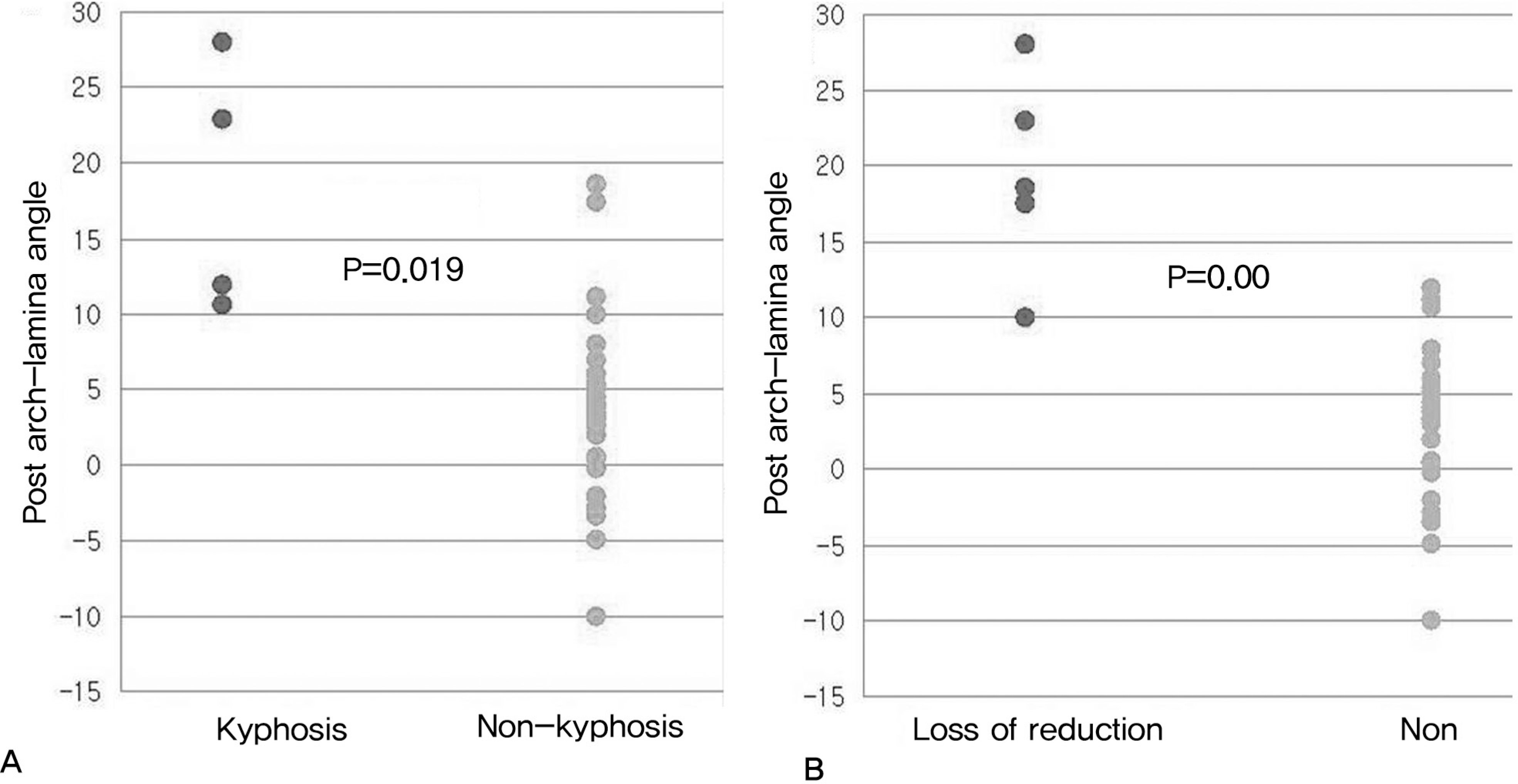
Fusion was achieved in all the patients within 3.5 months (range: 3-5 months). The radiologic findings in the 5 PWT patients showed a reduction loss and 3 patients showed subaxial cervical kyphosis (SCK). The TAS group had no reduction loss or SCK. The PSR group had no reduction loss and one patient showed SCK. A statistically significant reduction loss and SCK occurred in the group in which there was a posterior arch-laminar angle greater than 10 degrees before and after surgery.
REFERENCES
1.Song KW. Posterior Atlantoaxial Fusion of the Unstable Cervical Spine. J Korean Soc Spine Surg. 2000. 7:474–6.
3.Brooks AL., Jenkins EB. Atlanto-axial arthrodesis by the wedge compression method. J Bone Joint Surg Am. 1978. 60:279–84.

4.Margel F., Seeman PS., Gallen S. Stable posterior fusion of the atlas and axis by transarticular screw fixation. Cervical spine,. 1st ed.Berlin: Springer-Verlag;1987. p. 322–7.
5.Harms J., Melcher RP. Posterior C1-C2 Fusion With Polyaxial Screw and Rod Fixation. Spine. 2001. 26:2467–71.

6.Mechler RP., Puttlitz CM., Kleinstueck FS., Lotz JC., Harms J., Bradford DS. Biomechanical testing of posterior atlantoaxial fixationtechnique. Spine. 2002. 27:2435–40.
7.Mukai Y., Hosono N., Sakaura H, et al. Sagittal alignment of the subaxial cervical spine after C1-C2 transarticular screw fixation in rheumatoid arthritis. J Spinal Disord Tech. 2007. 20:436–41.

8.Chang H., Kim SK., Park JB., Chae JW., Lee SJ. Posterior Atlantoaxial Fusion with Posterior Wiring: Analysis of 19 Cases. J Kor Soc Spine Surg. 1997. 4:257–64.
9.Mukai Y., Hosono N., Sakaura H, et al. A retrospective radiographic analysis of subaxial sagittal alignment after posterior C1-2 fusion. Spine. 2004. 29:175–81.
10.Matsunnaga S., Onishi T., Sakou T. Significance of occipitoaxial angle in subaxial lesion after occipitocervical fusion. Spine. 2001. 26:161–5.
11.Nolan JP., Sherk HH. Biomechanical evaluation of the extensor musculature of the cervical spine. Spine. 1988. 13:9–11.

12.Iizuka H., Shimizu T., Tateno K, et al. Extensor musculature of the cervical spine after laminoplasty: morphologic evaluation by coronal view of the magnetic resonance image. Spine. 2001. 26:2220–6.
13.Yoshimoto H., Ito M., Abumi K, et al. A retrospective radiographic analysis of subaxial sagittal alignment after posterior C1-2 fusion. Spine. 2004. 29:175–81.
14.Dickman CA., Sonntag VKH. Surgical management of atlantoaxial nonunions. J Neurosurg. 1995. 83:248–53.

15.Crawford NR., Hurlbert RJ., Choi WG., Dickman CA. Differential biomechanical effects of injury and wiring at C1-C2. Spine. 1999. 24:1894–902.

16.Mimatsu K., Katoh F., Kawakami N., Nakagami W. Atlantoaxial fusion with posterior double wire fixation. Spine. 1992. 17:1409–13.

17.Matoshi S., Osamu K., Masanori I., Satoru S., Koki U., Ryouichi S. Atlantoaxial dislocation: A Follow-up study of surgical results. Spine. 1997. 22:759–63.
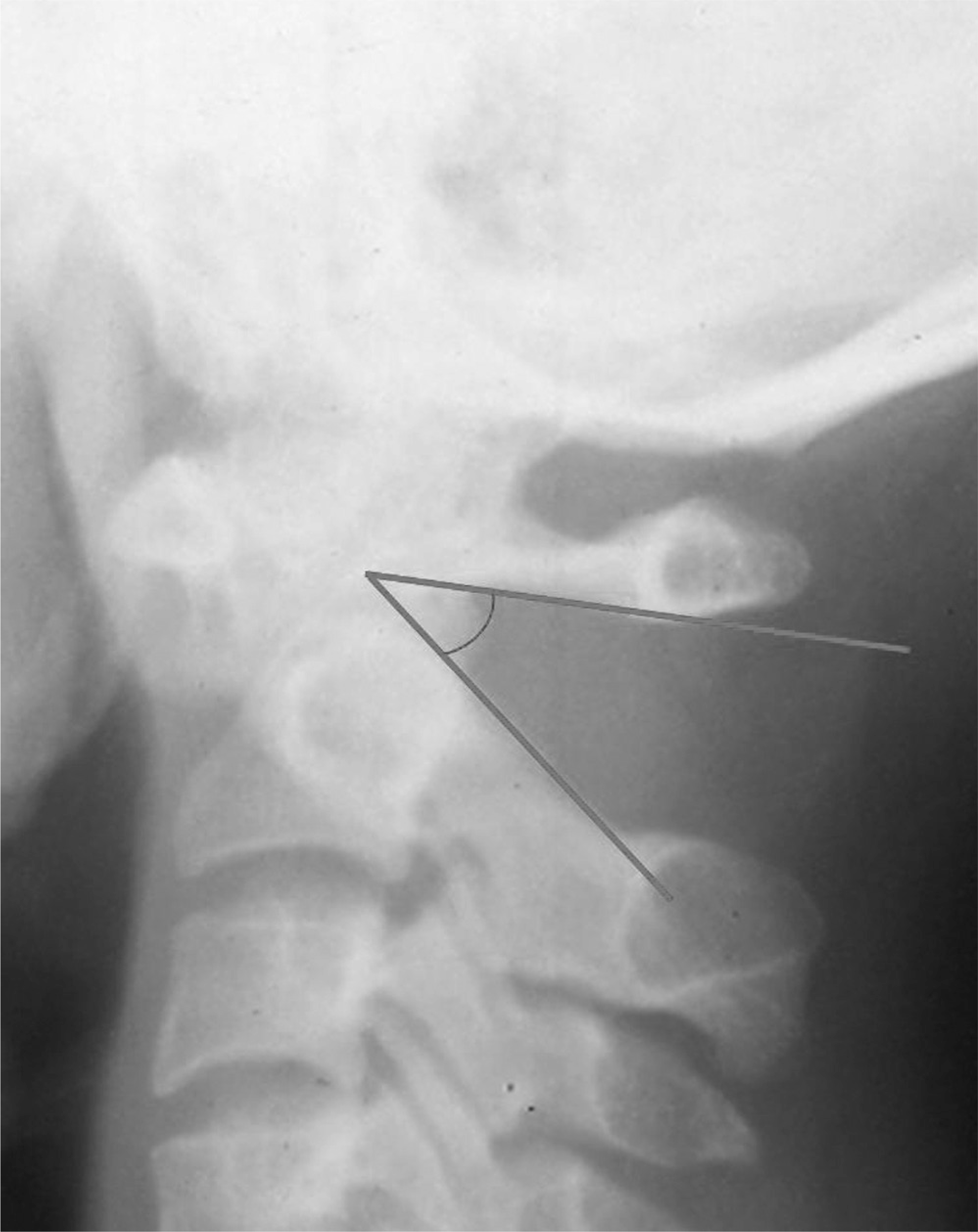
Fig 1.
Post arch-lamina angle is between inferior border point of anterior tubercle and posterior arch of atlas and upper border of lamina in axis.

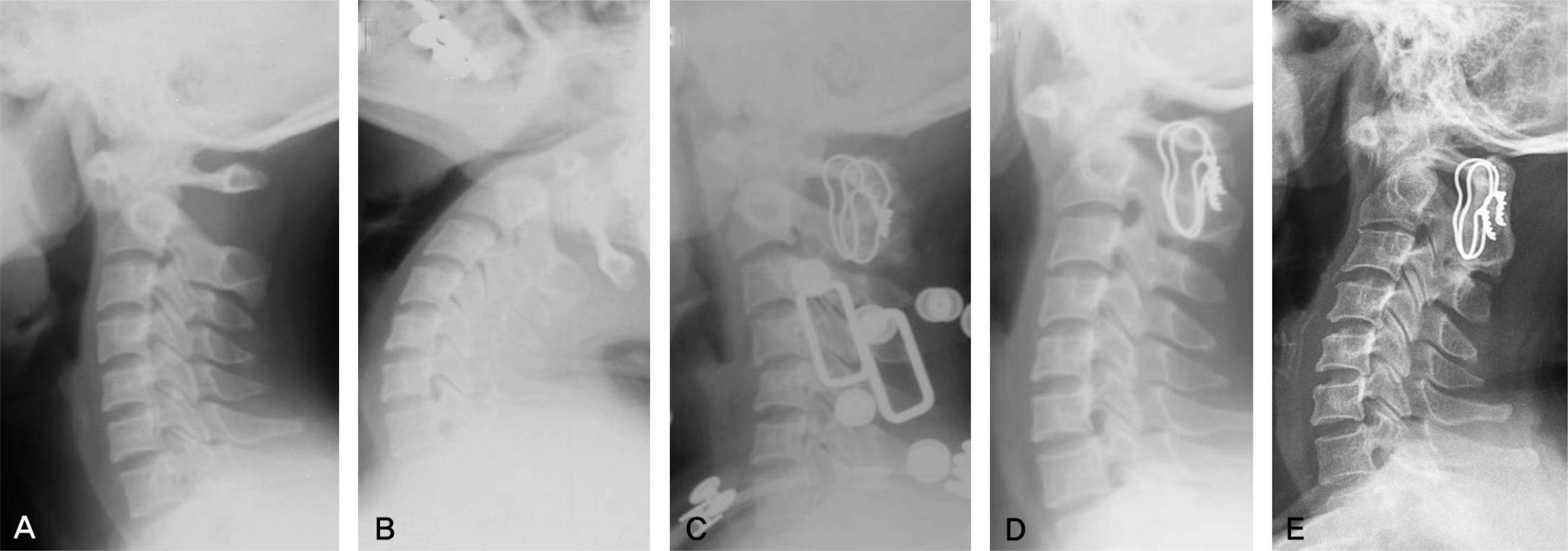
Fig 2.
Preoperative (A) flexion and (B) extension radiographs of a 31 year-old woman who underwent AAF using PWT for AAI caused by Os dontoideum.(ADI : 10 mm, posterior arch-lamina angle : 38°) (C) This film obtained immediately after surgery shows complete reduction (ADI :2mm, posterior arch-lamina angle : 15°) and subaxial kyphosis. (D) At postoperative 5 months, shows further loss of reduction (ADI : 8mm), AAF and alignment exchanged from kyphosis to lordosis. (E) Sixteen-year follow up radiograph shows that ADI (10mm) increase more than at postoperative 5 month after AAF. AAF: atlantoaxial fusion, ADI: atlanto-dental interval, PWT: posterior wiring technique, AAI: atlantoaxial instability

Fig 3.
52 year-old woman who underwent AAF using PSR for AAI caused by Rheumatoid arthritis. Preoperative (A) flexion, (B) extension. (C) Complete reduction was obtained but subaxial cervical kyphosis showed at 4 weeks postoperatively. (D) Subaxial cervical kyphosis maintained at 1 year after surgery. AAF: atlantoaxial fusion, PSR: posterior screw-rod fixation, AAI: atlantoaxial instability

Fig 4.
Correlation with pre-postoperative difference of posterior arch-lamina angle and (A) subaxial cervical kyphosis, (B) loss of reduction.

Table 1.
Radiological Results related to Procedure of Altantoaxial Fusion.
Table 2.
The Details of Reduction Loss(5 patients)
Table 3.
The Details of Subaxial Cervical Kyphosis(4 patients)


 PDF
PDF ePub
ePub Citation
 Print
Print


 XML Download