

 ePub
ePub Citation
 Print
Print


INTRODUCTION
Papillary thyroid carcinoma (PTC) is the predominant type of thyroid cancers and develops in the follicular cells of the thyroid. It is the most rapidly increasing cancer, probably because of the increased detection of small, low-risk PTC. The incidence of PTC rapidly increases in worldwide during the past 10 to 20 years. According to the data from the National Cancer Center in Korea (http://www.cancer.go.kr), among adolescents and adults between ages 15 years and 64 years, thyroid cancer is the first most common cancer, and PTCs are the most common of all thyroid cancers. Most PTC patients with thyroidectomy following the treatments of thyroid hormone suppression and radioactive iodine ablation have a good prognosis. However, PTC recurs in some patients and is the major one of endocrine cancer deaths [1,2]. To date, the tailored target therapies for the patients who fail to respond to the initial treatment paradigm and who have the progressive and refractory cancers are being investigated. Environmental factors such as radiation, hormones, diet, and smoking are risk factors of PTC, and genetic factor has also implicated as a risk factor for the development and progression of PTC [3-5].
The extracellular matrix (ECM) (i.e., thrombospondin 1 [THBS1, also known as TSP1] and fibronectin 1 [FN1]) plays a crucial role in the maintenance of cell and morphogenesis of tissue. ECM interacts with the cell adhesion molecules (CAMs) including integrins, CD36, CD44, and the immunoglobulin superfamily. CAMs provide a force of physical link between ECM and the cytoskeleton [6-9]. The ECM-CAM interactions also affect in the tumor microenvironments such as tumor cell adhesion, proliferation, migration, invasion, and metastasis [10-12]. Integrins are transmembrane proteins and compose of an alpha chain and a beta chain. Integrin, alpha 6 (ITGA6) is a member of ECM adhesion receptor, and interacts beta chains, making the heterodimeric complexes consisting of one alpha chain and one beta chain (α6β1 and α6β4) [9,13]. Both α6β1 and α6β4 may play critical roles in the progression of cancers and be involved in the initial formation of cancerous tumors. Abnormal expression of α6β4 in the suprabasal cell layers has been associated with an increased malignancy of squamous cell carcinomas [10-12]. Integrins participate in cellular signalings such as the mitogen-activated protein kinase (MAPK)/nuclear factor of kappa light polypeptide gene enhancer in B-cells (NFKB), phosphoinositide-3-kinase (PI3) kinase/v-akt murine thymoma viral oncogene homolog (AKT), and mothers against decapentaplegic homolog (SMAD) signalings [14-16]. In particular, α6β1 directly bind THBS1 in thyroid cancer cells and this complex then activates the MAPK signaling. These actions may remodel the ECM microenvironments and elicit tumor cell invasion into the lymph node and other tissues from the basement membrane, causing the progression of thyroid cancer [17]. Although integrins may be involved in PTC susceptibility, the genetic determinants have not yet been fully defined.
In this study, we explored the relationship between ITGA6 SNPs and PTC, and their clinicopathologic characteristics in Korean population.
METHODS
We enrolled 104 PTC patients (29 males and 75 females) and 318 control subjects (105 males and 213 females). PTC patients were selected among participants who visit at the Departments of Surgery and Otolaryngology-Head and Neck Surgery. Subjects with nodular hyperplasia, anaplastic carcinoma, follicular carcinoma, double primary of PTC and follicular carcinoma, and follicular variant of PTC were excluded. PTC was confirmed by pathologic examinations. Controls were recruited from healthy participants through a general health check-up program. Subjects with thyroid disease, cancers, and any severe diseases were excluded. Informed consent was obtained from all subjects. This study was conducted in accordance with the guidelines of the Helsinki Declaration. Patients were divided into subgroups in accordance to the size (<1 cm and ≥1 cm), number (unifocality and multifocality), location (one lobe and both lobes), extrathyroidal invasion (present and absent), lymph node metastasis (present and absent), and angiolymphatic invasion (present and absent).
For the selection of ITGA6 SNPs, we searched the promoter and coding regions of the ITGA6 gene in the SNP database of the National Center for Biotechnology Information (http://www.ncbi.nlm.nih.gov/SNP, BUILD 132). The SNPs with unknown heterozygosity or heterozygosity below 0.1 and unknown minor allele frequency (MAF) or MAF below 0.1 were excluded. Out of 14 promoter SNPs, there were 3 unknown heterozygosity, 2 heterozygosity below 0.1, 1 unknown MAF, and 7 MAF below 0.1. Among 12 missense SNPs, SNPs with unknown heterozygosity or heterozygosity below 0.1 were 9 and SNPs with unknown MAF were 2. Finally, one promoter SNP (rs2141698, -1687A/G) and one missense SNP (rs11895564, Ala380Thr) were selected.
Genomic DNA was extracted from peripheral blood using DNA Isolation Kit for Cells and Tissues (Roche Diagnostics Co., Indianapolis, IN, USA) and genotyping of each SNP was determined using direct sequencing (Macrogen Inc., Seoul, Korea). Polymerase chain reactions (PCRs) were conducted with the following primers: for rs2141698 (sense, 5'-GTTAACAGCTGGTGTGACATGG-3'; antisense, 5'-TTAGCCTTTTCCCTTCGTGTAA-3'; product size, 324 bp) and for rs11895564 (sense, 5'-GGAGCCCCACAGTATTTTGATA-3'; antisense, 5'-TAGTTTCTCCCATGTTGGTCAGG-3'; product size, 348 bp). PCR comprised 40 cycles at 94℃ for 30 seconds, 58℃ for 30 seconds, 72℃ for 30 seconds, and 1 cycle at 72℃ for 5 minutes for the final reaction. The PCR products were sequenced using the ABI PRISM 3730XL analyzer (Applied Biosystems, Foster City, CA, USA) and the sequencing data were analyzed by the SeqManII software (DNASTAR, Madison, WI, USA).
Hardy-Weinberg equilibrium (HWE) for each SNP in PTC patients and control subjects was estimated by the chi-square test. SNPStats (http://bioinfo.iconcologia.net/index.php?module=Snpstats), SNPAnalyzer Pro (Istech Inc., Goyang, Korea), and Helixtree (Golden Helix, Bozeman, MT, USA) were performed to obtain odds ratios (ORs), 95% confidence intervals (CIs), and P-values. Multiple logistic regression analysis and the Fisher's exact test were conducted using the following models: codominant1 (major allele homozygotes vs. heterozygotes), codominant2 (major allele homozygotes vs. minor allele homozygotes), dominant (major allele homozygotes vs. heterozygotes + minor allele homozygotes), recessive (major allele homozygotes + heterozygotes vs. minor allele homozygotes), and log-additive (major allele homozygotes vs. heterozygotes vs. minor allele homozygotes). Age and gender as covariates were adjusted to obtain statistical significance. Haploview ver. 4.2 (Daly Lab, Cambridge, MA, USA) was used to determine the linkage disequilibrium (LD) block and haplotypes between two SNPs. The data was also analyzed using SPSS ver. 18.0 (SPSS Inc., Chicago, IL, USA). The statistical significance level was set at P < 0.05.
RESULTS
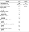
Table 1 shows the demographic and clinicopathologic features of PTC patients and control subjects. The age of PTC patients and control subjects was 53.2 ± 11.9 (mean ± SD) and 57.9 ± 12.1 years, respectively. In the subgroups of PTC, the number of PTC patients with cancer size ≥1 cm and <1 cm was 51 (50%) and 51 (50%), respectively. The number of PTC patients with unifocality and multifocality was 65 (64.4%) and 36 (35.6%), respectively. Out of PTC patients, 65 (65%) and 35 (35%) subjects displayed one lobe and both lobe, respectively, and 48 (47.5%) and 53 (52.5%) subjects displayed no extrathyroidal invasion and present extrathyroidal invasion, respectively. The number of PTC patients with cervical lymph node metastasis and without cervical lymph node metastasis was 65 (67%) and 32 (33%), respectively, and that with angiolymphatic invasion and without angiolymphatic invasion was 93 (93.9%) and 6 (6.1%), respectively. The total numbers of PTC patients among subgroups were different, because subjects with inappropriate and insufficient clinical data were excluded (Table 1).
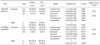
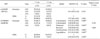
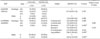
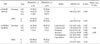
The genotype and allele distributions of the two examined SNPs are presented in Tables 2-5. Two SNPs (rs2141698, -1687A/G; rs11895564, Ala380Thr) of the ITGA6 gene were in HWE in the PTC and control groups, respectively (P > 0.05, data not shown). In Table 2, the genotype distribution of a missense SNP (rs11895564, Ala380Thr) was associated with the development of PTC (P = 0.018; OR, 2.01; 95% CI, 1.13 to 3.57 in codominant1 model; P = 0.011; OR, 2.11; 95% CI, 1.20 to 3.71 in dominant model; P = 0.024; OR, 1.97; 95% CI, 1.11 to 3.50 in overdominant model; P = 0.007; OR, 2.07; 95% CI, 1.23 to 3.51 in log-additive model). The A allele distribution of rs-11895564 was higher in the PTC group than in the control group (13.5% vs. 7.1%; P = 0.005; OR, 2.04; 95% CI, 1.24 to 3.37). The promoter SNP rs2141698 was not associated with the development of PTC in Korean population (Table 2). These results indicate that rs11895564 of the ITGA6 gene may be associated with the development of PTC, but rs2141698 not, and the A allele of rs11895564 might be a risk factor of PTC. All PTC patients were divided into subgroups according to the clinicopathologic characteristics. As shown in Table 3, the rs11895564 SNP was associated with the size of cancer (P = 0.028; OR, 0.34; 95% CI, 0.12 to 0.93 in codominant1 model; P = 0.016; OR, 0.31; 95% CI, 0.11 to 0.83 in dominant model; P = 0.040; OR, 0.36; 95% CI, 0.13 to 0.98 in overdominant model; P = 0.010; OR, 0.31; 95% CI, 0.12 to 0.81 in log-additive model). The A allele distribution of rs11895564 was lower in the size ≥1 cm group than in the size <1 cm group (6.9% vs. 19.6%; P = 0.010; OR, 0.30; 95% CI, 0.12 to 0.75). The results suggest that ITGA6 rs11895564 may contribute to the size of PTC. In Table 4, the rs11895564 SNP had significant differences between PTC patients with unifocality and PTC patients multifocality (P = 0.034; OR, 2.77; 95% CI, 1.08 to 7.11 in dominant model; P = 0.015; OR, 2.86; 95% CI, 1.20 to 6.81 in log-additive model). The A allele distribution of rs11895564 in the multifocality group (20.8%) was about 2.4-fold higher, compared to the unifocality group (8.5%). These data suggest that ITGA6 rs11895564 may be involved in the multifocality of PTC. In addition, rs11895564 was associated with the lymph node metastasis of PTC in only overdominant model (P = 0.029; OR, 0.27; 95% CI, 0.07 to 0.99) (Table 5). The rs2141698 and rs11895564 were not contributed to the location and extrathyroidal invasion of PTC, respectively (data not shown). We did not evaluate on the angiolymphatic invasion of PTC, because the number of patients with angiolymphatic invasion was little (n = 6). Haploview ver. 4.2 was used to estimate the LD block between rs2141698 and rs11895564. The LD block in the control group was not made (D' = 0.192). Therefore, the analysis of haplotype was not performed.
DISCUSSION
This study is first on the genetic relationships between ITGA6 SNPs and PTC in the Korean population. We analyzed the promoter SNP (rs2141698, -1687A/G) and missense SNP (rs11895564, Ala380Thr) in PTC patients and control subjects. We also investigated the association between two SNPs and the clinicopathologic characteristics of PTC. Our results revealed that a missense SNP (rs11895564) was associated with the development of PTC (P = 0.018 in codominant1, P = 0.011 in dominant, P = 0.024 in overdominant, and P = 0.007 in log-additive models; P = 0.005 in allele distributions). In recent, several researchers have reported the relationship between PTC and candidate genes including vascular endothelial growth factor A (VEGFA) [18], protein tyrosine phosphatase, receptor type, J (PTPRJ) [19], interleukin 6 (interferon, beta 2) (IL6) [20], IL10 [21], and collagen type XI α1 (COLIIAI) [22]. To our knowledge, there is no study that investigated for the association between ITGA6 and PTC. Only two studies were reported on the association between ITGA6 and cancers. Johnatty et al. [23] reported that an intron SNP (iSNP) rs13027811 of ITGA6 was related to ovarian cancer. The study by Cheng et al. [24] showed that the rs12621278 iSNP of ITGA6 was strongly associated with the progression of prostate cancer. Like above mentioned two literatures, our results showed that a missense rs11895564 SNP was associated with PTC. It is well-known that poor prognostic factors of PTC are thought to be large size, multifocality, multicentricity, extrathyroidal invasion, lymph node metastasis, age (>50 years), male, and high histologic grade [25-28]. When the maximum diameter of PTC is lesser 1 cm, papillary thyroid microcarcinoma (PTMC) is defined by the World Health Organization. PTMC generally appears good prognosis and therefore tend to have a less aggressive treatment, compared to conventional PTC. In contrast, some researchers have shown that the clinical feature of PTMC is similar pattern to PTC and they propose PTMC should be managed like the treatment of PTC [29,30]. In this study, the associations between a missense SNP (rs11895564) and the clinicopathologic characteristics of PTC were found. The rs11895564 SNP was associated with the size of cancer (<1 cm vs. ≥1 cm) (P = 0.028 in codominant1, P = 0.016 in dominant, P = 0.040 in overdominant, and P = 0.010 in log-additive models; P = 0.010 in allele distributions), the number of cancer (unifocality vs. multifocality) (P = 0.034 in dominant and P = 0.015 in log-additive models; P = 0.015 in allele distributions), and the lymph node metastasis (present vs. absent) (P = 0.029 in overdominant model). The A/G genotype and A allele distributions of rs11895564 in PTC with the cancer size ≥1 cm (A/G genotype, 13.7%; A allele, 6.9%) were about 2.3 and 2.8-fold lower than those in PTC with the cancer size <1 cm (A/G genotype, 31.4%; A allele, 19.6%), respectively. The rs11895564 SNP was also associated with the number of PTC. The A/G genotype and G allele distributions of rs11895564 in PTC with multifocality (A/G genotype, 30.6%; A allele, 20.8%) were about 1.8 and 2.4-fold higher, compared to PTC with unifocality (A/G genotype, 16.9%; A allele, 8.5%), respectively. The rs11895564 SNP was contributed to the lymph node metastasis of PTC. The A/G genotype and A allele distributions of rs11895564 in PTC with the lymph node metastasis (A/G genotype, 9.4%; A allele, 7.8%) were about 2.9 and 2.0-fold lower than those in PTC without the lymph node metastasis (A/G genotype, 27.7%; A allele, 15.4%), respectively. These results indicate that the A allele of rs11895564 has a protective effect to the size and lymph node metastasis of PTC, whereas that is a risk factor to the multifocality of PTC. Although we cannot exactly explain the difference effect of the A allele on each clinicopathologic characteristic of PTC, the change of amino acid in the rs11895564 SNP may independently related to each clinical phenotype of PTC. In recent studies, polymorphisms of certain genes, such as v-raf murine sarcoma viral oncogene homolog B1 (BRAF), cyclin-dependent kinase inhibitor 1B (CDKN1B), cholinergic receptor, nicotinic, alpha 3 (CHRNA3), and tumor necrosis factor (TNF-α) were associated with progression of cancers [31-35]. Chen et al. [34] reported that variant allele of rs1051730 in CHRNA3 gene was associated with larger tumor size at diagnosis of squamous cell carcinoma. They suggested that lung tumorigenesis may be driven not only by somatic mutations caused by tobacco carcinogens but also certain polymorphisms that may bring about physiological change. In present study, our results suggest one possibility that the SNP of ITGA6 may be a risk factor of PTC, and also contribute to the progression of PTC.
Our study has several limitations. Firstly, sample size is small. Secondly, controls did not examine thyroid ultrasonography for the detection of potential thyroid cancers. Thirdly, the follow-up study was not performed. Another limitation is that the multi-variate analysis and other risk factors including age, and gender were not conducted. For the exact correlation between IGTA6 and PTC, additional studies with larger sample size and based on the multivariate analysis could be needed.
In conclusion, our data suggest that a missense SNP (rs11895564, Ala380Thr) of the IGTA6 gene may be associated with the development of PTC and could be a useful marker for the cliniopathologic characteristics such as the size, number, and lymph node metastasis of PTC in Korean population.
 XML Download