

 PDF
PDF ePub
ePub Citation
 Print
Print


Min-Hye Kim1, Dong In Suh2 , Soo-Young Lee3, Yoon-Keun Kim4, Young-Joo Cho1, Sang-Heon Cho5
, Soo-Young Lee3, Yoon-Keun Kim4, Young-Joo Cho1, Sang-Heon Cho5
Abstract
Food allergy (FA) and atopic dermatitis (AD) are representative allergic diseases that begin early in life and result in considerable socioeconomic burden. While the pathophysiology and the optimal treatment modalities of these diseases are largely unknown, the role of microbes in health and disease are being highlighted. Recent advances in analyzing microbiome have enabled us to expand our research on impacts of the microbiome on the onset and course of FA and AD. Risk factors that are presumed to affect intestinal microbiome also modulate the onset of allergic diseases, which is more evident in AD than in FA. Considering animal studies, intestinal microbiota interacts with FA and the influence is bi-directional. The activation of regulatory T cell and the innate immune system is supposed to mediate the interaction. Regarding human studies, there exists the difference in the composition of microbiome between subjects with FA or AD and matched normal controls, which can further play as a predictive marker for later development of FA or AD. Probiotics are now investigated as a primary therapeutic agent or as an adjuvant tool for conventional therapies in preventing or modulating FA or AD. Currently, several reports on favorable outcomes become available, which should be replicated and backed up by large-scale studies with more detailed protocols.
Figures and Tables
Table 1
List of human microbiome studies on food allergy
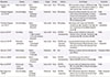
| Researcher | Disease | Subjects | Design | Sample | Method | Key findings | Comments |
|---|---|---|---|---|---|---|---|
| Apostolou et al. (2001)32 | Atopic dermatitis | Adults 8 (sensitive) 9 (control) | Case-control | Feces | FISH probing | LGG-consumption resulted in Bifidobacteria (▲) in healthy but not in milk-sensitive subjects, as well as general (▲) in bacterial numbers. | Profile, therapeutic effects |
| Thompson-Chagoyan et al. (2010)27 | Cows milk allergy | Infants 46 (allergic) 46 (control) | Clinical trial | Feces | FISH probing | Comparison of faecal samples from cows milk protein allergic infants (baseline/ 6 months) showed count and proportion of Lactobacilli (▲), counts and proportions of Enterobacteria (▼) and Bifidobacteria (▼). | Profile longitudinal |
| Thompson-Chagoyan et al. (2011)73 | Cows milk allergy | Infants 46 (allergic) 46 (control) | Case-control | Feces | FISH probing | Milk-allergic infant faeces had Clostridium cocoides group (▲), Atopobium cluster (▲), and sum of proportions of the different bacterial groups (▲). | Profile mechanisms |
| Ling et al. (2014)31 | Food allergy | Infants 17 (IgE-mediated) 17 (non-IgE-mediated) 45 (control) | Case-control | Feces | 16S rRNA pyrosequencing | Infants with IgE-mediated food allergy had Clotridium sensu stricto (▲), Anaerobacter (▲), and Bacteroides (▼), Clostridium XVIII (▼). | Profile |
| Azad et al. (2015)29 | Child cohort participants | Infants 12 (sensitized) 154 (control) | Case-control | Feces | 16S rRNA next generation sequencing | Low gut microbiota richness and an elevated Enterobacteriaceae to Bacteroidanceae ratio in early infancy are linked with subsequent food sensitization. | Profile longitudinal |
| Tang et al. (2015)33 | Peanut allergy | Children 31 (case) 31 (placebo) | Observative | N/A | N/A | Probiotics and peanut oral immunotherapy has good efficacy. | Therapeutic effects |
| Chen et al. (2016)74 | Food sensitization | Children 23 (sensitized), 22 (control) | Cohort | Feces | 16S rRNA pyrosequencing | In sensitized groups, the number of Bacteroidetes (▼) and that of Firmicutes (▲). | Profile |
| Hua et al. (2016)30 | American gut project participants | Adults 1,879 | Case-control | Feces | 16S rRNA sequencing | American adults with allergies have diversity (▼), Clostridiales (▼), and Bacteroidales (▲) in their gut microbiota. | Profile |
| Berni Canani et al. (2016)28 | Cows milk allergy | Infants 19 (allergic) 20 (control) | Clinical trial | Feces | 16S rRNA sequencing | EHCF+LGG promote tolerance in milk allergic infants, in part, by influencing the strain-level bacterial community structure. | Profile, therapeutic effects |
Table 2
List of human microbiome studies on atopic dermatitis
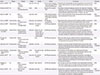
| Source | Disease | Subejcts | Design | Sample | Method | Key findings | Comments |
|---|---|---|---|---|---|---|---|
| Dekio et al. (2007)49 | AD | Adults 13 (case) 10 (control) | Case-control | Skin swab | Terminal restriction fragment length polymorphism (T-RFLP) analysis of 16S rRNA genes | Stenotrophomonas maltophilia (▲) was detected significantly more commonly in AD patients, whilst Dietzia maris was detected significantly more commonly in normal controls | Profile |
| Penders et al. (2007)55 | AD | Infants 957 (koala birth cohort) | Cohort | Feces | Real time-PCR for bacterial DNA | The presence of Escherichia coli (▲) was associated with a higher risk of developing eczema (OR, 1.87; 95% CI, 1.15–3.04). Infants colonised with Clostridium difficile (▲) were at higher risk of developing eczema (OR, 1.40; 95% CI, 1.02–1.91) and diagnosis of atopic dermatitis during the home visit (OR, 1.73; 95% CI, 1.08–2.78). | Profile longitudinal |
| Wang et al. (2008)75 | AD | Infants 15 (case) 20 (control) | Case-control | Feces | T-RFLP, Temporal temperature gradient gel electrophoresis (TTGE) analysis of 16S rRNA genes | There is a reduced diversity (▼) in the early fecal microbiota of infants with atopic eczema during the first 18 months of life. | Profile longitudinal |
| Fierer et al. (2008)40 | Normal skin flora | Adults 51 (healthy control) | Observative | Skin swab (hand) | 16S rRNA gene pyrosequencing | Propionibacteria (32%), Streptococcus (17%), Staphylococcus (8%), Corynebacterium (4%), Lactobacillus (3%) were the most abundant genera on palm surfaces. | Profile |
| Grice et al. (2009)39 | Normal skin flora | Adults 10 (healthy control) | Observative | Skin swab and scraping | 16S rRNA gene sequencing | Propionibacteria species and staphylococci species predominated in sebaceous sites. | Profile |
| Corynebacteria species predominated in moist sites, although staphylococci species were also represented. A mixed population of bacteria resided in dry sites, with a greater prevalence of β-Proteobacteria and Flavobacteriales. | |||||||
| Dominguez-Bello et al. (2010)76 | Normal skin flora | Mother & neonates 4 (vaginal delivery) 6 (cesarian section) | Observative | Mothers' skin, oral mucosa, vagina, neonates' skin, oral mucosa, nasopharyngeal aspirate | 16S rRNA gene sequencing | Vaginally delivered infants acquired bacterial communities resembling their own mother's vaginal microbiota, dominated by Lactobacillus, Prevotella, Sneathia spp., and C-section infants harbored bacterial communities similar to those found on the skin surface, dominated by Staphylococcus, Corynebacterium, Propionibacterium spp. | Profile |
| De Filippo et al. (2010)51 | Normal gut flora | Children 15 (from burkina faso, africa) 15 (from florence, italy) | Observative | Feces | 16S rRNA gene sequencing | Burkina Faso children showed a significant enrichment in Bacteroidetes (▲) and depletion in Firmicutes (▼). Also, Enterobacteriaceae (Shigella and Escherichia) (▼) were significantly underrepresented in BF than in EU children. | Profile |
| Capone et al. (2011)38 | Normal skin flora | Infants 31 (healthy infants) 5 (healthy mothers) | Observative | Skin swab | 16S rRNA gene sequencing | Composition of cutaneous microbial communities evolves over the first year of life, showing increasing diversity with age. Although early colonization is dominated by staphylococci, their significant decline contributes to increased population evenness by the end of the first year. | Profile longitudinal |
| Human Microbiome Project Consortium (2012)16 | Normal skin flora | Aldults 242 (human microbiome project cohort) | Cohort | Oral cavity, skin, stool, vagina | 16S rRNA gene sequencing | Skin communities were dominated by one of Staphylococcus (Firmicutes), Propionibacterium, or Corynebacterium (Actinobacteria) with a continuum of oral organisms (Streptococcus) appearing in nares communities. | Profile |
| Kong et al. (2012)46 | AD | Children 12 (case) 11 (placebo) | Case-control | Skin swab and scraping | 16S rRNA gene sequencing | In AD, the proportion of Staphylococcus sequences, particularly S. aureus (▲) was greater during disease flares than at baseline or post-treatment, and correlated with worsened disease severity. S. epidermidis (▲) also significantly increased during flares. | |
| Increases in Streptococcus, Propionibacterium, and Corynebacterium species were observed following therapy. | |||||||
| Gosalbes et al. (2013)37 | AD | Infants 20 (term newborns) 2 (infants) 7 (pregnant women) | Observative | Feces, meconium | 16S rRNA gene sequencing | One of the types was less diverse, dominated by enteric bacteria (▲) and associated with a history of atopic eczema in the mother, whereas the second type was dominated by lactic acid bacteria (▲) and associated with respiratory problems in the infant. | Profile |
| Seite et al. (2014)44 | AD | Children & adults 49 (case) | Observative | Skin swab | 16S rRNA gene sequencing | Overabundance of Staphylococcus species (▲) and a decrease in bacterial diversity were observed on affected skin. After 84-days of emollient treatment, increased overall diversity and a decrease in the Staphylococcus and Stenotrophomonas species were observed in treatment responders. | Profile longitudinal |
References
1. NIAID-Sponsored Expert Panel. Boyce JA, Assa'ad A, Burks AW, Jones SM, Sampson HA, et al. Guidelines for the diagnosis and management of food allergy in the United States: report of the NIAID-sponsored expert panel. J Allergy Clin Immunol. 2010; 126:6 Suppl. S1–S58.
2. Du Toit G, Santos A, Roberts G, Fox AT, Smith P, Lack G. The diagnosis of IgE-mediated food allergy in childhood. Pediatr Allergy Immunol. 2009; 20:309–319.

3. Thomas CL, Fernández-Peñas P. The microbiome and atopic eczema: More than skin deep. Australas J Dermatol. 2016; 01. 28. [Epub]. DOI: 10.1111/ajd.12435.
4. Rona RJ, Keil T, Summers C, Gislason D, Zuidmeer L, Sodergren E, et al. The prevalence of food allergy: a meta-analysis. J Allergy Clin Immunol. 2007; 120:638–646.
5. Hong SJ, Ahn KM, Lee SY, Kim KE. The prevalences of asthma and allergic diseases in Korean children. Korean J Pediatr. 2008; 51:343–350.
6. Lee JH, Han KD, Kim KM, Park YG, Lee JY, Park YM. Prevalence of atopic dermatitis in Korean children based on data from the 2008-2011 Korean National Health and Nutrition Examination Survey. Allergy Asthma Immunol Res. 2016; 8:79–83.
7. Gupta R, Holdford D, Bilaver L, Dyer A, Holl JL, Meltzer D. The economic impact of childhood food allergy in the United States. JAMA Pediatr. 2013; 167:1026–1031.
8. Kim C, Park KY, Ahn S, Kim DH, Li K, Kim DW, et al. Economic Impact of Atopic Dermatitis in Korean Patients. Ann Dermatol. 2015; 27:298–305.
9. Lack G. Update on risk factors for food allergy. J Allergy Clin Immunol. 2012; 129:1187–1197.
10. Pyun BY. Natural history and risk factors of atopic dermatitis in children. Allergy Asthma Immunol Res. 2015; 7:101–105.
11. Allen KJ, Koplin JJ. Prospects for Prevention of Food Allergy. J Allergy Clin Immunol Pract. 2016; 4:215–220.
12. Molloy J, Allen K, Collier F, Tang ML, Ward AC, Vuillermin P. The potential link between gut microbiota and IgE-mediated food allergy in early life. Int J Environ Res Public Health. 2013; 10:7235–7256.
13. Baker BS. The role of microorganisms in atopic dermatitis. Clin Exp Immunol. 2006; 144:1–9.
14. Palmer CN, Irvine AD, Terron-Kwiatkowski A, Zhao Y, Liao H, Lee SP, et al. Common loss-of-function variants of the epidermal barrier protein filaggrin are a major predisposing factor for atopic dermatitis. Nat Genet. 2006; 38:441–446.
15. Strachan DP. Hay fever, hygiene, and household size. BMJ. 1989; 299:1259–1260.
16. Human Microbiome Project Consortium. Structure, function and diversity of the healthy human microbiome. Nature. 2012; 486:207–214.
17. Kinross JM, Darzi AW, Nicholson JK. Gut microbiome-host interactions in health and disease. Genome Med. 2011; 3:14.
18. Choi S, Cho SH, Yi H. Human microbiome studies in Korea. Allergy Asthma Respir Dis. 2016; 4:311–320.
19. Kim BK, Rhee CK, Jung JY, Kang HR, Cho SH. Current status of microbiome research in asthma and chronic obstructive pulmonary disease. Allergy Asthma Respir Dis. 2016; 4:321–327.
20. NIH HMP Working Group. Peterson J, Garges S, Giovanni M, McInnes P, Wang L, et al. The NIH Human Microbiome Project. Genome Res. 2009; 19:2317–2323.
21. Noval Rivas M, Burton OT, Wise P, Zhang YQ, Hobson SA, Garcia Lloret M, et al. A microbiota signature associated with experimental food allergy promotes allergic sensitization and anaphylaxis. J Allergy Clin Immunol. 2013; 131:201–212.
22. Berni Canani R, Gilbert JA, Nagler CR. The role of the commensal microbiota in the regulation of tolerance to dietary allergens. Curr Opin Allergy Clin Immunol. 2015; 15:243–249.
23. Stefka AT, Feehley T, Tripathi P, Qiu J, McCoy K, Mazmanian SK, et al. Commensal bacteria protect against food allergen sensitization. Proc Natl Acad Sci U S A. 2014; 111:13145–13150.
24. Atarashi K, Tanoue T, Shima T, Imaoka A, Kuwahara T, Momose Y, et al. Induction of colonic regulatory T cells by indigenous Clostridium species. Science. 2011; 331:337–341.
25. Rodriguez B, Prioult G, Hacini-Rachinel F, Moine D, Bruttin A, Ngom-Bru C, et al. Infant gut microbiota is protective against cow's milk allergy in mice despite immature ileal T-cell response. FEMS Microbiol Ecol. 2012; 79:192–202.
26. Atarashi K, Tanoue T, Oshima K, Suda W, Nagano Y, Nishikawa H, et al. Treg induction by a rationally selected mixture of Clostridia strains from the human microbiota. Nature. 2013; 500:232–236.
27. Thompson-Chagoyan OC, Vieites JM, Maldonado J, Edwards C, Gil A. Changes in faecal microbiota of infants with cow's milk protein allergy--a Spanish prospective case-control 6-month follow-up study. Pediatr Allergy Immunol. 2010; 21(2 Pt 2):e394–e400.
28. Berni Canani R, Sangwan N, Stefka AT, Nocerino R, Paparo L, Aitoro R, et al. Lactobacillus rhamnosus GG-supplemented formula expands butyrate-producing bacterial strains in food allergic infants. ISME J. 2016; 10:742–750.
29. Azad MB, Konya T, Guttman DS, Field CJ, Sears MR, HayGlass KT, et al. Infant gut microbiota and food sensitization: associations in the first year of life. Clin Exp Allergy. 2015; 45:632–643.
30. Hua X, Goedert JJ, Pu A, Yu G, Shi J. Allergy associations with the adult fecal microbiota: Analysis of the American Gut Project. EBioMedicine. 2015; 3:172–179.
31. Ling Z, Li Z, Liu X, Cheng Y, Luo Y, Tong X, et al. Altered fecal microbiota composition associated with food allergy in infants. Appl Environ Microbiol. 2014; 80:2546–2554.
32. Apostolou E, Pelto L, Kirjavainen PV, Isolauri E, Salminen SJ, Gibson GR. Differences in the gut bacterial flora of healthy and milk-hypersensitive adults, as measured by fluorescence in situ hybridization. FEMS Immunol Med Microbiol. 2001; 30:217–221.
33. Tang ML, Ponsonby AL, Orsini F, Tey D, Robinson M, Su EL, et al. Administration of a probiotic with peanut oral immunotherapy: a randomized trial. J Allergy Clin Immunol. 2015; 135:737–744.e8.
34. Fredricks DN. Microbial ecology of human skin in health and disease. J Investig Dermatol Symp Proc. 2001; 6:167–169.
35. Mshvildadze M, Neu J, Shuster J, Theriaque D, Li N, Mai V. Intestinal microbial ecology in premature infants assessed with non-culture-based techniques. J Pediatr. 2010; 156:20–25.
36. DiGiulio DB, Romero R, Amogan HP, Kusanovic JP, Bik EM, Gotsch F, et al. Microbial prevalence, diversity and abundance in amniotic fluid during preterm labor: a molecular and culture-based investigation. PLoS One. 2008; 3:e3056.
37. Gosalbes MJ, Llop S, Vallès Y, Moya A, Ballester F, Francino MP. Meconium microbiota types dominated by lactic acid or enteric bacteria are differentially associated with maternal eczema and respiratory problems in infants. Clin Exp Allergy. 2013; 43:198–211.
38. Capone KA, Dowd SE, Stamatas GN, Nikolovski J. Diversity of the human skin microbiome early in life. J Invest Dermatol. 2011; 131:2026–2032.
39. Grice EA, Kong HH, Conlan S, Deming CB, Davis J, Young AC, et al. Topographical and temporal diversity of the human skin microbiome. Science. 2009; 324:1190–1192.
40. Fierer N, Hamady M, Lauber CL, Knight R. The influence of sex, handedness, and washing on the diversity of hand surface bacteria. Proc Natl Acad Sci U S A. 2008; 105:17994–17999.
41. Leyden JJ, Marples RR, Kligman AM. Staphylococcus aureus in the lesions of atopic dermatitis. Br J Dermatol. 1974; 90:525–530.
42. Ong PY, Ohtake T, Brandt C, Strickland I, Boguniewicz M, Ganz T, et al. Endogenous antimicrobial peptides and skin infections in atopic dermatitis. N Engl J Med. 2002; 347:1151–1160.
43. Gong JQ, Lin L, Lin T, Hao F, Zeng FQ, Bi ZG, et al. Skin colonization by Staphylococcus aureus in patients with eczema and atopic dermatitis and relevant combined topical therapy: a double-blind multicentre randomized controlled trial. Br J Dermatol. 2006; 155:680–687.
44. Seite S, Flores GE, Henley JB, Martin R, Zelenkova H, Aguilar L, et al. Microbiome of affected and unaffected skin of patients with atopic dermatitis before and after emollient treatment. J Drugs Dermatol. 2014; 13:1365–1372.
45. Huang JT, Abrams M, Tlougan B, Rademaker A, Paller AS. Treatment of Staphylococcus aureus colonization in atopic dermatitis decreases disease severity. Pediatrics. 2009; 123:e808–e814.
46. Kong HH, Oh J, Deming C, Conlan S, Grice EA, Beatson MA, et al. Temporal shifts in the skin microbiome associated with disease flares and treatment in children with atopic dermatitis. Genome Res. 2012; 22:850–859.
47. Seite S, Bieber T. Barrier function and microbiotic dysbiosis in atopic dermatitis. Clin Cosmet Investig Dermatol. 2015; 8:479–483.
48. Gallo RL, Nakatsuji T. Microbial symbiosis with the innate immune defense system of the skin. J Invest Dermatol. 2011; 131:1974–1980.
49. Dekio I, Sakamoto M, Hayashi H, Amagai M, Suematsu M, Benno Y. Characterization of skin microbiota in patients with atopic dermatitis and in normal subjects using 16S rRNA gene-based comprehensive analysis. J Med Microbiol. 2007; 56(Pt 12):1675–1683.
50. Ley RE, Peterson DA, Gordon JI. Ecological and evolutionary forces shaping microbial diversity in the human intestine. Cell. 2006; 124:837–848.
51. De Filippo C, Cavalieri D, Di Paola M, Ramazzotti M, Poullet JB, Massart S, et al. Impact of diet in shaping gut microbiota revealed by a comparative study in children from Europe and rural Africa. Proc Natl Acad Sci U S A. 2010; 107:14691–14696.
52. Watanabe S, Narisawa Y, Arase S, Okamatsu H, Ikenaga T, Tajiri Y, et al. Differences in fecal microflora between patients with atopic dermatitis and healthy control subjects. J Allergy Clin Immunol. 2003; 111:587–591.
53. Björkstén B, Sepp E, Julge K, Voor T, Mikelsaar M. Allergy development and the intestinal microflora during the first year of life. J Allergy Clin Immunol. 2001; 108:516–520.
54. Kalliomäki M, Kirjavainen P, Eerola E, Kero P, Salminen S, Isolauri E. Distinct patterns of neonatal gut microflora in infants in whom atopy was and was not developing. J Allergy Clin Immunol. 2001; 107:129–134.
55. Penders J, Thijs C, van den Brandt PA, Kummeling I, Snijders B, Stelma F, et al. Gut microbiota composition and development of atopic manifestations in infancy: the KOALA Birth Cohort Study. Gut. 2007; 56:661–667.
56. Ismail IH, Oppedisano F, Joseph SJ, Boyle RJ, Licciardi PV, Robins-Browne RM, et al. Reduced gut microbial diversity in early life is associated with later development of eczema but not atopy in high-risk infants. Pediatr Allergy Immunol. 2012; 23:674–681.
57. Abrahamsson TR, Jakobsson HE, Andersson AF, Björkstén B, Engstrand L, Jenmalm MC. Low diversity of the gut microbiota in infants with atopic eczema. J Allergy Clin Immunol. 2012; 129:434–440. 440.e1–440.e2.
58. Nylund L, Satokari R, Nikkilä J, Rajilić-Stojanović M, Kalliomäki M, Isolauri E, et al. Microarray analysis reveals marked intestinal microbiota aberrancy in infants having eczema compared to healthy children in at-risk for atopic disease. BMC Microbiol. 2013; 13:12.
59. Ouwehand AC, Isolauri E, He F, Hashimoto H, Benno Y, Salminen S. Differences in Bifidobacterium flora composition in allergic and healthy infants. J Allergy Clin Immunol. 2001; 108:144–145.
60. Sanders ME. Probiotics: definition, sources, selection, and uses. Clin Infect Dis. 2008; 46:Suppl 2. S58–S61.
61. Kalliomäki M, Salminen S, Arvilommi H, Kero P, Koskinen P, Isolauri E. Probiotics in primary prevention of atopic disease: a randomised placebo-controlled trial. Lancet. 2001; 357:1076–1079.
62. Kalliomäki M, Salminen S, Poussa T, Arvilommi H, Isolauri E. Probiotics and prevention of atopic disease: 4-year follow-up of a randomised placebo-controlled trial. Lancet. 2003; 361:1869–1871.
63. Isolauri E, Arvola T, Sütas Y, Moilanen E, Salminen S. Probiotics in the management of atopic eczema. Clin Exp Allergy. 2000; 30:1604–1610.
64. Wang IJ, Wang JY. Children with atopic dermatitis show clinical improvement after Lactobacillus exposure. Clin Exp Allergy. 2015; 45:779–787.
65. Drago L, De Vecchi E, Toscano M, Vassena C, Altomare G, Pigatto P. Treatment of atopic dermatitis eczema with a high concentration of Lactobacillus salivarius LS01 associated with an innovative gelling complex: a pilot study on adults. J Clin Gastroenterol. 2014; 48:Suppl 1. S47–S51.
66. Niccoli AA, Artesi AL, Candio F, Ceccarelli S, Cozzali R, Ferraro L, et al. Preliminary results on clinical effects of probiotic Lactobacillus salivarius LS01 in children affected by atopic dermatitis. J Clin Gastroenterol. 2014; 48:Suppl 1. S34–S36.
67. Boyle RJ, Bath-Hextall FJ, Leonardi-Bee J, Murrell DF, Tang ML. Probiotics for treating eczema. Cochrane Database Syst Rev. 2008; (4):CD006135.
68. Kim SO, Ah YM, Yu YM, Choi KH, Shin WG, Lee JY. Effects of probiotics for the treatment of atopic dermatitis: a meta-analysis of randomized controlled trials. Ann Allergy Asthma Immunol. 2014; 113:217–226.
69. Panduru M, Panduru NM, Sălăvăstru CM, Tiplica GS. Probiotics and primary prevention of atopic dermatitis: a meta-analysis of randomized controlled studies. J Eur Acad Dermatol Venereol. 2015; 29:232–242.
70. Gueniche A, Knaudt B, Schuck E, Volz T, Bastien P, Martin R, et al. Effects of nonpathogenic gram-negative bacterium Vitreoscilla filiformis lysate on atopic dermatitis: a prospective, randomized, double-blind, placebo-controlled clinical study. Br J Dermatol. 2008; 159:1357–1363.
71. Volz T, Skabytska Y, Guenova E, Chen KM, Frick JS, Kirschning CJ, et al. Nonpathogenic bacteria alleviating atopic dermatitis inflammation induce IL-10-producing dendritic cells and regulatory Tr1 cells. J Invest Dermatol. 2014; 134:96–104.
72. Mahe YF, Perez MJ, Tacheau C, Fanchon C, Martin R, Rousset F, et al. A new Vitreoscilla filiformis extract grown on spa water-enriched medium activates endogenous cutaneous antioxidant and antimicrobial defenses through a potential Toll-like receptor 2/protein kinase C, zeta transduction pathway. Clin Cosmet Investig Dermatol. 2013; 6:191–196.
73. Thompson-Chagoyan OC, Fallani M, Maldonado J, Vieites JM, Khanna S, Edwards C, et al. Faecal microbiota and short-chain fatty acid levels in faeces from infants with cow's milk protein allergy. Int Arch Allergy Immunol. 2011; 156:325–332.
74. Chen CC, Chen KJ, Kong MS, Chang HJ, Huang JL. Alterations in the gut microbiotas of children with food sensitization in early life. Pediatr Allergy Immunol. 2016; 27:254–262.
75. Wang M, Karlsson C, Olsson C, Adlerberth I, Wold AE, Strachan DP, et al. Reduced diversity in the early fecal microbiota of infants with atopic eczema. J Allergy Clin Immunol. 2008; 121:129–134.
76. Dominguez-Bello MG, Costello EK, Contreras M, Magris M, Hidalgo G, Fierer N, et al. Delivery mode shapes the acquisition and structure of the initial microbiota across multiple body habitats in newborns. Proc Natl Acad Sci U S A. 2010; 107:11971–11975.
 XML Download