

 PDF
PDF ePub
ePub Citation
 Print
Print


INTRODUCTION
Asthma is a chronic relapsing disease in which the airways in the lungs become constricted. The condition may be life-long, and the degree of obstruction changes over time. It is the commonest chronic lung disease and the UK has the highest prevalence world-wide: it affects 1 in 5 children and 1 in 12 adults. When I entered research in 1973, asthma was regarded as a disorder of "bronchospasm" treated symptomatically with unrestricted use of inhaled "bronchodilators". Since such treatment was linked to a dramatic increase in mortality linked to loss of bronchodilator responsiveness,1 there was an urgent need to understand more about its underlying mechanisms to provide a sound basis for its prevention and control.
Asthma is triggered by mast cells
Most asthma is associated with sensitisation of the airways to common allergens such as those from house dust mites. First we showed that, upon exposure to allergen, rapid airway narrowing (early response) was the result of IgE-dependant activation of airway mast cells with release of the rapidly acting granule-associated preformed mediators histamine and tryptase along with the newly generated products prostaglandin D2, cysteinyl leukotrienes (LTC4 and LTD4)2 and adenosine3 acting together to contract airway smooth muscle, promote vascular leakage and stimulate mucus secretion. Airway mast cells were also shown to be a key source of small proteins (cytokines and chemokines) associated with the allergic response such as interleukins (IL)-4, IL-5, IL-6 and TNFα4 which, in concert with the inflammatory mediators, stimulate the recruitment and activation of secondary effector cells, starting with neutrophils, followed by eosinophils and then T lymphocytes to cause late phase airway narrowing and airway hyperresponsiveness.5 Mast cell mediator release was also shown to be responsible for acute airway narrowing in response to other stimuli such as exercise, cold air, and fog but without a late phase inflammatory response.
Mechanisms of asthma chronicity and severity
While allergen and other forms of bronchoprovocation gave us insight into selected inflammatory events, there was a need to uncover the mechanisms of asthma persistence. To do this we used fibreoptic bronchoscopy, tissue biopsy and lavage of the airways. Even in its mildest form, asthma displayed all the features of a chronic inflammatory disorder involving persistent mast cell, eosinophil and T-lymphocyte activation facilitated through the phased expression of adhesion molecules on microvessels by ongoing inflammatory mediator and cytokine release.6 The beneficial anti-inflammatory effect of corticosteroids was shown to be through depletion of mast cells, eosinophils and T-cells by depriving them of cytokines and chemokines required for their recruitment, activation and survival.7 As we investigated more severe disease, aspirin intolerance became an increasingly prominent. This subtype was caused by generation of high levels of LTC4 from enhanced activity of the terminal enzyme in leukotriene synthesis LTC4 synthase both in airway mast cells and eosinophils8 and helped explain the selective efficacy of leukotriene modifiers in this form of the disease. Other forms of severe asthma were characterised by high levels of IgE production. We showed that a chimeric IgG monoclonal antibody (mAb) directed to that part of the IgE molecule which bound with high affinity to the mast cell receptor (FcεR1) could effectively clear IgE by forming small immune complexes without triggering the allergic response.9 This laid the ground for the development of a humanised anti-IgE mAb, omalizumab, now used worldwide as the first biologic for the treatment of severe asthma. However, with a significant proportion of asthmatic patients experiencing incomplete or no benefit from allergen reduction strategies or anti-inflammatory treatment, it was becoming increasingly apparent that the disease was more than allergic airway inflammation.
Asthma as an epithelial disease
In 1989 we were the first to draw attention to the remodelling of the subepithelial basement membrane (BM) unique to asthma, and to suggest that this was the consequence of sustained epithelial injury, loss of cell attachment and activation of subepithelial myofibroblasts to secrete repair-type collagens.10 Using selective markers of epithelial injury (epidermal growth factor receptor, polyADP-ribose-polymerase p85 cleavage peptide), cell cycling (P21waf) and repair (Ki67) applied to airway biopsies and in vitro primary epithelial cell cultures, we discovered that the asthmatic epithelium in children and adults adopted features of a chronic epithelial wound in being more susceptible to injury and failing to repair adequately (summarized in ref. 11). Additional evidence for this has been our demonstration that the integrity of tight junctions, which control epithelial permeability and confer epithelial columnar cell stability and survival, is severely compromised in asthma. This barrier defect persists after passage in tissue culture indicative of an inherent defect independent of airway inflammation.12 Such a defect in physical barrier facilitates breaching of the epithelium by proinflammatory environmental insults such as allergens, microorganisms and pollutants to enhance local immune activation and inflammation. Thus, in contrast to a normal epithelium that heals by "primary intention" with no scar formation, that of asthmatics is not only more susceptible to oxidant injury, but also responds in healing by "secondary intention" with excess secretion of growth factors to drive mucous metaplasia, fibrosis, angiogenesis and increased smooth muscle that characteristic of remodelling. To capture this concept, we propose that there is persistent activation of the EMTU involved in fetal branching morphogenesis.11 This novel approach to asthma connects the early life origins of the disease with the emergence of different endotypes, responses to treatment and natural history over the lifecourse.
A key question that these studies raised is whether the epithelial "set point" for to environmental injury and repair in asthma is altered by activation of fundamental transcription factors such as SAM pointed domain-containing Ets transcription factor (spdef) and thyroid transcription factor 1 (TTF-1) involved in the regulation of goblet cell metaplasia and lung morphogenesis. In collaboration with Jeffrey Whittset at the Children's Hospital in Cincinnati, we showed that TTF-1 was persistently suppressed in airway asthmatic epithelial cells which, in conditional TTF-1 deficient mice, translated into goblet cell induction and enhanced mucus secretion. In contrast, mucous metaplasia induced by aeroallergen was inhibited by epithelial over-expression of TTF-1.13 Application of transcriptomics to the airways of antigen sensitised and challenged TTF-1 over-expressing mice not only showed inhibition of genes controlling mucus production (e.g. spdef, calcium-activated chloride channel regulator 1 and 3, mucin-5AC), but also those involved in airway remodelling (e.g. trefoil factor, metalloprotease 12) and T cell-regulated allergic-type inflammation (e.g. IL-4, IL-13, chemokine (C-C motif) ligand 17). Ingenuity® pathway analysis of the differentially expressed genes revealed that TTF-1 was an integral part of a gene network that controls mucous cell metaplasia, airway remodelling and allergic-type inflammation and has provided further support for the critical role played by the airway epithelium in orchestrating the multiple cellular events of asthma.
Remodelling of the airways characteristic of chronic asthma has been largely considered to be secondary to inflammation and yet anti-inflammatory drugs have only a partial beneficial effect. A further possibility is that distortion of a chronically damaged epithelium by repeated bronchoconstriction could drive remodelling as initially, a protective response. When early bronchoconstriction was provoked by inhaled allergen or methacholine in asthma, both produced similar increases in biomarkers of remodelling - thickening of the BM with collagen III deposition, epithelium production of transforming growth factor-β and mucous metaplasia despite only the former challenge triggering an inflammatory response of eosinophil influx and accompanying late phase airway narrowing. Prevention of methacholine-induced bronchoconstriction by prior administration of the β2-agonist salbutamol prior to a methacholine challenge completely inhibited all three indices of remodelling.14 Thus, repeated airway narrowing is a sufficient stimulus for inducing reactive airway wall remodelling and that, in addition to controlling airway inflammation to prevent remodelling in the management of chronic asthma, prevention of repeated bronchoconstriction should also be a therapeutic objective.
In a search for factors that could further contribute to the enhanced activity of the EMTU in asthma, we uncovered A Disintegrin Metalloprotease 33 (ADAM33) on chromosome 20p13 by positional cloning.15 This was the first novel asthma susceptibility gene to be described and has been replicated in over 33 different population samples worldwide. ADAM33 encodes a multifunctional 120kD protein which is selectively expressed in airway fibroblasts, myofibroblasts and smooth muscle. Polymorphism of ADAM33 is associated with reduced lung function in childhood, airway hyper-responsiveness and accelerated decline in lung function over time, all features of chronic asthma. Our discovery of a soluble 55kD fragment of ADAM33 with enzymatic properties capable of driving new blood vessel formation and myogenesis has provided a further mechanistic link between airway morphogenesis and remodelling in this disease.
Mechanisms and treatment of asthma exacerbations
An important component of asthma, accounting for most of the disease morbidity and mortality, is the exacerbation in which deterioration in disease control occurs over several days. After establishing PCR-based detection methods for human rhinoviruses (RV), we used longitudinal cohorts to first establish the importance of respiratory viruses in causing asthma exacerbations both in children (>85%)16 and in adults (>65%)17 with RV major and minor subtypes dominating. Controlled nasal infection of asthmatics with RV16 revealed a mixed eosinophilic and lymphocyte (CD4+ and CD8+) airways inflammation of the lower airways in association with increased bronchial hyperresponsiveness which returned to baseline during convalescence.18 It was then important to understand why asthmatic airways were so susceptible to, what are usually regarded, as innocuous common cold viruses. After first showing that RV preferential infected the airway epithelium in asthma, we used primary epithelial cell monolayer cultures infected with major subtype RV16 subtype to pursue mechanisms. While bronchial epithelial cells (BECs) from normal subjects could effectively inhibit viral replication and eliminate remaining virus through activation of programmed cell death (apoptosis), BECs derived from asthmatic airways not only enabled the virus to survive but also to replicate eventually resulting in cytotoxic cell death, inflammatory mediator release and new virus shedding. This asthma-related epithelial defect was the result of impaired induction of interferon (IFN)-β.19 In monolayer BEC cultures, the asthmatic defect in clearing virus could be corrected by applying a very low dose of exogenous IFNβ and was interferon regulatory factor (IRF)-3-dependant. Thus the asthma "lesion" appeared to be in impaired signaling of viral RNA via microsomal toll-like receptor 3 to the initial induction of endogenous primary IFNs (step 1), leaving step 2 (the IRF-7-dependant induction of the anti-viral response via autacoid activation of the common IFNα/β receptor) intact. In a separate study transcriptomic and pathway analyses of circulating mononuclear cells before, during and after asthma exacerbations there occurs a distinct and systemic innate Type1 IFN response, but weaker B-cell antigen receptor and IL-4 adaptive immune response as compensation for the defect in mucosal innate immunity.20
In translating this to the clinic, a small amount of inhaled IFNβ would create a novel therapeutic approach to manage severe asthma exacerbations by restoring antiviral innate immunity to the epithelium. After first showing that inhaled rhIFN-β1a activates the airway antiviral pathways in asthma, we have now completed a successful Phase 2 clinical trial showing that inhaled rhIFN-β1a given at the start of a common cold was highly efficacious at blocking virus-related exacerbations in severe asthma in parallel to reducing viral load without any treatment-related side effects.
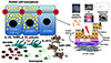
A new paradigm for asthma pathogenesis
Taken together, this research has created a new paradigm for asthma that places the airway epithelium at the center of disease pathogenesis (Figure).21 In orchestrating how the airways respond to a variety of environmental exposures, the epithelium and underlying mesenchyme are well positioned to control not only the inflammatory response, but also structural changes in the airways associated with different asthma phenotypes, treatment responses and disease evolution over the lifecourse. These discoveries provide a strong basis for creating novel therapies that are more directed towards increasing the lung's resistance to the inhaled environment rather than concentrating all efforts on trying to suppress inflammation once it is established.
 XML Download