

 PDF
PDF ePub
ePub Citation
 Print
Print


INTRODUCTION
Cockroach (CR) is an important source of inhalant allergens, and sensitization to CR is associated with asthma exacerbation.1 Moreover, epidemiologic data show a close relationship between CR sensitization and the prevalence of allergic asthma.2 Standardization of allergen extracts is important for diagnosis and immunotherapy.3 The German CR Blattella germanica is the most commonly found CR in Korean homes.4,5 However, CR extracts have not been standardized. The levels of protein and major allergens (Bla g 1 and Bla g 2) vary in the CR extracts that are commercially available in the USA.6 The concentration of CR extract supplied by the manufacturers is usually expressed in weight to volume (w/v) units. A designation of 1:10 w/v indicates that the solution contains the extractable material from 1 g of raw material added to 10 mL of buffer solution. The biological potencies of commercial German CR extracts have been estimated at 10-8570 bioequivalent allergy units (BAU)/mL in the USA.6,7 Moreover, protease activity is known to play an important role in the pathogenesis of CR allergy.8 CR extracts contain various proteases, which can degrade proteins, including allergens, in the extracts.9 Therefore, it is desirable to produce CR extracts that retain considerable protease activity without any significant degradation of the IgE-reactive components.
In the present study, we used a standardized method to produce extracts of German CRs, which were reared at the Korea National Arthropods of Medical Importance Resource Bank, Yonsei University College of Medicine, Seoul, Korea. The concentrations of the major allergens (Bla g 1 and Bla g 2) in the Korean extracts were compared with those of an extract obtained from a US company, i.e., the Hollister-Stier (HS) extract. The allergenic activities of these extracts were compared in an in vitro inhibition analysis.
MATERIALS AND METHODS
Allergen extraction
Lyophilized CR (20 g) was pulverized using a mortar and pestle. The powdered CR (10 g) was defatted with ethyl ether (1:5, w/v). The allergen was extracted for 48 hours at 4℃ in phosphate-buffered saline (PBS; pH 7.4) that contained 0.2% phenol. The extract was centrifuged at 13,000 ×g for 15 minutes at 4℃, and the supernatant was dialyzed (cut-off, 3,500 Da; Spectrum, Houston, TX, USA) extensively against distilled water. The dialyzed sample was filtered (0.22-µm pore; Millipore, Bedford, MA, USA) and lyophilized once again. The protein concentration was determined by the Bradford assay (Bio-Rad, Hercules, CA, USA) after reconstitution in buffers. Thereafter, the extract was aliquoted, lyophilized, and stored at -80℃ until use.
Protein analyses
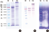
The protein profiles of the CR extracts were examined by SDS-PAGE. Samples (10 µg), which were reconstituted in PBS (pH 7.4) that contained 50% glycerol and 0.03% human serum albumin (HSA), were run on 12.5% gels under reducing conditions. Proteins were visualized by staining with Coomassie Brilliant Blue R250 or transferred onto nitrocellulose membrane (Amersham, Buckinghamshire, UK). The membranes were incubated with 1:4 dilutions of sera (pooled serum from five patients or five healthy controls) after blocking with 3% skim milk in TBST (50 mM Tris [pH 7.5], 0.05% Tween-20). Subsequently, IgE-reactive proteins were probed with alkaline phosphatase-conjugated goat anti-human IgE (1:1,000; Sigma-Aldrich, St. Louis, MO, USA) for 1 hour, and the color was developed with nitro blue tetrazolium and 5-bromo-4-chloro-3-indolyl-phosphate (Promega, Madison, WI, USA). The membrane was washed three times with TBST between each incubation step.
In vitro specific IgE binding inhibition assay
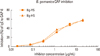
Allergen potencies were compared using the competitive specific IgE binding CAP inhibition system with the HS allergen extract (Hollister-Stier Laboratories, Spokane, WA, USA). The Korean extract dissolved in distilled water was used for the inhibition study. The serum samples (diluted 1:4) were pre-incubated with various concentrations of inhibitors (0.001 µg/mL to 20 µg/mL), and anti-human IgE reactivity was measured using the UniCAP system (Phadia, Uppsala, Sweden), according to the manufacturer's instruction. The percentage of inhibition was calculated as: (1-Ai/A0)×1,000, where Ai is the IgE value (kU/mL) with inhibitor and A0 is the IgE value without inhibitor.
Measurements of levels of major CR allergens and endotoxin
The levels of the CR allergens, Bla g 1 and Bla g 2, in the CR extracts reconstituted in distilled water were determined by two-site ELISA (Indoor Biotechnologies Inc., Charlottesvillle, VA, USA). Endotoxin content was measured using QCL-1000 (Lonza, Walkersville, MD, USA).
Assessment of proteolytic activity
For the analysis of gelatinolytic proteases, samples (200 ng/well), which were reconstituted in PBS (pH 7.4) that contained 50% glycerol and 0.03% HSA, were run on 10% SDS-PAGE gels that contained 0.1% gelatin (Invitrogen). After electrophoresis, the gel was incubated in renaturing buffer (Invitrogen) for 1 hour at room temperature, and then placed in developing buffer (Invitrogen) overnight at 4℃. The gel was stained with Coomassie Brilliant Blue R250.
RESULTS
Allergenic activities of the CR extracts
The protein concentration of the Korean extract was very low (410 µg/mL) compared with that of the HS extract (2,300 µg/mL). However, the concentrations of Bla g 1 and Bla g 2 in the Korean extract (405 U/mg and 273 ng/mg, respectively) were higher than those in the HS extract (187 U/mg and 56 ng/mg, respectively) (Table).
The allergenic activities of the CR extracts were examined using an in vitro specific IgE binding inhibition assay. The Korean extract showed 94.2% inhibition of IgE reactivity, as compared with the HS extract (Fig. 2).
The Korean extract had more apparent bands in the SDS-PAGE analysis (Fig. 1A). The presence of a protein band close to the well in the HS extract lane implies some aggregation of the extract. No IgE-reactive bands were detected when sera from healthy controls were used for the immunoblotting (data not shown).
Protease and endotoxin activities of the CR extracts
A broad range of gelatinolytic activity was detected for the proteins in the range of 10-100 kDa in both the Korean and HS extracts (Fig. 1C). In both extracts, the primary proteolytic activity was detected for proteins of 10-30 kDa.
The endotoxin levels of the Korean and HS extracts were determined to be 3,440 EU/mL and 6,580 EU/mL, respectively (Table). However, the endotoxin level per mg of protein was much higher in the Korean extract (8,390 EU/mg) than in the HS extract (2,861 EU/mg).
DISCUSSION
We produced a German CR extract using a standardized procedure and characterized the extract by comparing it with the HS extract.
In the electrophoretic analysis, the Korean extract showed multiple bands with molecular masses that ranged from 15 kDa to 100 kDa, including the 66-kDa band of HSA, which is generally added to allergen extracts to increase stability (Fig. 1A). However, the HS extract had less-pronounced bands and some aggregation of proteins around the well. The overall patterns of IgE-reactive proteins in the IgE immunoblots were similar for the Korean and HS extracts, with the exception of the aggregated proteins (Fig. 1B). The proteolytic activities of the two extracts, as revealed by gelatin zymography, were similar (Fig. 1C). The Korean German CR extract was able to inhibit 94.2% of the IgE reactivity, as compared with the HS extract (Fig. 2). Overall allergenicity is essentially identical for the extracts from HS and Korea. Further characterization of the allergenicities of the extracts using an in vivo method is needed.
The levels of Bla g 1 and Bla g 2 in the two extracts were found to be quite different (Table). Polymorphisms of mite allergen sequences are thought to influence the amount of allergens in mite extracts.10 However, no sequence variability was found for the recognition sites on Bla g 2 for the monoclonal antibodies 7C11 and 4C3 (capture and detection antibodies for Bla g 2; Indoor Biotechnologies Inc.).11
Bla g 1 (25-30 kDa) and Bla g 2 (36 kDa) have not been confirmed as the major IgE binding proteins in Korean patients with allergic diseases.12 The IgE reactivities of purified Bla g 1 and Bla g 2 have been described as not being strong,13,14 and the major allergens that dominate the allergenicity of the CR extract need to be identified. Components with molecular masses of less than 14, 38, 50, 64, and 76 kDa have been described as important in Korea.12 The Bla g 2 levels of the extracts were found to be non-proportional to the overall allergenicities.7 It is very interesting to observe strongly IgE-reactive proteins of approximately 60 kDa and 70 kDa in both the Korean and HS extracts (Fig. 1B). The observed similarity of the patterns of IgE-reactive bands in the immunoblots indicates the presence of similar IgE-reactive components in the extracts. Identification of these allergens, which are important in Korea, is necessary for enhanced characterization and standardization of the CR extract.
CR extract is rich in proteases,9 which can activate the various cell types that are important in allergic airway inflammation.15 Special care is needed, although not recommended, when combining fungal and CR extracts for immunotherapy, given the stability of the allergens in the mixtures.16
High levels of endotoxin were detected in the CR extracts, as compared to house dust mite extracts (1-8,485 EU/mL).17 The average levels of endotoxin detected in standardized house dust mite extracts were: 4,619 EU/mL (range, 849-8,485 EU/mL) for seven Dermatophagoides farinae extracts; and 11 EU/mL (range, 1-34 EU/mL) for seven D. pteronyssinus extracts. However, up to 33,805 EU/mL of endotoxin was detected in the standardized cat pelt extract.17 CRs are known to carry various microorganisms, including bacteria.18,19 The endotoxin present in the CR extract is believed to be mainly derived from the flora or commensals in the CR gut.
The German CR extract prepared in the present study could be useful for the development of allergy diagnostics and immunotherapeutics in Korea.

 XML Download