

 PDF
PDF ePub
ePub Citation
 Print
Print


References
1. Batson OV. The function of the vertebral veins and their role in the spread of metastases. 1940. Clin Orthop Relat Res. 1995; (312):4–9.
2. Rodríguez Salas N, González Paz C, Rivera T, López Alfonso A, Martín Marino A, Lara Alvarez MA. Colonic anastomosis and colonic polyp mucosal metastasis of signet ring cell gastric adenocarcinoma. Clin Transl Oncol. 2010; 12:238–239.

3. Ogiwara H, Konno H, Kitayama Y, Kino I, Baba S. Metastases from gastric adenocarcinoma presenting as multiple colonic polyps: report of a case. Surg Today. 1994; 24:473–475.
4. Katon RM, Brendler SJ, Ireland K. Gastric linitis plastica with metastases to the colon: a mimic of Crohn's disease. J Clin Gastroenterol. 1989; 11:555–560.
5. Gao B, Xue X, Tai W, et al. Polypoid colonic metastases from gastric stump carcinoma: a case report. Oncol Lett. 2014; 8:1119–1122.
6. Metayer P, Antonietti M, Oumrani M, Hemet J, Lemoine F, Basuyau J. Metastases of a gastric adenocarcinoma presenting as colonic polyposis. Report of a case. Dis Colon Rectum. 1991; 34:622–623.
7. Lee IH, Lee JE, Byeon SW, et al. A case of advanced gastric cancer presenting as multiple colonic lymphoid hyperplasia. Korean J Gastroenterol. 2015; 66:221–226.
8. Tiszlavicz L. Stomach cancer metastasizing into a solitary adenomatous colonic polyp. Orv Hetil. 1990; 131:1259–1261.
9. Tiszlavicz L. Metastasis of a stomach carcinoma in a solitary adenomatous cecal polyp. Zentralbl Allg Pathol. 1990; 136:277–282.
10. Park SY, Kim HS, Hong EK, Kim WH. Expression of cytokeratins 7 and 20 in primary carcinomas of the stomach and colorectum and their value in the differential diagnosis of metastatic carcinomas to the ovary. Hum Pathol. 2002; 33:1078–1085.
Fig. 1.
Initial abdominal CT finding. The bilateral masses (right ovary, 22.3×21.4×16.9 cm; left ovary, 6.2×6.2×5.5 cm) and large amount of ascites suggested advanced ovary cancer.
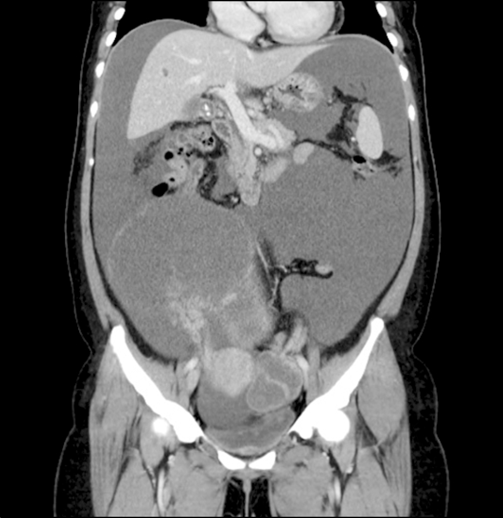
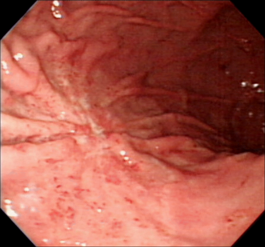
Fig. 2.
Esophagogastroduodenoscopic finding. A 1.8 cm depressed lesion with mucosal convergence is seen in the great curvature side of the midbody.

 XML Download