

 PDF
PDF ePub
ePub Citation
 Print
Print


Abstract
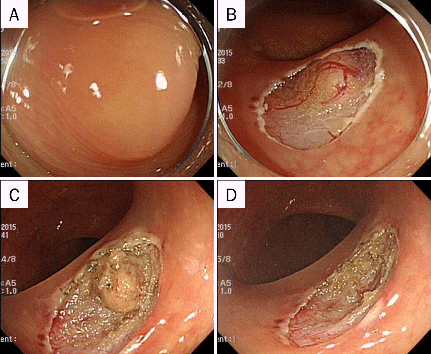
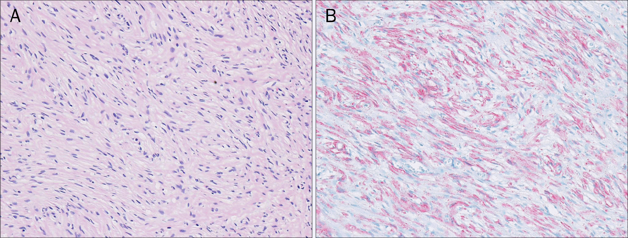
Neurofibromas are benign, slow-growing nerve sheath tumors of the peripheral nervous system, arising from Schwann cells, and classically associated with neurofibromatosis type 1 (Nf1, von Recklinghausen's disease). They occur rarely in the gastrointestinal tract as isolated neoplasms, outside the classical clinical feature of neurofibromatosis. We herein present an isolated colonic neurofibroma without any systemic signs of neurofibromatosis. A 59-year-old female came to our hospital for constipation. On physical examination, general appearance showed no definite skin lesions. A subepithelial tumor measuring 0.8 cm was detected at the distal descending colon on colonoscopy. The lesion was removed completely by endoscopic resection. Microscopic examination showed proliferation of spindle cells in the mucosa and infiltration of inflammatory cells. Immunohistochemical staining was positive for S-100 protein. The above morphological and immunohistochemical characteristics were consistent with a diagnosis of a solitary neurofibroma of the sigmoid colon.
References
1. Ferner RE, Gutmann DH. International consensus statement on malignant peripheral nerve sheath tumors in neurofibromatosis. Cancer Res. 2002; 62:1573–1577.
2. Korf BR. Neurofibromatosis. Handb Clin Neurol. 2013; 111:333–340.

3. Feinstat T, Tesluk H, Schuffler MD, et al. Megacolon and neurofibromatosis: a neuronal intestinal dysplasia. Case report and review of the literature. Gastroenterology. 1984; 86:1573–1579.
4. Reynolds RM, Browning GG, Nawroz I, Campbell IW. Von Recklinghausen's neurofibromatosis: neurofibromatosis type 1. Lancet. 2003; 361:1552–1554.
5. Riddle ND, Gorden L, Rojiani MV, Hakam A, Rojiani AM. CD44 and p53 immunoexpression patterns in NF1 neoplasms – indicators of malignancy and infiltration. Int J Clin Exp Pathol. 2010; 3:515–521.
6. Hochberg FH, Dasilva AB, Galdabini J, Richardson EP Jr. Gastrointestinal involvement in von Recklinghausen's neurofibromatosis. Neurology. 1974; 24:1144–1151.
7. Fisher C. Immunohistochemistry in diagnosis of soft tissue tumours. Histopathology. 2011; 58:1001–1012.
8. Turner MS, Goldsmith JD. Best practices in diagnostic immunohistochemistry: spindle cell neoplasms of the gastrointestinal tract. Arch Pathol Lab Med. 2009; 133:1370–1374.
9. Cichowski K, Jacks T. NF1 tumor suppressor gene function: narrowing the GAP. Cell. 2001; 104:593–604.
10. Bononi M, De Cesare A, Stella MC, et al. Isolated intestinal neurofibromatosis of colon. Single case report and review of the literature. Dig Liver Dis. 2000; 32:737–742.
11. Amin MB, Ma CK, Linden MD, Kubus JJ, Zarbo RJ. Prognostic value of proliferating cell nuclear antigen index in gastric stromal tumors. Correlation with mitotic count and clinical outcome. Am J Clin Pathol. 1993; 100:428–432.
12. Franklin A, Uff JS, Spencer J. Anal neurofibrosarcoma treated with fast neutron irradiation. Br Med J. 1978; 1:959–960.
13. Raszkowski HJ, Hufner RF. Neurofibromatosis of the colon: a unique manifestation of von Recklinghausen's disease. Cancer. 1971; 27:134–142.
14. Kim KO, Jang BI, Moon HJ, et al. A solitary colonic neurofibroma in a patient without neurofibromatosis. Korean J Gastrointest Endosc. 2008; 36:44–47.
15. Hindy P, Parvin R, Hanna K, Andrawes S, Gress F, Goodman A. An isolated neurofibromal polyp of the colon. Case Rep Gastroenterol. 2012; 6:58–62.
16. Wiesen A, Davidoff S, Sideridis K, Greenberg R, Bank S, Falkowski O. Neurofibroma in the colon. J Clin Gastroenterol. 2006; 40:85–86.
17. Panteris V, Vassilakaki T, Vaitsis N, Elemenoglou I, Mylonakou I, Karamanolis DG. Solitary colonic neurofibroma in a patient with transient segmental colitis: case report. World J Gastroenterol. 2005; 11:5573–5576.
 XML Download