

 PDF
PDF ePub
ePub Citation
 Print
Print


Abstract
We report an extremely rare case of metastatic common bile duct cancer from pulmonary adenocarcinoma presenting as obstructive jaundice. The patient was a 76-year-old male, who presented with generalized weakness and right upper quadrant pain. Plain chest X-ray noted multiple small nodules in both lung fields. Abdominal computed tomography scan showed a stricture of the mid common bile duct along with ductal wall enhancement. Endoscopic retrograde cholangiography revealed a concentric, abrupt narrowing of the mid-common bile duct suggestive of primary bile duct cancer. However, pathology comfirmed metastatic common bile duct cancer arising from pulmonary adenocarcinoma with immunohistochemical study with thyroid transcriptional factor-1 (TTF-1).
References
1. Smith HJ. Extrahepatic bile duct obstruction in primary carcinoma of the lung: incidence, diagnosis, and nonoperative treatment. J Natl Med Assoc. 1980; 72:215–220.
2. Moon SG, Han JK, Kim TK, Kim AY, Kim TJ, Choi BI. Biliary obstruction in metastatic disease: thin-section helical CT findings. Abdom Imaging. 2003; 28:45–52.

3. Lee YJ, Kim SH, Lee JY, et al. Differential CT features of intraductal biliary metastasis and double primary intraductal polypoid cholangiocarcinoma in patients with a history of extra-biliary malignancy. AJR Am J Roentgenol. 2009; 193:1061–1069.
4. Guazzi S, Price M, De Felice M, Damante G, Mattei MG, Di Lauro R. Thyroid nuclear factor 1 (TTF-1) contains a homeodomain and displays a novel DNA binding specificity. EMBO J. 1990; 9:3631–3639.
5. Lazzaro D, Price M, de Felice M, Di Lauro R. The transcription factor TTF-1 is expressed at the onset of thyroid and lung morpho-genesis and in restricted regions of the foetal brain. Development. 1991; 113:1093–1104.
6. Bohinski RJ, Huffman JA, Whitsett JA, Lattier DL. Cis-active elements controlling lung cell-specific expression of human pulmonary surfactant protein B gene. J Biol Chem. 1993; 268:11160–11166.
7. Fabbro D, Di Loreto C, Beltrami CA, Belfiore A, Di Lauro R, Damante G. Expression of thyroid-specific transcription factors TTF-1 and PAX-8 in human thyroid neoplasms. Cancer Res. 1994; 54:4744–4749.
8. Fabbro D, Di Loreto C, Stamerra O, Beltrami CA, Lonigro R, Damante G. TTF-1 gene expression in human lung tumours. Eur J Cancer. 1996; 32A:512–517.
9. Folpe AL, Gown AM, Lamps LW, et al. Thyroid transcription factor-1: immunohistochemical evaluation in pulmonary neuroendocrine tumors. Mod Pathol. 1999; 12:5–8.
10. Kaufmann O, Dietel M. Expression of thyroid transcription factor-1 in pulmonary and extrapulmonary small cell carcinomas and other neuroendocrine carcinomas of various primary sites. Histopathology. 2000; 36:415–420.
11. Dennis JL, Hvidsten TR, Wit EC, et al. Markers of adenocarcinoma characteristic of the site of origin: development of a diagnostic algorithm. Clin Cancer Res. 2005; 11:3766–3772.
12. Roh MS, Hong SH. Utility of thyroid transcription factor-1 and cytokeratin 20 in identifying the origin of metastatic carcinomas of cervical lymph nodes. J Korean Med Sci. 2002; 17:512–517.
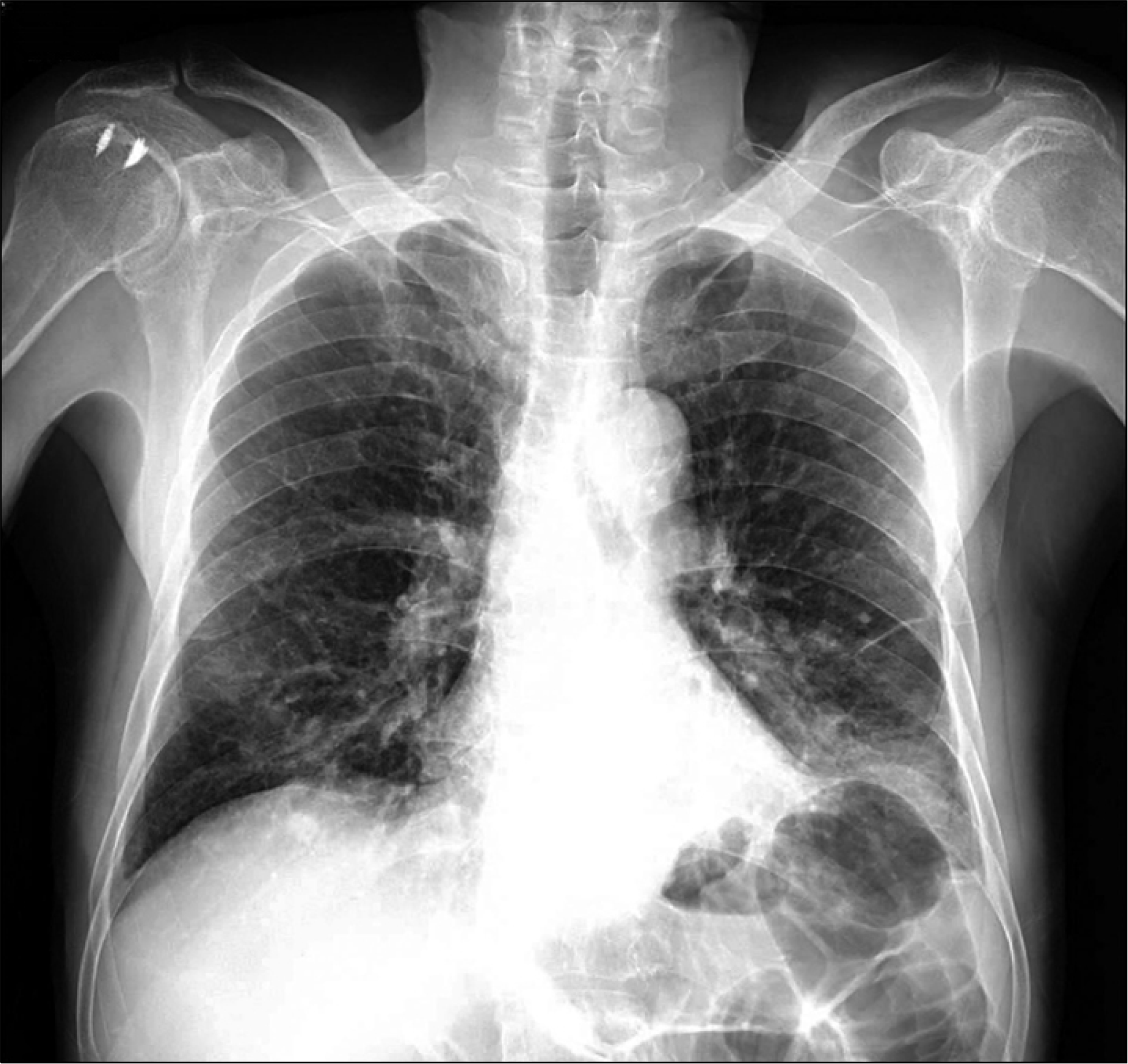
Fig. 2.
Abdominal CT scan. (A) Ductal wall enhancement (arrow) was seen on axial image. (B) Note the stricture at the mid-common bile duct along with shouldering and ductal wall enhancement (arrow) on coronal image.
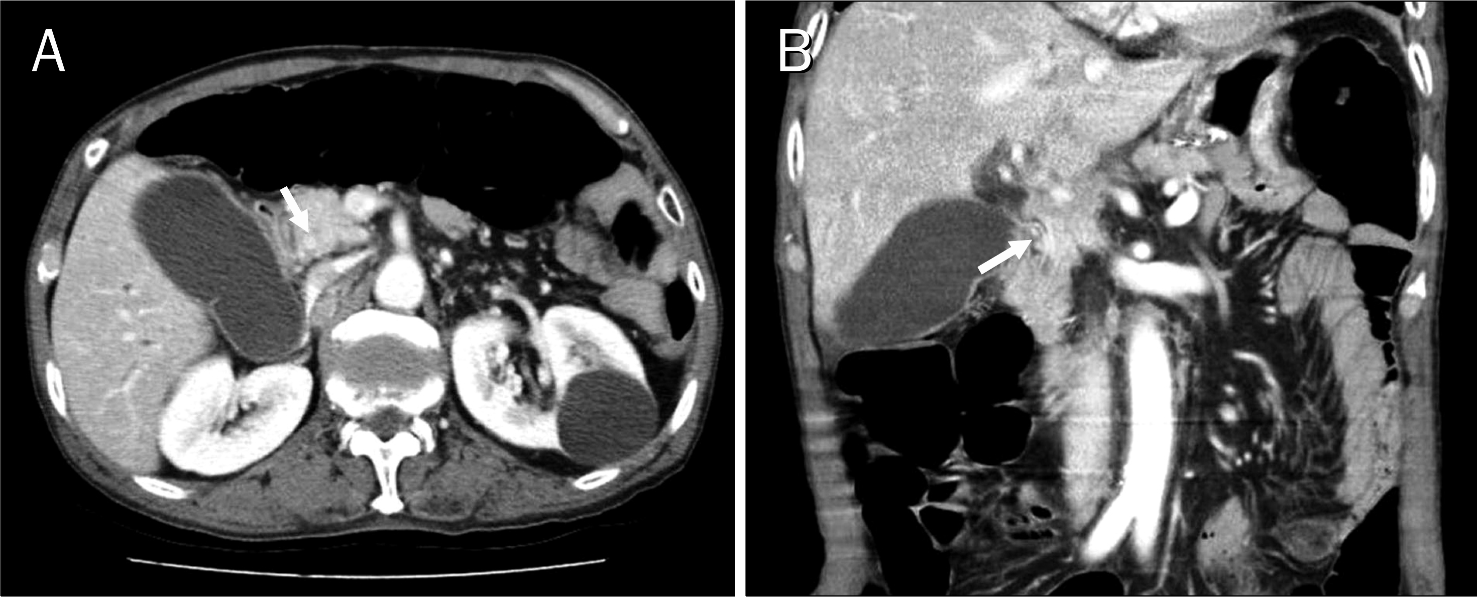
Fig. 3.
Imaging and histologic findings of the common bile duct. (A) Endoscopic retrograde cholangiography revealed a concentric, abrupt narrowing of the mid-common bile duct. (B) Biopsy exhibited clusters of malignant columnar cells with prominent nucleoli and intracytoplasmic mucin vacuoles (H&E, ×400). (C) Adenocarcinoma cells demonstrated strong nuclear immunoreactivity for thyroid transcriptional factor-1 (×400).
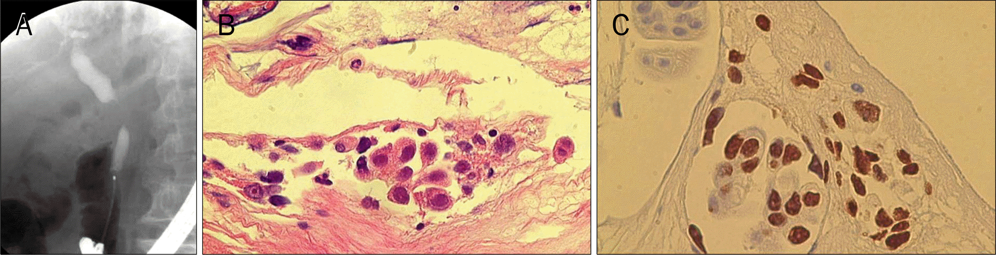
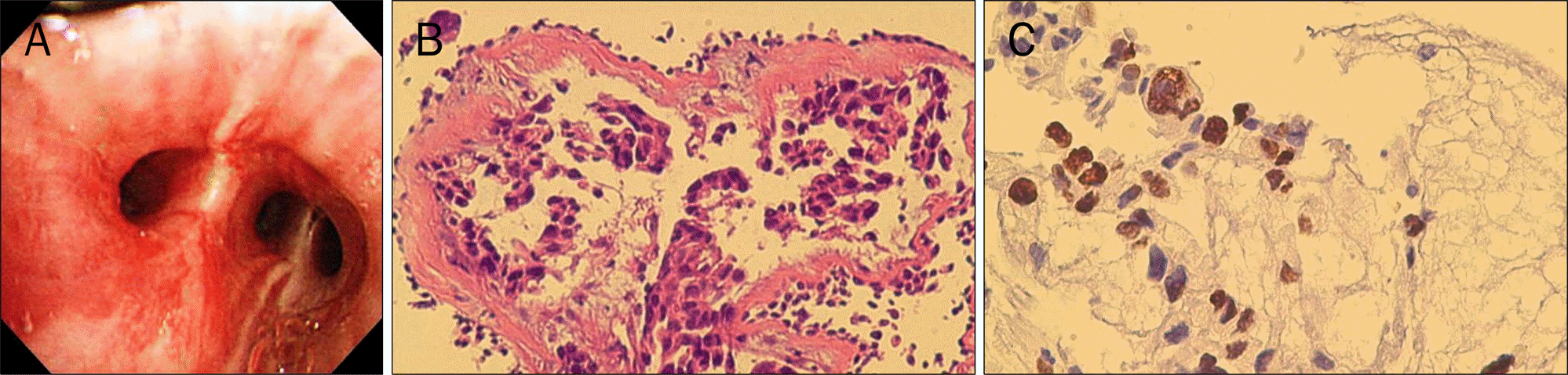
Fig. 4.
Bronchoscopic findings and bronchial biopsy specimen. (A) Bronchoscopy showed hyperemic mucosal change, finding suspicious of cancer cell infiltration. (B) Biopsy revealed haphazardly infiltrating nests of adenocarcinoma cells with intracytoplasmic mucin production (H&E,×200). (C) Adenocarcinoma cells were also reactive for thyroid transcriptional factor-1 (×400).
 XML Download