

 PDF
PDF ePub
ePub Citation
 Print
Print


INTRODUCTION
Colonic muco-submucosal elongated polyp (CMSEP) was first described by Matake et al.1 in 1998. It is a clinical and morphologic term describing an elongated projection of normal mucosa and submucosa and also called elongated non-neoplastic colonic polyp. Histopathologically, CMSEP is covered by normal mucosa and shows a dense submucosal layer containing edematous loose connective tissue and dilated vascular and lymphatic structures.2 Although CMSEP can be manifested with hemorrhage,2 most remains asymptomatic and is diagnosed incidentally. We report a case of adenocarcinoma arising from a CMSEP in a patient with lower gastrointestinal bleeding. To our knowledge, this is the second case report of CMSEP associated with a cancerous transformation.
CASE REPORT
A 53-year-old man presented with a history of repeated hematochezia for 2 days. He was an active smoker with a 30-pack-year smoking history and had consumed 60 g of alcohol a day for several years. He did not have family history of cancer. One month prior to his current visit, he underwent upper gastrointestinal endoscopy for dyspepsia where small Helicobacter plyori (HP)-positive gastric ulcer was found at the antrum and eradicated with a one week course of tripe anti-HP regimen. Physical examination on admission revealed a mildly obese Korean man in no acute distress. He had anemic conjunctiva of mild degree and the vital sign was stable. His abdomen was soft with normal bowel tones. Hemoglobin level on admission was 7.0 g/dL with a mean corpuscular volume of 81.4 fL. Tumor markers and coagulation studies were within normal limits. A repeat upper gastrointestinal endoscopy confirmed that the previous ulcer had healed and failed to find any source of recent bleed.
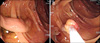
At subsequent colonoscopy performed after bowel preparation, an elongated polyp and several small diverticula were noted in the distal and proximal ascending colon, respectively. While diverticula in the proximal ascending colon showed no recent bleeding stigmata in their bases, the elongated polyp (35 mm in length, 8 mm in diameter) had a conspicuous erythema on the tip and therefore was considered an origin of recent hemorrhage (Fig. 1). An arteriovenous malformation was clinically suspected for its unusual appearance and therapeutic snare polypectomy was performed after pre-application of hemoclip.
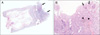
On histopathological examination, the elongated thick stalk of the polyp was covered by normal mucosa with muscularis mucosae and had a submucosal layer that consisted of loose edematous stroma with dilated blood vessels and lymphatics (Fig. 2A), which were consistent with CMSEP. On the tip of the CMSEP, there was a small area of well differentiated adenocarcinoma invading submucosal layer. The cancerous lesion was a non-polypoid type with central depression (Fig. 2B), which referred to early colon cancer type IIc according to Japanese classification of colorectal carcinoma.3 The depth of submucosal invasion measured 0.22 cm. The adenocarcinoma coexisted with a small focus of adenoma with high grade dysplasia in the lesion. Many hemosiderin pigments were noted in the adjacent submucosal tissue and considered as a sign of recent bleeding. Despite the presence of submucosal invasion, an additional bowel resection was not performed because the polyp was structurally pedunculated, and resection margin was far away from the lesion. After the procedure, he was discharged without further bleeding episode. He has been well without any signs of recurrence or bleeding for 5 years.
DISCUSSION
This case is unique in that an early invasive cancer of non-polypoid type developed in a polyp with very long stalk. Since tubular elongation is unusual shape in commonly encountered pedunculated polyps in which head part is usually larger than stalk, we prefer to diagnose this case as 'cancer arising from a CMSEP' rather than simply calling it as 'pedunculated form of early colon cancer'. As the name implies, CMSEP is a clinical term describing abnormal finger like projection of normal mucosa and submucosa. Because its original meaning is non-neoplastic, discussing tumorous association in CMSEP may not be appropriate. Nonetheless, we considered CMSEP as an underlying polyp associated with early invasive cancer for following reasons. Firstly, CMSEP is less defined yet and not enough to be categorized into conventional polyp classification. Since Matake et al.1 first reported clinical characteristics in 15 patients with CMSEP, only 50 cases so far including Japanese literature or meeting abstract have been reported.4 Based on previous studies, about one-third is associated with concomitant colonic neoplasia. Therefore, it is difficult to say CMSEP is non-neoplastic polyp that does not even have chance to develop cancer or to harbor it by chance. Secondly, previously reported CMSEP cases share common clinical features with our cases. As in the presented case, CMSEPs often manifest with bleeding. According to the previous studies, half of the cases are related to occult and/or gross lower gastrointestinal bleeding.4 One of key histologic features of CMSEP is dilated submucosal vessels thereby predisposing to lower intestinal bleeding. The rupture of dilated submucosal vessels into eroded surface epithelium could be the possible explanation.4 In addition to lower intestinal bleeding, colonic diverticulum, another finding shown in this case, is frequently associated with CMSEP. Some authors tried to link CMSEP to 'polypoid prolapsing mucosal folds in diverticular disease' and proposed that both diseases are same entity as they share similar pathology, clinical characteristics, and underlying mechanism.5 Last but not least reason for our assumption is, of course, morphologic similarity mentioned above. Furthermore, relatively small neoplastic cell mass better support the idea that elongation might not be secondarily induced against gravity. Despite these supportive data, there is little direct evidence on whether CMSEP is etiologically related to colonic neoplasia beyond coincidence. Only one report with 2 cases so far has demonstrated neoplastic associations in CMSEPs in the literature.6 In that study, well-differentiated adenocarcinomas/adenoma with non-polypoid downward growth pattern was demonstrated in an elongated polyp just as in our case. Striking similarity between two reports prompted us to assume that there is, to some extent, an etiologic connection between CMSEP and colorectal neoplasia. Non-polypoid cancer with downward growth type is defined as those in which the tumors have a central depression and the marginal area of the cancerous lesion is clearly bordered by noncancerous mucosa.7 In summary, the present case is diagnosed as non-polypoid depressed type of early colon cancer arising from a CMSEP. Long term mechanical stress such as hypoxia or spilled blood product might have influence on the promotion of existing tumor or possible tumor initiation.
 XML Download