References
1. Chang S, Lim JH, Choi D, et al. Hepatic visceral larva migrans of Toxocara canis: CT and sonographic findings. AJR Am J Roentgenol. 2006; 187:W622–629.
2. Beaver PC, Jung RC, Cupp EW. Clinical parasitology. 9th ed.Philadelphia: Lea & Febiger;1984. p. 320–329.
3. Kaplan KJ, Goodman ZD, Ishak KG. Eosinophilic granuloma of the liver: a characteristic lesion with relationship to visceral larva migrans. Am J Surg Pathol. 2001; 25:1316–1321.
5. Kwon NH, Oh MJ, Lee SP, Lee BJ, Choi DC. The prevalence and diagnostic value of toxocariasis in unknown eosinophilia. Ann Hematol. 2006; 85:233–238.

6. Despommier D. Toxocariasis: clinical aspects, epidemiology, medical ecology, and molecular aspects. Clin Microbiol Rev. 2003; 16:265–272.

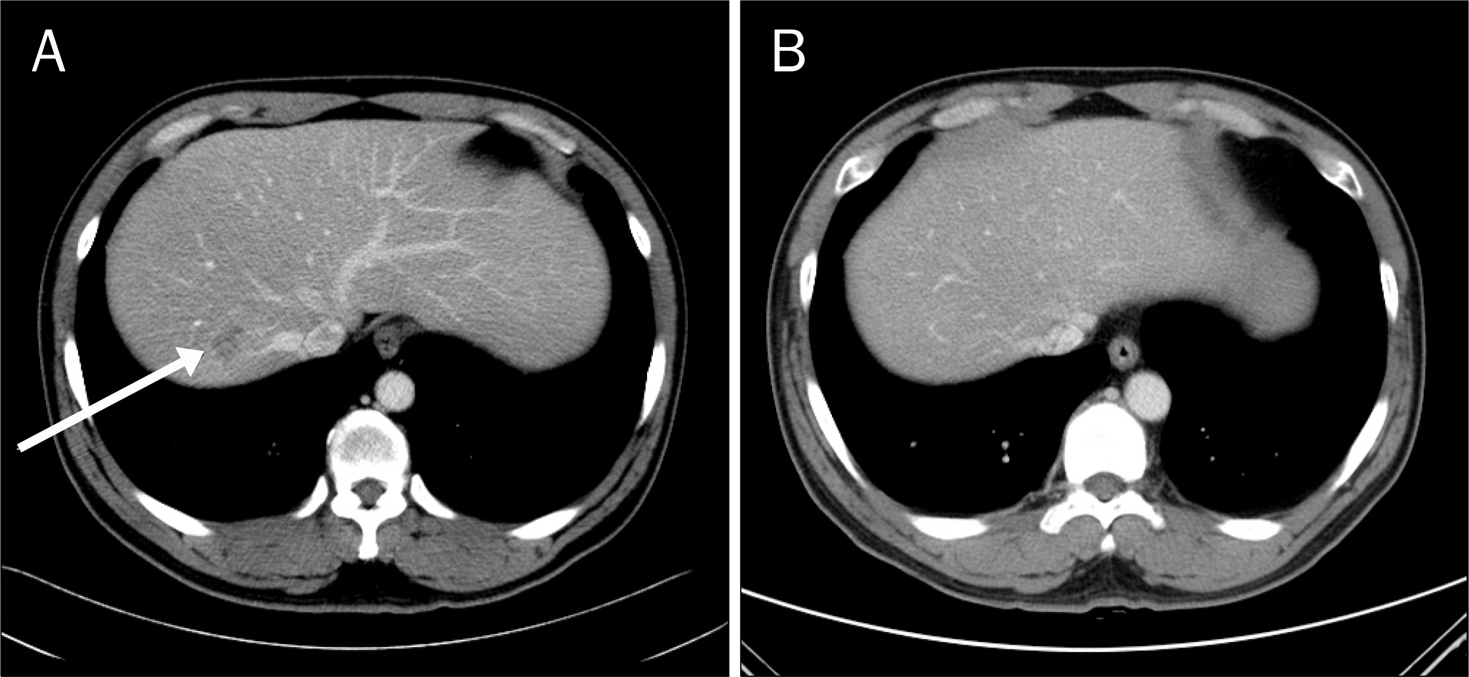
Fig. 1.
(A) CT showed 20 mm sized, well-defined, and faintly low attenuated mass in the S8 of the liver. (B) CT showed 12 mm sized, ill-defined, and faintly low attenuated lesion in the S7 of the liver. (C) Follow up CT (after 8 weeks) showed decreased size of (20 mm → 13 mm) well-defined, and faintly low attenuated mass in the S8 of the liver.


 PDF
PDF ePub
ePub Citation
 Print
Print


 XML Download