Abstract
Acute cholangitis usually develops in congenital hepatic fibrosis (CHF), accompanied by cystic dilated bile ducts. However, it can also develop in simple CHF and may lead to critical course. A 30-year old man presented with recurrent acute cholangitis without bile duct dilatation. He visited the hospital for febrile sense and abdominal pain in the right upper quadrant. He had been admitted several times for hepatosplenomegaly and cholangitis since childhood and received a liver biopsy 15 years ago. Abdominal computed tomography (CT) and endoscopic retrograde cholangiopancreatography (ERCP) revealed hepatosplenomegaly and a mildly dilated bile duct without stones or biliary cysts. His condition improved after conservative treatment. However, during a two-month follow up period, the patient experienced three episodes of acute cholangitis. A liver biopsy was performed and showed periportal fibrosis and intrahepatic ductular dysplasia, characteristics of congenital hepatic fibrosis. The periportal fibrosis and the infiltration of inflammatory cells were aggravated compared to 15 years ago. There was no evidence of hepatic cirrhosis. He was diagnosed with congenital hepatic fibrosis with recurrent acute cholangitis without intrahepatic duct dilatation, and conservatively treated with antibiotics.
REFERENCES
1. Murray-Lyon IM, Shilkin KB, Laws JW, Illing RC, Williams R. Non-obstructive dilatation of the intrahepatic biliary tree with cholangitis. Q J Med. 1972; 41:477–489.
2. Lieberman E, Salinas-Madrigal L, Gwinn JL, Brennan LP, Fine RN, Landing BH. Infantile polycystic disease of the kidneys and liver: clinical, pathological and radiological correlations and comparison with congenital hepatic fibrosis. Med. 1971; 50:277–318.
3. Choi CS, Oh HJ, Kim BS, et al. A case of congenital hepatic fibrosis presented with symptom of acute cholangitis. Korean J Gastroenterol. 2005; 46:237–241.
4. Kerr DN, Harrison CV, Sherlock S, Walker RM. Congenital hepatic fibrosis. Q J Med. 1961; 30:91–117.
6. Sanzen T, Harada K, Yasoshima M, Kawamura Y, Ishibashi M, Nakanuma Y. Polycystic kidney rat is a novel animal model of Caroli's disease associated with congenital hepatic fibrosis. Am J Pathol. 2001; 158:1605–1612.

7. Teufel J, Farack UM. Hepatobiliary fibropolycystic diseases. Two cases of Caroli's disease. Scand J Gastroenterol Suppl. 1987; 139:76–80.

8. Yönem Ö, Özkayar N, Balkanci F, et al. Is congenital hepatic fibrosis a pure liver disease? Am J Gastroenterol. 2006; 101:1253–1259.

9. McCarthy LH, Baggenstoss AH, Logan GB. Congenital hepatic fibrosis. Gastroenterology. 1965; 49:27–36.

10. Erlinger S, Sakellaridis DA, Maillard J, Benhamou JP. The angiocholitic forms of the congenital hepatic fibrosis. Presse Med. 1969; 77:1189–1191.
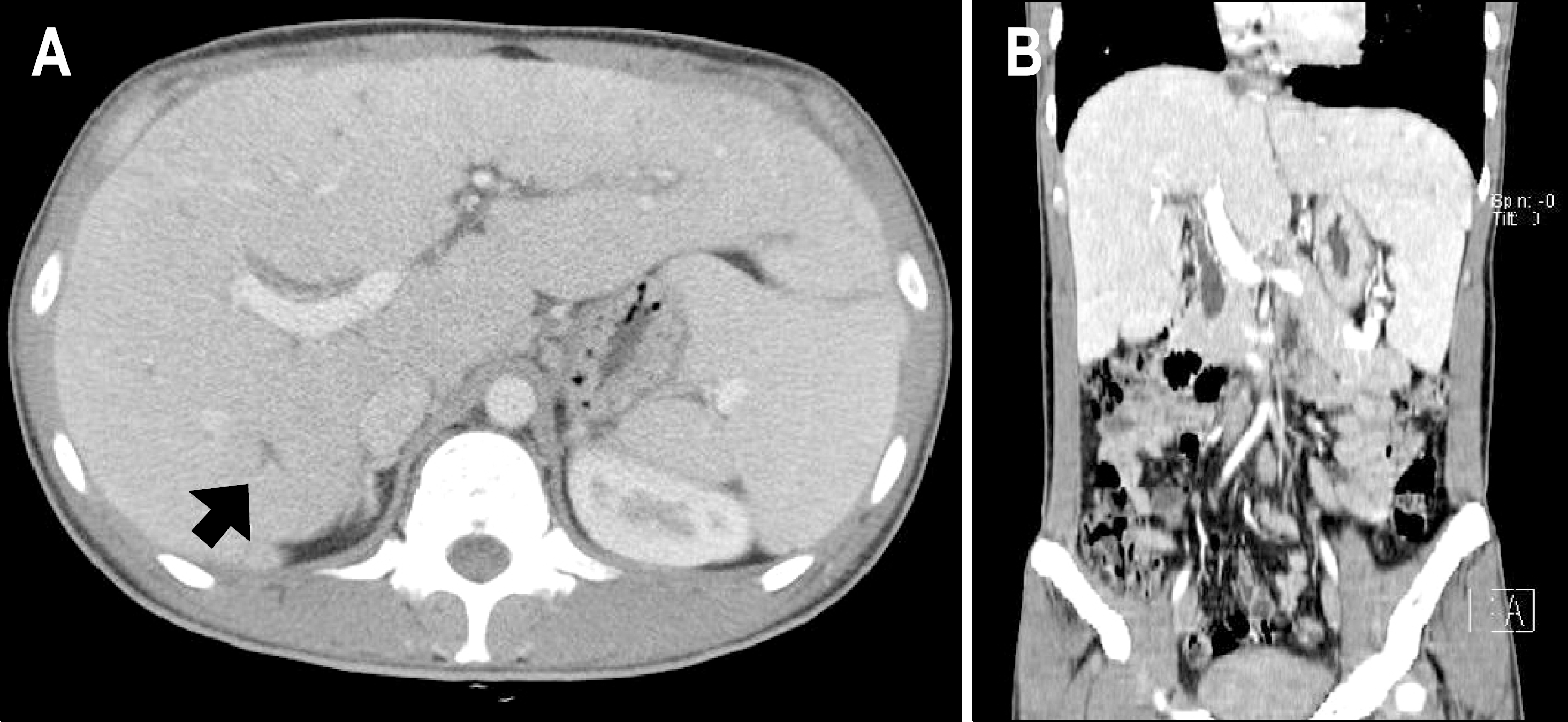
Fig. 1.
Abdominal CT scan finding (A, axial scan; B, coronal multiplanar reformation). It showed diffuse hepatic enlargement, splenomegaly, and mild focal intrahepatic bile duct dilatation (arrow).

Fig. 2.
Endoscopic retrograde cholangiopancreatography findings. It demonstrated normal papilla (A), mild dilatation of extrahepatic bile duct (B) and minimal irregularities of the intrahepatic bile ducts in the left lobe (C, arrow).

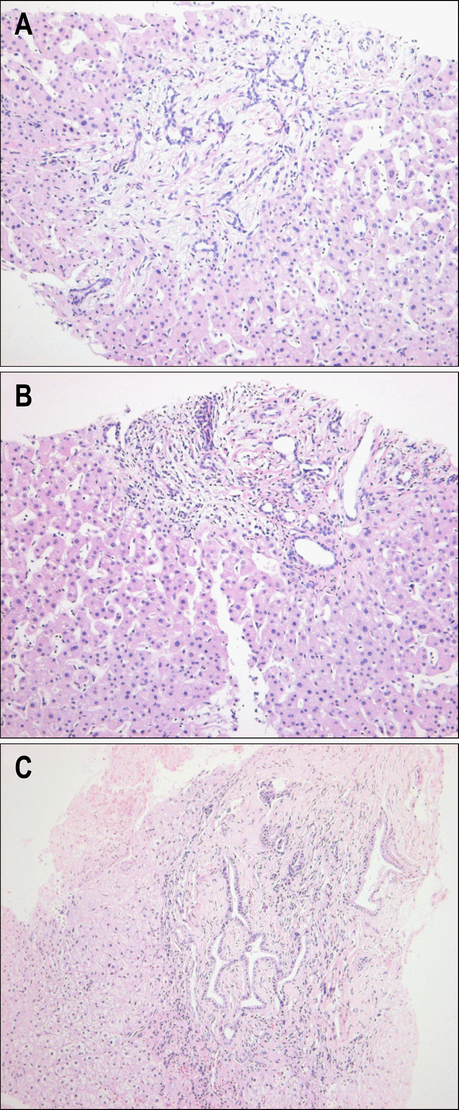
Fig. 3.
(A) The liver biopsy showed marked portal fibrosis containing many anastomosing ducts without regenerative nodule formation of the adjacent hepatocytes (H&E, ×200). (B) Some portal tracts exhibited fibrosis, anastomosing ducts, and ductal neutrophilic infiltration (H&E, ×200). (C) The liver biopsy taken 15 years ago demonstrated extensive portal fibrosis containing many dilated anastomosing ducts (H&E, ×200).


 PDF
PDF ePub
ePub Citation
 Print
Print


 XML Download