

 PDF
PDF ePub
ePub Citation
 Print
Print


INTRODUCTION
Inflammation is a protective response to pathogens such as bacteria, viruses, and parasites [1]. In inflammatory responses, macrophages are known to be one of the major immune cells involved [1,2], which are activated by several stimuli such as cytokines and viruses, and secrete pro-inflammatory mediators including nitric oxide (NO), prostaglandin E2 (PGE2), tumor necrosis factor-α (TNF-α), interleukin-1β (IL-1β), and IL-6 [1,2]. These inflammatory mediators are critically associated with pain, fever, edema, and recruiting additional immune cells to the site of inflammation [1]. However, overproduction of inflammatory mediators is associated with tissue damage [3]. Moreover, several studies have been reported that chronic inflammatory response could increase risk of chronic inflammatory diseases (CIDs) such as cancer, diabetes, and cardiovascular disease [4,5,6]. Thus, inhibition of pro-inflammatory mediator release could be beneficial in attenuating the inflammatory response in chronic diseases.
Expression of pro-inflammatory mediators, including inducible NO synthase (iNOS), cyclooxygenase-2 (COX-2), and pro-inflammatory cytokines, is regulated by activation of nuclear factor-κ B (NF-κB) and mitogen-activated protein kinases (MAPK) [2]. NF-κB exists as a heterodimer consisting of p65 and p50 subunits which are associated with inhibitors of κB (IκB) in the cytoplasm as an inactive form [2,7,8]. When macrophages are exposed to stimuli such as lipopolysaccaride (LPS), IκB is phosphorylated by IκB kinase (IKK) and NF-κB is separated from the phosphorylated IκB [2,3,7]. Separated NF-κB translocates to the nucleus and induces transcription of a variety of target genes such as iNOS, COX-2, IL-1β, and IL-6 [3,9,10]. Therefore, activation of NF-κB leads to inflammatory responses. LPS also stimulates MAPK pathways which increase pro-inflammatory cytokines expression through activator protein-1 (AP-1) activation [11,12]. MAPK activates AP-1 by phosphorylation then activated AP-1 binds to DNA to induce transcription of pro-inflammatory cytokines [12].
Rubus coreanus Miquel (RCM) has been used as one of the traditional Korean medicine for treatment of prostatism, diabetes mellitus, impotence, spermatorrhea, enuresis, and asthma [13]. Recent studies have shown that RCM reduces prostate tumor growth, kidney and stomach diseases, and rheumatoid arthritis because of high amounts of polyphenols [14,15,16,17]. Several studies reported that RCM contains high amounts of phenolic compounds including ellagic acid, gallic acid, cinnamic acid, protocatechuic acid, sangiin H-4, sanguiin H-6, 23-hydroxytormentic acid, and nigaichgoside F1 [18,19,20,21]. Other studies have shown that dietary polyphenols reduce inflammatory responses by reducing oxidative stress [22,23,24]. High polyphenol containing food such as strawberries, cherries, grapes, wine, or tea, decreased PGE2 and NO production by suppression of COX-2 and iNOS expressions through reduction of oxidative stress [22,23,24]. Ripe and unripe RCM also has been shown to reduce inflammatory cytokines production such as TNF-α, and IL-6 in mast cells and macrophages [16,25]. In addition, ellagitannins in ripe Rubus berries decreased gastric inflammation through suppression of NF-κB pathway [26]. RCM has been used as a medicine or food depending on its maturation. Unripe RCM has been used as a traditional remedy for some chronic diseases but ripe RCM has been taken as a fruit. Both RCM has been shown to have anti-carcinogenic, anti-diabetic, and anti-inflammatory effects. However, few studies have compared their anti-inflammatory effects depending on their maturation and none of them elucidated its mechanism. Therefore, in this study, we determine the effect of RCM on inflammatory responses and its possible mechanism depending on its maturation.
MATERIALS AND METHODS
Preparation of RCM extracts
Ripe and unripe fruits of RCM were collected from May to June of 2010 in the Gokseong area of South Korea and freeze-dried. Four different extracts were prepared from the fruits: i) 50% ethanol extracts of unripe fruit (UE), ii) aqueous extracts of unripe fruit (UH), iii) 50% ethanol extracts of ripe fruits (RE), and iv) aqueous extracts of ripe fruit (RH). Freeze-dried unripe or ripe fruits were pulverized and extracted in solutions of 30 grams of berries/L in distilled water or 50% ethanol by heating at 45℃ for 1.5 h. Supernatants were collected and and frozen dried with a lyophilizer. The obtained RCM extracts were dissolved in dimethyl sulfoxide (DMSO) and kept at -20℃ until it was used.
Cell culture
The murine macrophage cell line RAW 264.7 was purchased from the Korea Cell Line Bank (Seoul, Korea). Cells were cultured in DMEM containing 10% fetal bovine serum (v/v), 5% penicillin (100 U/ml), and streptomycin (100 µg/ml; Gibco TM Invitrogen, Grand Island, NY, USA) at 37℃ in a humidified atmosphere of 5% CO2. RCM (400 µg/ml) was added on plated cells for 24 h, and then stimulated with 1 µg/ml of LPS (Sigma-Aldrich, St. Louis, MO, USA) for the indicated time.
Cell viability assay
Cells were treated with increasing doses of RCM extracts for 24 h then stimulated with 1 µg/ml of LPS for 18h. Cell survival was determined by adding 500µg/ml of 3-(4,5-Dimethylthiazol-2-yl)-2,5-diphenyltetrazolium bromide (MTT) to each well and incubated for another 2 h at 37℃. After removal of medium, cells were lysed with isopropanol. The absorbance of the colored solution was quantified by measuring at 450nm with a microplate reader (Molecular Devices, Spectra MAX 340).
Measurement of NO and PGE2 productions
RAW 264.7 cells were pretreated with 400 µg/ml of RCM extract for 24h then stimulated with LPS (1 µg/ml). After 18h, NO and PGE2 levels in the culture media were determined using Nitrate/Nitrite Colorimetric Assay kits and PGE2 EIA kits (Cayman Chemical, Ann Arbor, MI, USA), respectively.
Measurement of pro-inflammatory cytokine production
RAW 264.7 cells were incubated with RCM extracts for 24 h then stimulated with LPS for 18 h. Cell-free supernatants were subsequently employed in pro-inflammatory cytokine assays using mouse enzyme-linked immunosorbent assay (ELISA) kits (R&D systems, Minneapolis, MN, USA), following protocol of the manufacturer.
RNA preparation and RT-PCR
RAW 264.7 cells were incubated with RCM extracts for 24 h then induced by LPS (1 µg/ml) for 4 h. Total RNA from LPS-induced RAW 264.7 cells was prepared using RNeasy mini kits (QIAGEN, Valencia, CA, USA) according to manufacturer's protocol and stored at -80℃ before use. Total RNA was reverse-transcribed with Superscript First strand synthesis systems kits (Invigrogen, Carlsbad, CA, USA) to obtain cDNA. All PCR analyses using a thermal cycler (Bioer Technology Co., LTD, Hangzhou, China) were subsequently carried out in a 20 µl volume containing 10 µl of SYBR Green, 100 pmol of 5' and 3' primers, and cDNA. PCR primers used in this study are the following: iNOS: 5'-AGTGGTGTTCTTTGCTTC-3' (forward) and 5'-GCTTGCCTT ATACTGGTC-3' (reverse); COX-2: 5'-GGTCTGGTGCCTGGTCTG-3' (forward) and 5'-CTCTCCTATGAGTATGAGTCTGC-3' (reverse); TNF-α: 5'-ACGGCATGGATCTCAAAGAC-3' (forward) and 5'-AGATAG CAAATCGGCTGACG-3' (reverse); IL-1β : 5'-AATCTATACCTGTCCT GTGTAATG-3' (forward) and 5'-GCTTGTGCTCTGCTTGTG-3' (reverse); IL-6: 5'-CTTCCATCCAGTTGCCTTCTT-3' (forward) and 5'-ACG ATTTCCCAGAGAACATGT-3' (reverse); GAPDH : 5'-CCATCACCATC TTCCAGGAGCG-3' (forward) and 5'-AGAGATGATGACCCTTTTG GC-3' (reverse). After amplification, a portion of the PCR products were electrophoresed on 1% agarose gel and visualized under UV after ethidium bromide staining.
Western blot analysis
After RCM treatments, cells were stimulated with LPS and lysated in RIPA lysis buffer containing cocktail protease inhibitors and phosphatase inhibitors (Sigma-Aldrich, St. Louis, USA). Immunoblot assay was carried out as described previously [27] with antibodies against iNOS, COX-2, p-IκB, ERK 1/2, p-ERK 1/2, p38, p-p38, JNK, p-JNK, and β-actin from Santa Cruz Biotechnology (Santa Cruz, CA, USA).
Nuclear extraction and NF-κB transcriptional activity assay
RAW 264.7 cells were harvested after 24h RCM treatment and subsequent LPS stimulation for 20 min. Nuclear proteins were extracted using a Nuclear Extraction kit (Cayman, Ann Arbor, MI, USA), and NF-κB transcriptional activity was measured using an NF-κB transcriptional factor assay kit (Cayman, Ann Arbor, MI, USA). Two micrograms of extracted nuclear proteins were incubated overnight at 4℃ and p65 primary antibody was incubated. After wash, secondary antibodies were incubated and developed with transcription factor developing solution. The absorbance of the colored solution was quantified by measuring at 450nm with a microplate reader (Molecular Devices, Spectra MAX 340).
Statistical analysis
All data are presented as means ± standard error (SE) from at least three independent experiments. Data were statistically analyzed using one-way analysis of variance (ANOVA), followed by Duncan's multiple range test using SPSS Statistics V.17.0. A value of P < 0.05 was considered statistically significant.
RESULTS
Non-cytotoxic level of RCM on RAW 264.7 cells
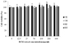
To avoid any cytotoxic effect caused by RCM, we investigated the effects of RCM extracts on cell viability in RAW 264.7 using MTT assays (Fig. 1). RCM extract had no effect on cell viability except for UE, which slightly increased cell viability at 200 µg/ml. RAW 264.7 were treated with 400 µg/ml of RCM for subsequent analyses.
Effects of RCM on LPS-induced NO production and iNOS expressions in RAW 264.7 cells
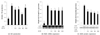
To investigate the effects of RCM on LPS-induced NO production and iNOS expression, cells were treated with 400 µg/ml of RCM extract for 24 h then stimulated with 1 µg/ml of LPS for 18 h. As shown in Fig. 2A, RCM treatment significantly reduced NO production compared to LPS-stimulated control. In addition, RCM pretreatments significantly reduced mRNA as well as protein expression of iNOS compared to LPS-stimulated control (Fig. 2B and 2C).
Effects of RCM on LPS-induced PGE2 production and COX-2 expressions RAW 264.7 cells
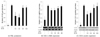
We attempted to determine whether RCM extracts modulated LPS-induced productions of PGE2 and COX-2 expression. Unripe RCM treatment significantly reduced PGE2 production by about 50% compared to LPS-stimulated control, but ripe RCM treatment did not (Fig. 3A). Moreover, unripe RCM significantly reduced COX-2 mRNA and protein expressions (Fig. 3B and 3C). However, ripe RCM did not modulate COX-2 expressions (Fig. 3B and 3C).
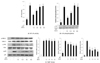
Effects of RCM on LPS-induced TNF-α, IL-1β, and IL-6 productions and expressions
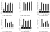
Next, we determine the effect of RCM on pro-inflammatory cytokine expression (Fig. 4). In TNF-α production, only UE treatment significantly decreased mRNA (Fig. 4A) and protein expression compared to LPS-stimulated control (Fig. 4B). However, UH, RE, and RH treatment had no effects on TNF-α production. RCM extract significantly decreased IL-1β and IL-6 expressions compared to LPS-stimulated control (Fig. 4C, 4D, 4E, and 4F). These results suggest that the inhibitory effects of RCM on the LPS-induced production of pro-inflammatory cytokines are regulated at the transcriptional level.
Effects of RCM on LPS-induced NF-κB and MAP kinase activities
Since inflammatory mediator expressions are activated by NF-κB and MAP kinase activities, we tested whether these activities are modulated by RCM extract (Fig. 5). RAW 264.7 cells were treated with RCM for 24h before 20min of LPS stimulation. LPS-induced NF-κB activities were significantly decreased when cells were pretreated with unripe RCM (Fig. 5A). Especially, UE treatment reduced NF-κB activity by about 50% compared to LPS-stimulated control. However, ripe RCM treatment had no such effect. Furthermore, considering LPS-induced NF-κB activation requires degradation of IκB, we investigated cytosolic phosphorylated IκB protein expression in RAW 264.7 cells (Fig. 5B). Cytosolic p-IκB proteins were less detected in UE- and UH-treated macrophage cells, but RE and RH treatment had no effect on IκB phosphorylation. Since pro-inflammatory cytokines are transcriptionally regulated by MAP kinases activities, we also measured 3 different MAP kinases, extracellular signal-regulated kinase 1/2 (ERK 1/2), MAPK p38, and c-Jun N-terminal kinase (JNK), phosphorylation levels (Fig. 5C). Unripe and ripe RCM significantly reduced JNK and p38 phosphorylation but had no effects on ERK1/2 activation. Unripe and ripe RCM treatment showed about 20~25% reduction of JNK phosphorylation and 60% reduction of p38 phosphorylation. These data indicated that unripe RCM reduced pro-inflammatory mediator expression through suppression of NF-κB and MAP kinase activities but ripen RCM only decreased two MAP kinases, JNK and p38, phosphorylation.
DISCUSSION
Since RCM has been shown to have high amounts of polyphenols and has been used as a traditional remedy for some CIDs, we determined the anti-inflammatory effects of RCM and their possible mechanisms using LPS-induced RAW 264.7 cells. In this study, we showed that unripe RCM (UE and UH) significantly reduced pro-inflammatory mediator expressions through suppression of NF-κB activity and MAP kinase activity.
We demonstrated that unripe RCM (UE and UH) showed stronger reduction of pro-inflammatory mediator production compared to ripe RCM (RE and RH). Several studies have reported that during maturation of RCM, amounts of polyphenols decreased while sugar increased [21,28,29]. We previously have reported that unripe RCM contained more of cinnamic acid (16 times), ferulic acid (6 times), epicatechine (6 times), protocatechuic acid (4 times), gallic acid (3 times), and vanillic acid (2.5 times) compared to ripe RCM [21]. Moreover, active compounds of unripe RCM, nigaichigoside F1 was present at approximately 2.5% (w/dried w). However, ripe RCM contained more of anthocyanins (4.1g/100g of dried weight) and cyaniding compared to unripe RCM [21]. Polyphenols are known as excellent anti-oxidants and have been shown to have anti-inflammatory effects [19,28,30]. Therefore, unripe RCM, containing higher amounts of polyphenolic compounds, showed stronger anti-inflammatory response than ripe RCM.
In this study, we also showed that unripe RCM reduced pro-inflammatory mediator expressions through suppression of NF-κB activity, and MAP kinases, JNK and p38, activities. NF-κB, a transcriptional factor, regulates expression of pro-inflammatory mediators including iNOS, COX-2, and proinflammatory cytokines, and NF-κB activity is regulated by phosphorylation of IκB [2]. Unripe RCM significantly reduced phosphorylation of IκB, which blocked NF-κB translocation into nucleus and reduced pro-inflammatory mediators and cytokines expressions. In addition, unripe RCM significantly reduced MAP kinase phosphorylation. Several studies have shown that pro-inflammatory mediator expressions are regulated by phosphorylation of MAP kinase families such as ERK 1/2, p38, and JNK [31]. Phosphorylated MAP kinase activates AP-1 consisting of c-Jun and c-Fos, and then activates AP-1, a transcriptional factor that regulates inflammatory responses via modulation of expression of inflammatory mediator [31,32,33]. Other studies also showed that RCM inhibited inflammatory response through reduction of NF-κB and MAPK activity in mast cells and gastric cells [25,26]. Shin et al. reported that ripe RCM attenuated allergic inflammation such as histamine release, passive cutaneous anaphylaxis, and cytokine productions through inhibition of NF-κB and MAPK activities [25]. Moreover, Agli et al. showed that ellagitannins in Rubus berries was the anti-inflammatory compound and ellagitannins suppressed gastric inflammation through decrease of NF-κB translocation [26]. In this study, we showed that unripe RCM reduced pro-inflammatory mediator expression through suppression of NF-κB activity as well as MAP kinases, JNK and p38, activities. However, even though ripe RCM (RE and RH) reduced IL-1β and IL-6 production, it only modulated JNK and p38 phosphorylation but not NF-κB activities in macrophages. These results suggest that effects of anti-inflammatory response of RCM are differently modulated depending on cell lines or tissues.
Unripe RCM significantly reduced NO and PGE2 production, and iNOS and COX-2 mRNA and protein expressions. NO is a high reactive molecule that is synthesized from L-arginine in a reaction catalyzed by an NOS. Three different NOS isoforms have been identified [34], among which iNOS is known to be the most important in regulation of inflammatory responses [34,35]. Activated macrophages generate large amounts of NO by iNOS induction until the enzyme is degraded [35,36,37]. NO generated in the body has cytotoxicity activity and is involved in the induction biological response by activation of NO-sensitive enzymes such as guanylate cyclase [35,36,37]. However, overproduction of NO could be one of the causes of inflammatory disorder by suppressing growth of lymphocytes and damaging other normal cells and tissues [34,35,37]. PGE2 is also an inflammatory mediator and is produced by COXs. COX exists in two forms; COX-1 is expressed in almost all tissues and associated with blood flow, whereas COX-2 is dramatically up-regulated during inflammation and induces inflammatory responses such as fever and vasodilatation by generating PGE2 [38,39]. COX-2 converts arachidonic acid to prostanoids such as prostaglandins, prostacyclins and thromboxanes [40], and over production of PGE2 has been implicated in the pathology of several CIDs [41]. Since iNOS and COX-2 are key mediators of inflammation, unripe RCM could be used as an anti-inflammatory reagent.
Ethanol extract of unripe RCM significantly reduced proinflammatory cytokine expressions. Other RCM extracts significantly decreased IL-1β and IL-6 expressions but not TNF-α expression. TNF-α, IL-1β and IL-6 are key cytokines for inflammation induction in macrophages [42]. These pro-inflammatory cytokines are involved in inflammatory responses such as generation of fever, induction of COX-2 expression, increase of PGE2 production, and promoting of immune cell differentiation [43]. However, sustained production of pro-inflammatory cytokines recruit immune cells to the site of inflammation and amplify the inflammatory state, thereby increasing risk of CIDs [3]. In this study, we found that LPS highly stimulated pro-inflammatory cytokines but RCM extracts selectively inhibited cytokine expression. These results suggest that RCM, especially unripe RCM, possesses useful anti-inflammatory activity through inhibition of pro-inflammatory cytokine production.
In this study, we showed that RCM can reduce expression and production of several inflammatory mediators, and unripe RCM (UE and UH) more effectively inhibits inflammatory responses compared to ripe RCM (RE and RH) via suppression of NF-κB activation and MAP kinases phosphorylation. Therefore, these results suggest that unripe RCM might be used for treatment of CIDs. Since sulfasalazine or glucocorticoids are widely used for treatment of CIDs, but are known to have side-effects such as fatigue, stomach pain, diarrhea, and osteoporosis [44,45,46]. Thus, many studies have been focused on natural products to safely suppress inflammation via suppression of NF-κB activation and/or MAP kinases activities [47]. Therefore, RCM which has been used as a traditional remedy, might be used as a potential functional material for prevention and treatment of CIDs because of its anti-inflammatory effects.
 XML Download