

 PDF
PDF ePub
ePub Citation
 Print
Print


Introduction
Diabetes mellitus is defined as a condition in which the body does not produce enough, or properly respond to, insulin. This causes glucose to accumulate in the blood, often leading to various complications (Rother, 2007). The total number of people with diabetes mellitus worldwide was estimated to rise from 171 million in 2000 to 366 million by 2030 (Wild et al., 2004). The prevalence of diabetes in Asian populations has increased rapidly in recent decades. In 2007, more than 110 million individuals in Asia were living with diabetes. Unlike in the West, where older populations are most affected, the burden of diabetes in Asian countries is disproportionately high in the young and middle-aged. Asian diabetes is also characterized by low body mass index (Chan et al., 2009).
The major goal in the treatment of diabetes mellitus is keeping both short-term and long-term blood glucose levels within acceptable limits to reduce the risk of long term complications. Although optimizing both fasting blood glucose and postprandial glucose levels is important in achieving near-normal glucose levels, it has been reported that postprandial glucose levels could be a better marker of glycemic control than fasting blood glucose levels (Avignon et al., 1997). Some drugs have been developed to improve postprandial hyperglycemia by inhibiting intestinal α-glucosidase activity. Acabose is a competitive inhibitor of sucrase, glucoamylase, and isomaltase. Voglibose has stronger α-glucosidase inhibitory activity against maltase and sucrase. Miglitol is a strong inhibitor of glucoamylase, sucrase, and isomaltase (Goda et al., 2007). However, because the chronic use of synthetic α-glucosidase inhibitors can have undesirable side effects, such as flatulence, diarrhea, and abdominal cramping, their use may be limited. Therefore, attention has focused on alternative hypoglycemic agents from natural products with effective and safe α-glucosidase inhibition activities and fewer side effects (Lee et al., 2007). Mulberry leaves have been known as traditional medicine to cure and prevent diabetes (Asano et al., 2001). In particular, 1-deoxynojirimycin (DNJ), which is abundant in mulberry leaf (Morus alba L.), is believed to be a typical naturally occurring imino sugar with potent biological activity (Asano et al., 2001; Kimura et al., 2004). Many studies have been limited to determine DNJ contents in mulberry leaf using ranges of analytical methods and to provide a mechanism of DNJ with emphasis on the inhibition of α-glucosidase. Moreover, there is lack of information on the preparations of test materials in many cases. It should be noted that wide variations in bioactive constituents can occur in various preparations.
The present study was designed to perform two separate experiments to clarify whether mulberry leaf extract (MLE), an aqueous extract of dried mulberry leaf powder (MLP) shows postprandial hypoglycemic effect via inhibition of α-glucosidase activity, to test whether MLE also inhibits glucose transport in small intestine and to examine the chronic effect of MLP on hyperglycemia and other related metabolic abnormalities. These were tested using Goto-Kakizaki (GK) rats since this animal model resembles typical Asian type-2 diabetes, which characteristically presents in non-obese individuals, along with counterpart Wistar normal rats.
Materials and Methods
Reagent
Maltose was purchased from Sigma Chemical Co. (St. Louis, MO, USA). DNJ and 9-fluorenylmethyl chloroformate were purchased from Wako (Osaka, Japan). All other reagents used were analytical grade.
Preparations of MLP and MLE
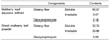
To prepare the MLP, mulberry leaves (Morus alba L.) were procured from a farm in Naju, Korea. Healthy plants were washed thoroughly under running tap water, shade dried, and ground to a fine powder using an air mill. To prepare the MLE, the MLP was infused in hot water at 60℃ for 1 h two times. The whole mass was centrifuged, and then the supernatants were freeze-dried. The DNJ was analyzed by a reversed-phase high-performance liquid chromatography equipped with a fluorescence detector after derivatization with 9-fluorenylmethyl chloroformate (FMOCCl) (Kim et al., 2003). Dietary fibers were also measured using the Prosky method (Prosky et al., 1984; Prosky et al., 1992). The dietary fiber and DNJ contents in the MLP and MLE are shown in Table 1.
Animals
Diabetic GK male rats (SLC, Inc., Hamamatsu, JAPAN) and their counterpart control Wistar rats were purchased from Jung-Ang Lab Animals Inc. (Seoul, KOREA). The rats were housed in individual cages in an air-conditioned room (22-24℃) with a 12 h light/dark cycle and 45 ± 5% humidity in compliance with the Guide for the Care and Use of Laboratory Animals (Nutrition Research Council, 1997). Feed (chow or experimental diets) and water (boiled and filtered) were provided ad libitum at all times. All animals were used in the experiments after 1~4 weeks of acclimatization and were killed humanely after the experiments.
Effect of a single dose of MLE using either maltose or glucose as substrate
The GK and Wistar rats (7 weeks old) were divided into 2 groups (n=5, each) based on body weight and oral glucose tolerance tests (OGTT) were performed. The rats were fasted overnight for 12 h and then given a single dose of maltose (2 g/kg body weight) with or without MLE (3.75 g/kg body weight; 6 mg/kg body weight as DNJ) by gavage feeding. This dose was selected taking into account the study that evaluated plasma DNJ concentration after oral administration of mulberry DNJ (purity > 95%) in the rats (Nakagawa et al., 2007). DNJ level in MLE was determined by HPLC before use. Blood glucose levels were measured in tail blood samples using an Accu-Check (Roche Diagnostics, Manheim, Germany) at 0, 15, 30, 45, 60, 90, and 120 min after each treatment. The incremental area under the curve (IAUC) was calculated using the trapezoidal rule with fasting levels as the baseline. A week later, the overnight fasted rats were administered a single dose of glucose (2 g/kg body weight) with or without MLE (same dose as above) by gavage feeding, respectively. After 30 minutes, the rats were euthanized and blood was collected from the heart for the determination of glucose and DNJ.
Effect of 8-week MLP administration
The GK and Wistar rats (10 weeks old) were stratified according to body weight and randomly assigned to control or treatment diet (n=10/diet) respectively. The experimental diets were formulated based on the nutrient content of the 93G diet of the American Institute of Nutrition and MLP inclusion level was determined based on the published data (Andallu & Varadacharyulu, 2003; Andallu & Varadacharyulu, 2007). The MLP diet was adjusted for carbohydrate, protein, and fat to create an equivalent energy density as that of the control diet. The dietary fiber content of the MLP diet was also adjusted to provide the same level as that of the control diet (Table 2). Body weights, fasting blood glucose (FBG) levels, and food intake were measured every week. After the 8-week period, the rats were sacrificed by exsanguinations from the heart under light ether anesthesia. The plasma was separated by centrifugation and stored at -80℃ until further analysis.
Biochemical assays
Serum glucose was measured using an enzymatic method with a test kit from Kodak Ektachem (Rochester, NY, USA). Insulin was determined by ELISA with a kit from Mercodia (Uppsala, Sweden). Insulin sensitivity was estimated by the homeostasis model assessment of insulin resistance (HOMA-IR) [Fasting plasma insulin (µU/ml) × fasting blood glucose (mmol/l)/22.5]. Hemoglobin A1c (HbA1c) and advanced glycation end products (AGEs) were measured using kits from BioSystems (Barcelona, Spain) and CycLex Co (Nagano, Japan), respectively. Determinations of triglycerides and free fatty acids were performed by enzymatic methods using kits from Asan Pharmaceuticals Ltd. (Seoul, Korea) and BioVision (Woburn, MA, USA), respectively. C-reactive protein (CRP) was determined by the ELISA method using a kit from BioVendor Co. (Modrice, Czech Republic).
Results
OGTT with MLE, using either maltose or glucose as substrate
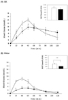
Fig. 1 shows that blood glucose concentrations were markedly elevated after maltose-loading and reductions in blood glucose concentration tended to be delayed in the GK rats compared to the normal Wistar rats. Oral administration of MLE significantly decreased peak heights of the glucose response at 45 min in the GK and at 30 min in Wistar rats by 28% and 62%, respectively, compared to the untreated groups (P < 0.05). IAUC, integrated over a period of 2 h (ΔAUC0-2h) after maltose-loading, was reduced by 11% in the GK rats (not significant statistically), but by 49% in the Wistar rats (P < 0.01), as compared to their respective controls.
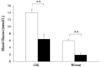
To examine whether MLE also inhibit the glucose uptake in the small intestine, blood glucose level was measured at 30 min, Tmax after a single dose of glucose-loading. The data presented in Figure 2 shows that blood glucose increases were significantly inhibited by the oral administration of MLE in both the GK and Wistar rats (P < 0.01). DNJ was detected in the blood 30 min after oral MLE administration in the range of 0~1.14 µmol/L (data not shown).
Eight-week treatment with MLP
There were no differences in body weight or food intake for either the GK or Wistar rats by MLP treatment. Based on the average food intake data and the specifics of the MLP (Table 1), DNJ consumption was calculated as 3.15 ± 0.11 mg/kg. Physical signs, glutamic oxaloacetic transaminase (GOT), glutamic pyruvic transaminase (GPT), total protein, and albumin were normal in all groups throughout the treatment period (data not shown).
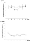
The FBG levels were determined every week at fixed times from the 2nd week of the experimental period. In response to MLP treatment, hyperglycemia was decreased significantly in the GK rats at weeks 4 and 5 (P < 0.05), but then glucose returned to levels similar to those of the controls towards the end of the 8-week experimental period. The FBG levels of the Wistar rats were not significantly affected in response to MLP treatment during the experiment (Fig. 3).
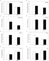
High FBG, insulin, HOMA-IR, glycated proteins (HbA1c and AGEs), and CRP were characteristics of the GK rats. The levels of FBG, insulin, HOMA-IR, and CRP were only decreased by 4%, 12%, 18%, and 25% respectively, with the inclusion of MLP in the diet without statistical significances in the GK rats. Wistar rats showed no changes in these parameters (Fig. 4, A~C, F). Eight week treatment of MLP tended to increase AGEs both in the GK and Wistar rats. However, the other glycated protein, HbA1c was increased statistically in Wistar rats (Fig. 4, D~E). Compared to Wistar rats, the GK rats showed lower blood TG and FFA levels. By treating MLP for 8 weeks, TG level was decreased by 74% but the difference was not statistically significant between two groups in the GK rats (Fig. 4, G~H).
Discussion
Two separate experiments were performed to investigate the antihyperglycemic effects of acute and chronic oral administration of mulberry leaf (Morus alba L.) in GK rats, the spontaneous non-obese animal model for type II diabetes, and their counterpart Wistar rats. First, we measured blood glucose levels using a single dose of maltose or glucose as substrate to clarify the effects of MLE on postprandial blood glucose responses as a result of inhibition of α-glucosidase or inhibition of glucose uptake in the small intestine. The results clearly show that MLE treatment at a dose of 3.75 g/kg body weight (6 mg/kg body weight as DNJ) decreased peak glucose responses and the ΔAUC0-2h of postprandial blood glucose after maltose-loading in the treated rats compared with the non-treated controls. Using wide ranges of DNJ concentrations from single pure chemical to mulberry leaf extracts of different preparations, several studies (Kimura et al., 2007; Miyahara et al., 2004; Oku et al., 2006; Yatsunami et al., 2008) have reported that DNJ, an N-containing sugar abundant in mulberry leaf, inhibited the activity of α-glucosidase. α-Glucosidase is a key enzyme to metabolize non-absorbable oligosaccharides into absorbable monosaccharides in the small intestine. Therefore, the glycemic control exerted by MLE might have been, at least in part, attributable to α-glucosidase inhibition.
The results also indicate that postprandial blood glucose responses at 30 min after glucose loading were significantly decreased by the oral administration of MLE in the GK and Wistar rats. Since DNJ is structurally similar to glucose, the intestinal absorption of DNJ can be regulated via a glucose transporter at the small intestinal brush-border (Voss et al., 2007). This beneficial effect shown for glucose absorption is postulated to be attributed to the activity of specific constituents in the MLE, such as quercetin, which is recognized as an inhibitor of glucose absorption (Kwon et al., 2007). In addition, the nutrient composition of the MLE (soluble dietary fiber) may have also played a role in its effects (Wheeler & Pi-Sunyer, 2008; Wong & Jenkins, 2007).
Next, given the role of MLE in postprandial glucose control, we examined the chronic effects of mulberry leaf on hyperglycemia and other metabolic abnormalities in GK rats for 8 weeks. The MLP was mixed into the diet at 10% (w/w) with adjustments of carbohydrate, protein, and fat to provide similar energy density and dietary fiber content for all experimental groups. Daily DNJ consumption was calculated based on the average food intake data and the specifics of MLP and it was about 3 mg/kg body weight. MLP administration for 4 and 5 weeks improved FBG levels in the GK rats, but these levels returned to values that were similar to those of the controls by the end of the 8-week treatment. Such discordant results may be partly attributable to aging, since it is known that insulin resistance gradually increases in GK rats with age. At the end of the experiment, the rats were 18 weeks old. Therefore, diabetes may have progressed to a more severe insulin-resistant stage that could not be mitigated by the level of MLP used in this experiment (Igarashi et al., 2007).
The GK rat is an animal model of spontaneous non-insulin-dependent diabetes mellitus. The pathogenesis of diabetes in the GK rat includes insulin resistance and abnormal glucose metabolism (Goto & Efendic, 1993; Janssen et al., 1999) In contrast to many other rodent models of type 2 diabetes, GK rats do not become obese and do not develop hyperlipidemia (Andallu & Varadacharyulu, 2003; Zhou et al., 1995) Accordingly, we confirmed that the GK rats showed characteristically higher FBG, insulin, HOMA-IR, glycated proteins, CRP and lower TG and FFA in the blood compared to those in Wistar rats respectively. Eight week treatment of MLP did not show statistically significant decreases, but tended to decrease in most parameters. Attributing reason of these results seems to be related with the lower contents of 1-deoxynojirimycin in MLP and/or compensation of dietary fiber of the experimental diet. The dietary fiber content of the MLP diet was adjusted to provide the same level as that of the control diet. HbA1c was increased statistically in Wistar rats. However, it does not provide any biological meaning.
In summary, the present study clearly demonstrates that preparations of mulberry leaf with focus on DNJ concentration can improve postprandial hyperglycemia and some metabolic abnormalities in GK rats. We propose that these effects might be mediated through the inhibition of glucose transporter as well as inhibition of α-glucosidase at the gut brush-border by specific principal phytochemicals and nutrients such as DNJ, quercetin, and soluble dietary fiber. Further investigations are in progress to elucidate the mechanisms of action of MLE enriched with DNJ.
 XML Download