

 PDF
PDF ePub
ePub Citation
 Print
Print


Introduction
Most studies suggested that exercise could be viewed as an effective antioxidant and antiatherogenic therapy. However, evidence is accumulating that strenuous exercise induces an imbalance between free radical production and the body's antioxidant defense systems (Ji, 1999; Lovlin et al., 1991; Maxwell et al., 1993; Sahlin et al., 1991). It has been reported that less the experience one has in training, higher the stress level was gotten (Powers & Hamilton, 1999). The contribution of free radical damage to the development of atherosclerosis is also established (Schwenke, 1998).
Vitamin B6 seems to be associated in some defense mechanisms especially against lipid peroxidation in tissues, since its deficiency increased this process when animals totally lacked in vitamin B6 diet (Ravichandran & Selvam, 1990; Ravichandran & Selvam, 1991). Marginal vitamin B6 contents increased lipid peroxidation and considerably stimulated the activity of glutathione-dependent enzyme (Cabrini et al., 1998). Increased plasma and tissue lipid peroxidation has been reported in rats receiving a vitamin B6 deficient diet (Benderitter et al., 1996). Pyridoxal 5' phosphate (PLP), the active form of vitamin B6, is essential as a cofactor for the metabolism of homocysteine to the amino acid, cysteine (Selhub, 1999). Vitamin B6 deficiency is a risk factor for coronary artery disease by elevated homocysteine levels. In addition, the antioxidative properties of vitamin B6 have recently been discovered (Jain & Lim, 2001; Matxain, 2006). However, the direct evidence that vitamin B6 deficiency affects the body antioxidative status with exercise has not been reported. So it is important to study the potential role of vitamin B6 deficiency on the effects of oxidative stress associated with exercise.
Therefore, the goal of this study was to determine whether vitamin B6 deficiency has effects on antioxidant enzyme activities and lipid profile under exercise-induced oxidative stress.
Materials and Methods
Experimental diets
Forty eight male weanling Sprague-Dawley rats (Daehanbiolink Co., Korea) were divided into 2 groups: group 1 (control, 24 rats), group 2 (vitamin B6 deficient, B6-, 24 rats). Rats were received a vitamin-free casein based semi synthetic diet which met AIN-93 recommendation (Reeves, 1997) with the exception of vitamin B6.
Exercise and sample collection
At the end of week 4, animals in each dietary group were subdivided into 3 exercise groups: pre-exercise (PreE); post-exercise (PostE); recess after exercise (recessE). PreE groups were sacrificed without exercise at the end of week 4. Exercised groups were exercised on a treadmill (10° incline, 0.5-0.8 km/h) with fasting state for 1 hour; animals in the recessE groups were allowed to take a rest for 1 hour after exercise. At the respective time points, animals were sacrificed by decapitation under the light ether anesthesia. Immediately following decapitation, plasma and liver were rapidly removed and stored at -40℃ until analyzed.
Biochemical analysis
The activity of plasma catalase (EC.1.11.1.6) was determined with a commercial kit based on the method of Zamocky (Bioxytech Catalase-520). The activity of superoxide dismutase (SOD, EC 1.15.1.1), the ratio of reduced glutathione and oxidized glutathione, and the level of malondialdehyde were determined in liver cytosol. Liver was homogenized in cold Tris-KCl buffer (0.1 M). The homogenized solution was centrifuged (8,000×g, 4℃, 30 min). The supernatant was then centrifuged (10,000×g, 4℃, 30 min. Again the supernatant was ultra-centrifuged (105,000×g, 4℃, 90 min) and separated the cytosol. SOD activity was determined with a commercial kit based on the method of Nebot (Bioxytech SOD-525). The ratio of reduced glutathione/oxidized glutathione (GSH/GSSG) was determined with a commercial kit based on the method of Anderson (Bioxytech GSH/GSSG-412). The level of malondialdehyde (MDA) was determined with a commercial kit based on the method of Gerard-Monnier (Bioxytech MDA-586).
Plasma Triglyceride (TG) was analyzed with a commercial kit based on the Trinder method (Youngdong Pharmaceutical Co., Korea). Total cholesterol (TC) was analyzed with a commercial kit based on enzymatic method (Youngdong Pharmaceutical Co., Korea). High-density lipoprotein-cholesterol (HDL-C) was analyzed with a commercial kit based on the same analytical method as total cholesterol after the precipitation of very low-density lipoprotein-cholesterol (VLDL-C), low-density lipoprotein-cholesterol (LDL-C) and chylomicron with polyethyleneglycol (International Reagent Co., Japan). Atherosclerotic index was calculated as (TC-HDL-C)/HDL-C.
Pyridoxal 5'-phosphate (PLP) was measured by HPLC method (Kimura et al., 1996) which was modified as follows: The mobile phase (0.1 M potassium dihydrogen phosphate containing 0.1 M sodium perchlorate, 0.5 g/l sodium bisulfite, pH 3) was pumped at a flow rate of 1.0 ml/min into the column (µBondpack ODS column, 3.9×300 mm, 10 µm porous packing, C18, Waters). Tissue samples were homogenized in cold sodium phosphate buffer (80 mM, pH 7.4). Aliquots of the tissue homogenates and plasma were added to perchrolic acid (1 M) and allowed to sit for one hour to release PLP from protein. This mixture was then centrifuged (18000×g, 4℃, 15 min) and the supernatants were removed. Fifty µl aliquot of supernatant was loaded in the sample loop and then injected onto the column. Samples for vitamin B6 analysis were prepared under yellow fluorescent lighting to prevent photodegradation of the vitamers.
Statistical analysis
All data were subjected to the analysis of variance and tested for significant differences by Duncan's multiple range tests (SAS Institute, Cary, NC). A p value < 0.05 was considered to be significant. The significance of difference between control group and B6- group was tested using independent two-sample t-test at P < 0.05.
Results
Table 1 demonstrates that the feed efficiency ratio (FER) and the final body weight of B6- were significantly lower than those of the control group. Plasma PLP concentration of B6- group was also significantly lower than those of the control group. Thus, it was considered that rats fed B6- diets became deficient in vitamin B6 by the 4th week.
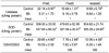
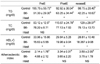
Table 2 demonstrates the effect of vitamin B6 deficiency on catalase activities. Compared to those of control group, the catalase activity of B6- group was significantly lower regardless of exercise. SOD activity of B6- group was also lower regardless of exercise. SOD activity of B6- group was decreased with exercise and was significantly lower than that of control groups in post-exercise and recess after exercise. Compared to those of control group, GSH/GSSG ratio was significantly lower in vitamin B6 deficient rats with pre-exercise. However, there was no significant difference between control and B6- groups in post-exercise and recess after exercise because GSH/GSSG ratio of control group was decreased with exercise but those of B6- groups was not significantly changed in post-exercise and recess after exercise. Table 3 demonstrates the effect of vitamin B6 deficiency on MDA levels. Compared to those of control group, MDA levels were significantly higher in vitamin B6 deficient rats in pre- and post-exercise and there was no difference between control and B6- groups in recess after exercise. Table 4 demonstrates the effect of vitamin B6 deficiency on plasma lipid profile. Compared to those of control group, the triglyceride level was significantly low in vitamin B6 deficient rats regardless of exercise and the tendency of decrease in post-exercise and recess after exercise was similar in both control and B6- groups. HDL-C level was significantly low in vitamin B6 deficient rats regardless of exercise and the tendency of no change in post-exercise and recess after exercise was similar in both control and B6- groups. Compared to those of control group, atherosclerotic index was significantly higher in vitamin B6 deficient rats in pre-exercise. However, there was no significant difference between control and B6- groups in post-exercise and recess after exercise.
Discussion
This study demonstrated that a reduction in antioxidative status caused by vitamin B6 deficiency may be aggravated under exercise-induced oxidative stress. At various points during the study, the antioxidative status in rats was evaluated using the ratios of GSH/GSSG and the activities of catalase and SOD as a direct measure and the level of MDA and lipid profile as an indirect, long-term measure. The vitamin B6 deficiency in rats was verified by the lowered plasma PLP levels as a direct measure and lowered body weight and FER as an indirect, long-term measure.
The hypothesis that vitamin B6 deficiency cannot react effectively to stress and decreases the antioxidative status was verified by results from two different measurements. First, GSH/GSSG ratio was significantly lower in vitamin B6 deficient rats in pre-exercise. This decreased activity of antioxidant enzymes has been also reported in vitamin B6 deficiency (Bordoni et al., 2006; Selvam & Ravichandran, 1993). Decreased GSH/GSSG ratio suggests a degraded antioxidant protection, which may have contributed to the higher exercise-induced ROS following vitamin B6 deficient diet. There was no significant differences between control group and vitamin B6 deficient group in post-exercise and recess after exercise because GSH/GSSG ratio of control group was decreased with exercise but those of vitamin B6 deficient groups was not significantly changed in post-exercise and recess after exercise. Therefore it is assumed that vitamin B6 deficiency induced an increase of plasma glutathione peroxidase activity and a decrease of plasma total antioxidant status under pre-exercise conditions and was accompanied by a decreased ratio of reduced glutathione and oxidized glutathione. It is generally reported that glutathione peroxidase activity after regular exercise training is increased in rats (Powers et al., 1999) and resting GSH/GSSG levels is increased 61% following isometric exercise training in humans (Peters et al., 2006). It is also reported that antioxidant nutrient status and exercise training have an interactive effect on oxidative stress and antioxidant enzyme activities (Benderitter et al., 1996; Chang et al., 2007). Although, compare to control group, the catalase activity of vitamin B6 deficient group was significantly lower regardless of exercise, this difference was decreased in post-exercise and recess after exercise because exercise-induced oxidative stress induced the decrease of plasma catalase activity in control group but exercise-induced oxidative stress did not affect the catalase activity in vitamin B6 deficient group. Exercise increases oxygen consumption and generation of reactive oxygen species such as superoxide and hydrogen peroxide. The SOD activity of control group remained stable with exercise-induced oxidative stress but the SOD activity of vitamin B6 deficient groups was decreased with exercise and was significantly lower in post-exercise and recess after exercise. Thus, it is suggested that vitamin B6 deficient rats did not react effectively to stress and decreased antioxidant enzymes activity in this study.
Second, an increased susceptibility to lipid peroxidation is facilitated by the decreased activities of antioxidant enzymes. Previous studies reported that the concentration of thiobarbituric acid reactive substances (TBARS) in liver was high in vitamin B6 deficient rats (Benderitter et al., 1996) and the liver glutathione concentration was increased in rats fed excess vitamin B6 (Mahfouz & Kummerow, 2004). Increased susceptibility to lipid peroxidation in rat liver and heart (Cabrini et al., 1998), rat plasma (Ravichandran & Selvam, 1991) and cells of Fusarium species (Kayali & Tarhan, 2006) was also reported. Because the MDA level in this study was significantly higher in vitamin B6 deficient rats in pre- and post-exercise and tended to be higher in recess after exercise although the difference was not significant, it is assumed that vitamin B6 deficiency leads to an increase of lipid peroxidation in rats.
Compared to that of control group, atherosclerotic index was significantly higher in vitamin B6 deficient rats in pre-exercise. However, there was no significant difference between control group and vitamin B6 deficient group in post-exercise and recess after exercise because atherosclerotic index of control group was increased in post-exercise and recess after exercise but that of vitamin B6 deficient group was not significantly changed with exercise. Also, HDL-C level was significantly low in vitamin B6 deficient rats regardless of exercise. Thus, it is suggested that vitamin B6 deficiency has a negative effect on atherosclerotic index but does not aggravate it further under exercised induced oxidative stress.
Therefore, despite the many uncertainties regarding the mode of action, these results suggest that vitamin B6 deficient animal do not react effectively to oxidative stress and a reduction in antioxidative status caused by vitamin B6 deficiency may be aggravated under exercise-induced oxidative stress.


 XML Download