

 PDF
PDF ePub
ePub Citation
 Print
Print


Introduction
Cefepime (a molecular formula of C19H25ClN6O5S2HClH2O and a molecular weight of 571.5) is a parenteral fourth-generation cephalosporin antibiotic with a wide spectrum of antimicrobial activity and a pharmacokinetic profile similar to ceftazidime [18]. It is active against many Gram-positive and Gram-negative bacteria, such as Staphylococcus aureus, Streptococcus pneumoniae, Escherichia coli, Klebsiella pneumoniae, Proteus mirabilis and Pseudomonas aeruginosa [5,25], with less susceptibility to extended-spectrum-lactamases [13]. The chemical structure of cefepime allows it to bind to the penicillin-binding proteins and penetrate the outer membrane of Gram-negative bacteria more rapidly than most cephalosporins. In humans, cefepime has been approved for the treatment of lower respiratory tract, intra-abdominal, complicated and uncomplicated urinary tract infections as well as uncomplicated skin and skin structure infections [18]. It has also been shown to be therapeutically equivalent to cefotaxime and ceftriaxone in the treatment of pediatric meningitis [20].
Fever, which may be associated with many bacterial and viral diseases, changes various physiological parameters such as the heart rate, renal blood flow, hepatic and total splanchnic blood flow, diuresis, and enzyme activities [14], which can alter the pharmacokinetics of certain drugs [3]. For example, an increased volume of distribution for penicillin-G has been reported in rabbits, pigs and dogs during endotoxin-induced fever [26]. However, pigs, dogs and rabbits showed higher blood concentrations of sulphathiazole, sulphadimidine and gentamicin during endotoxin-induced fever [26]. Therefore, animals suffering from fever may require a modified dose regimen.
The pharmacokinetics of cefepime have previously been examined in monkeys and rats [7], adult horses [10], dogs [8,24], neonatal foals [8] calves [11] and ewes [12]. However, there is little information of the pharmacokinetics of cefepime after administration via the intramuscular route, which is the most popular and convenient route of administration.
Rabbits are quite prone to abscesses formation and respiratory diseases. The bacteria most often involved in these complications include Pasteurella multocida and Staphylococcus aureus. However, there are few antibiotics that can provide a safe and effective therapy for such conditions particularly those caused by resistant strains. Cefepime can be used to treat bacterial infections caused by strains resistant to other antibiotics. Furthermore, animals suffering from fever may require a modified dosage regimen. Therefore, the main objective of this study was to determine the effects of experimentally induced fever on the disposition kinetics of cefepime administered intramuscularly to rabbits. The dose used in this experimental model is somewhat lower than that used in previous studies in a meningitis model [9].
Materials and Methods
Antimicrobial agent administration
Cefepime hydrochloride powder (Maxipime; Bristol-Meyers Squibb, USA) was reconstituted with sterile normal saline to a final concentration of 10% according to the manufacturer's guidelines, and was administered intramuscularly to healthy and febrile rabbits at a dose of 75 mg/kg BW.
Animals and husbandry
The study was performed in accordance with the guidelines for animal care of the Faculty of Veterinary Medicine, Cairo University, Egypt. Six healthy male rabbits, weighing 2,100-2,500 g, were obtained from the Laboratory Animal Farm, Faculty of Veterinary Medicine, Cairo University, Egypt. The rabbits were housed individually in cages under a 12-h light/dark cycle and fed good quality hay (alfalfa) and/or a pelleted feed concentrate (fiber 18%, protein 14%, calcium >1 and fat 2%) with free access to water. The room temperature and relative humidity were maintained at 20 and 22℃, and between 30 and 60%, respectively. The animals were allowed to acclimatize and did not receive any drug treatment for at least 15 days preceding the study. The same rabbits were used in subsequent experiments after observing a minimum washout period of 2 weeks.
Experimental protocol
The study comprised of the following 2 phases:
Phase 1: The animals were individually weighed immediately be administering the drug in order to determine the precise dose. Prior to the intramuscular injection, each rabbit was placed in a restraining device. All the rabbits were injected with freshly prepared cefepime (75 mg/kg BW) into the left semimembranous muscle. Heparinized blood samples (0.5 ml) were obtained from the right auricular vein. Blood samples were collected immediately before (pre-treatment, 0 h) and 10, 20, 30, 45 min as well as 1, 2, 4, 6, 8, 10, 12, 24, and 48 h after drug administration. Each blood sample was gently inverted a few times in order to allow for complete mixing with the anticoagulant and stored on ice. Within 30 min of collection, each sample was centrifuged for 15 min at approximately 1,500 × g to separate the plasma. The plasma samples were stored in -20℃, and assayed on the same day of sampling.
Phase 2: The clinically isolated E. coli strains (obtained from Department of Microbiology, Faculty of Veterinary Medicine, Cairo University, Egypt) were stored at -80℃ until needed. These strains were recovered by inoculating a small portion of the stock into heart infusion agar (Difco, USA) overnight at 37℃ to obtain logarithmic-phase growth. A single colony was selected and suspended in a pyrogen-free phosphate buffered solution. The concentrations of bacteria after the overnight culture were estimated from the optical density as well as by serial dilution. This suspension was then diluted to achieve a concentration of 107~108 colony-forming units (CFU)/ml. An infection was induced by an intravenous inoculation of 5 × 10⁸ CFU, 24 h before the pharmacokinetic investigation. Cefepime was administered intramuscularly at the same dose and the sampling procedures were performed as in phase 1.
Analytical method
The plasma cefepime concentrations were determined using a microbiological assay method described elsewhere [2,17], with Bacillus subtilis ATCC 6633 as the indicator strain growing on Mueller-Hinton agar (Mast Group, UK). Briefly, six wells, 8 mm in diameter, were cut at equal distances into a (120 × 120 mm) petri plate containing 25 ml of seeded agar. The wells were filled with the test samples and/or a cefepime standard solution (prepared from a commercial solution). Standard curve of cefepime was prepared in pooled antibacterial-free plasma. The standards and samples were tested in duplicate. The plates were kept at room temperature for 2 h before being incubating at 37℃ for 18 h. The mean inhibition zone diameters were measured and the concentrations in the plasma samples were calculated from the standard curve. The standards were included in each assay plate in order to compensate for any plate-to-plate variations. There was a linear relationship between the zone of inhibition and the logarithm of the plasma cefepime concentration with a correlation coefficient of 0.990. The intra-day coefficient of variation was <8%. The reproducibility of this method was excellent and the inter-assay variability was <5%. The limit of quantitation was 0.1 µg/ml. A standard curve was considered acceptable if the quality control samples were within 15% of the nominal concentration. This assay failed to distinguish between cefepime and its antibacterial metabolite(s). Therefore, results are expressed as the plasma cefepime equivalent activity. However, in order to simplify the presentation, the term, concentrations, are used throughout the text.
Pharmacokinetic analysis
The pharmacokinetic parameters of cefepime were estimated by noncompartmental method using WinNonlin V2.0 (Pharsight, USA). The rate constant associated with the log/linear part of the curve (λ) was determined using linear regression. The total area under the plasma concentrationtime curve (AUC) was calculated using the linear trapezoidal rule. The AUC from 0 to infinity [AUC0~∞] was calculated as the AUC (0, ∞) = AUC + Ct/λ (where Ct is the last plasma concentration measured). The elimination half-life (t1/2λ) of cefepime was calculated using the following equation: t1/2λ =ln 2/λ.
Statistical analysis
The results are expressed as the mean ± SD. The differences in the pharmacokinetic values obtained before and after the infection were compared using a paired t-test (two-tailed). All the data was analyzed using the statistical program, Sigmastat (version 2.0; SPSS, USA). The differences were considered statistically significant at p < 0.05.
Results
Response to infection and drug
The intravenous inoculation of 5 × 10⁸ CFU of Escherichia coli caused an increase in body temperature. Twenty-four hour after the injection, the temperature increased to 1.0℃ above the basal value. With the exception of mild anorexia, there were no abnormal findings of appearance in the infected rabbits, such as corneal hyperemia or increased lassitude.
No serious adverse events of cefepime were observed throughout the study. Most animals experienced none to mild pain and only minimal discomfort at the injection site.
Pharmacokinetic analysis
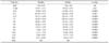
The standard curve for cefepime in plasma was linear within the concentration ranges of 0.1 to 200 µg/ml. Table 1 shows the mean plasma concentration-time data of cefepime after intramuscular administration in rabbits before and after the infection. The infection caused a marked increase in the plasma cefepime concentration from 0.166 to 24 h after administration, with the exception of 0.5 h. The drug was not detected in the plasma 48 h after drug administration (Table 1). Table 2 shows the pharmacokinetic parameters . There was a significant decrease in the elimination half-life (p = 0.002) compared with healthy animals. In addition, the infection significantly increased the peak plasma concentrations (p = 0.022), the mean residence time (p = 0.002), the area under the plasma-concentration time curve (p < 0.0001) and the area under the moment curve (p = 0.0001).
Discussion
The interpretation of the data considered the assay method used (microbiological) and the sensitivity of the assay method. This is because cefepime and its putative metabolites might have different antimicrobial activities, the ratio of the parent to its metabolites might not remain constant throughout the dosing interval and the movement of the metabolites from the blood may not be the same as for the parent drug. Therefore, there might be some error in interpreting the concentrations derived from the microbiological assay for the purpose of establishing minimally effective concentrations in plasma. The bioassay method was chosen because of cost constraints and that other researchers have reported non-significant differences between the results of the microbiological assay and HPLC methods [17]. On the other hand, Bächer et al. [2], reported a good correlation coefficient between the HPLC and the bioassay in human serum (r = 0.950), but only presented the data from the bioassay method.
The intravenous inoculation of the E.coli suspension into the rabbits simulates some of the pathological effects of septicemia in humans [31]. The increase in body temperature, cardiac and respiratory rate and peripheral resistance are followed by secondary acidosis and decreases in cardiac output. These rheological alterations correspond to the changes in circulation observed in septicemic humans [22]. Furthermore, the changes in the acid-base balance also influence the physicochemical properties of cephalosporins. The changes in pH lead to sigmoidal changes in the degree of ionization, which in turn leads to changes in the mean residence time [21,29].
The acute phase response (APR) is defined as a pathophysiological condition induced by many causal factors, i.e. infection, inflammation and tissue. The APR induces many systemic changes, which include fever, increased lassitude, loss of appetite as well as the synthesis and secretion of acute phase hepatic proteins [27]. Significant concentrations of proinflammatory cytokines, such as tumor necrosis factor-α, interleukins and interferons, are produced during APR, which leads to the direct suppression of the microsomal cytochrome P450 (CYP)-dependent activity in the liver [1,4,30]. This might in turn alter the pharmacokinetic profile of the some drug. Furthermore, studies in experimental animals have shown that the maximum depression of CYP occurs 12~24 h after an LPS injection [15]. Cephalosporins have neither inhibitory nor stimulatory effects [16]. Therefore, the decreases in the elimination of cefepime in the infected rabbits might be related to the suppression of the CYP isoforms by the microbial suspension. The process of enzyme inhibition usually begins with the first dose of the inhibitor, and the onset and offset of inhibition correlate with the half-lives of the drugs used [6]. Similar findings of a decrease in elimination had been reported for theophylline and phenytoin in humans during APR as follows: a decrease of theophylline elimination due to a viral respiratory infection [3], bacterial pneumonia [23,28] and influenza vaccine inoculation [19]. There are few reports on the in vivo metabolism of cefepime in animals. In humans, cefepime is metabolized to N-methylpyrrolidine (NMP), which is rapidly converted to the N-oxide (NMP-N-oxide). To our knowledge, there is no evidence of the production of active metabolites in rabbits.
The significantly higher AUC, area under first moment of plasma concentration-time curve and mean residence time values in febrile rabbits observed in this study show that the drug remains in the body for a comparatively longer duration in the febrile condition.
In conclusion, the cefepime dose used in this study produced therapeutically useful concentrations in the plasma. The endotoxin-induced febrile state produced significant changes in the plasma levels and some of the pharmacokinetic variables of cefepime. Nonetheless, these changes are not likely to adversely affect the desirable properties of cefepime in rabbits.

 XML Download