

 PDF
PDF ePub
ePub Citation
 Print
Print


Hermaphrodite means an animal or a human being in which male and female sex organs are present simultaneously, or in which the sex organs contain both ovarian and testicular tissue [3]. Sometimes one gonad becomes a testis while the other becomes an ovary; sometimes the gonads become ovotestes containing a mixture of male and female components [1]. Although hermaphroditism is often associated with infertility, that is not always the case. Hermaphroditism occurs rarely in human and animal populations [5]. Only few cases of hermaphrodite have been reported in various breeds of dogs such as Basset hound [2], Cocker spaniel [7] and Pug [8]. Here, we report a very rare case of a hermaphrodite dog.
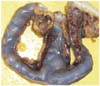
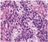
Hermaphroditism was identified in a 3-year-old American Cocker spaniel with external female appearance. The dog was presented due to vaginal discharge, loss of appetite. The bitch showed a reddish finger-like structure protruding about 6 cm from the vulva. The owner had noticed the appearance of abnormal genitalia since its purchase. The urethral orifice was located cranially to the base of the os clitoridis. Abdominal sonography and radiography demonstrated abnormal enlargement of uterus. The bitch was gonadohysterectomised and a clitorisectomy was performed. The finger-like structure was about 3 cm in diameters and revealed an enlarged os clitoridis including bone. The uterus was enlarged and filled with cheese-like sticky materials (Fig. 1). The gonads were situated caudal to the kidneys at the cranial tips of the uterine horns, and were composed mainly of ovarian follicles. However, in some area, seminiferous tubules and interstitial cells were observed (Fig. 2). The trimmed tissues were fixed in 10% neutral buffered formalin, and embedded in paraffin. Four µm sections were made and stained with hematoxylin and eosin for histopathological examination. The histology of both gonads was similar, being ovotestes. There was a compact arrangement of seminiferous tubules lined by Sertoli cells. Germ cells were absent, but large numbers of interstitial cells were present near the capsule of both gonads. Both were covered with a cuboidal epithelium characteristic of ovarian surface epithelium. Oviductal tissue and incompletely developed epididymis were found adjacent to each gonad. Each gonad had efferent tubules, a pampiniform plexus, fimbriae, and a uterine tube. Clinicopathological features of the dog suggested hermaphroditism with bilateral ovotestes and pyometra.
The present study describes a hermaphrodite dog with pyometra. We thought that vaginal discharge in this case was probably related with her pyometra. The bitch made a complete recovery following an ovariohysterectomy. Abnormalities in sexual development can arise from a defect at any step in sexual development, the chromosomal, gonadal or phenotypic level of differentiation [4]. Abnormalities of gonadal sex occur in an individual if the chromosomal and gonadal sex does not agree [2]. The pig is one of the species in which hermaphrodite has been well described. In this species, there is a high incidence of this condition, which affects around 0.1-0.5% of XX females [6]. However, hermaphroditism is very rare in dog and human. Furthermore, a hermaphrodite dog with pyometra has not been reported until now. This report describes the first hermaphrodite case with pyometra in an American Cocker spaniel dog in Korea.
 XML Download