

 PDF
PDF ePub
ePub Citation
 Print
Print


INTRODUCTION
Transurethral resection of bladder tumor (TURBT) is well-established modality for treating non-muscle-invasive bladder cancer. Furthermore, it is essential for evaluating the pathological depth of bladder tumors in resected specimens. Despite improvements in endoscopic instruments and imaging systems, however, TURBT remains a challenging technique for unskilled surgeons to master.
There are well-recognized complications associated with TURBT, such as bladder perforation, irrigation fluid absorption, bleeding, infection, damage to the ureteric orifices, obturator nerve stimulation, and tumor cell implantation [1]. Among these, bladder perforation is one of the most frequent complications of TURBT and is sometimes associated with obturator reflex. Several researchers have reported that the risk of obturator nerve stimulation is from 10.6% to 11% [2,3].
Several advances in instruments and techniques, including modifications of resectoscope design and snare resection, have been proposed to remove pedunculated bladder tumors [4,5]. However, difficulties remain in safely collecting adequate specimens for flat tumors. We introduce an easy technique for superficial bladder cancer that uses a standard monopolar resectoscope loop.
In this study, we demonstrate the easy resection technique for superficial bladder tumors that we have termed the "grasp and bite" TURBT technique and compare it with conventional TURBT.
MATERIALS AND METHODS
1. Patients
This was a retrospective study in the Department of Urology of Chonnam National University Hospital (Gwangju, Korea) between January 2012 and April 2013. This study included a total of 29 men and 6 women who had bladder tumors with superficial lesions and who underwent TURBT. Patients with a huge unresectable mass, an invasive tumor shown during preoperative imaging, or an irradiated bladder were excluded. Bladder tumors were diagnosed preoperatively with cystoscopy under local anesthesia. Group 1 patients were treated by use of the conventional TURBT technique, and group 2 patients underwent the grasp and bite technique. We applied each technique during a distinct time period. Conventional TURBT was performed in 2012 and grasp and bite TURBT was performed in 2013. All procedures were performed by a single experienced operator. This study was approved by the Institutional Review Board of Chonnam National University Hospital (IRB No. CNUH-2014-025).
2. TURBT technique
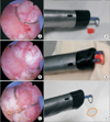
Transurethral resection was performed with a monopolar resectoscope system (24-Fr Karl Storz, Tuttlingen, Germany). For the grasp and bite technique, the operator initially defined the tumor morphology and safe margin. In small and flat lesions, the tumor and surrounding mucosal lesions were positioned between the resection loop and the end portion of the resectoscope sheath. The resectoscope sheath was then moved backward carefully. The tumor could be held tightly with the loop electrode and sheath. We call this the "grasp" step (Fig. 1; Video clip, Supplementary material). Using the grasping technique, the tumor lesions were lifted up. In the "bite" step, the surgeon maintained a tight hold and resected the tumor while grasping. A cystoscopic view and schematic illustrations are shown in Fig. 1. For resection during the bite step, the movement of the electrode loop was straight backward in a linear direction. Resection was performed carefully so as not to cause bladder perforation without excessive bladder wall distention. Poorly located tumors were accessed by suprapubic pressure and nearly emptying the bladder. When necessary, cold cup biopsy was done at the tumor base and edges. The resection site was carefully inspected for bleeding and coagulation. In bulky or pedunculated tumors, the protruding mass was resected rapidly with the grasp and bite technique. After removal of the bulging mass, the base lesion and surrounding mucosa were resected carefully by using grasp and bite TURBT as mentioned earlier. Also, the tumor margin could be resected in the same manner repeatedly.
Resected specimens were reviewed by a pathologist to evaluate the depth of resection. Specimens were measured by hematoxylin and eosin (H&E) staining and immunohistochemical smoothelin stain to determine the muscularis mucosae.
3. Statistical analysis
Independent t-tests were used to compare the mean values of two independent parametric continuous variables. The Mann-Whitney test was used to compare the median values of two nonparametric continuous variables. Categorical variables were verified by Pearson chi-square or Fisher exact test. p<0.05 was considered to indicate statistical significance. Statistical analyses were performed with IBM SPSS Statistics ver. 20.0 (IBM Co., Armonk, NY, USA).
RESULTS
From January to April 2012, 16 patients were enrolled who had undergone conventional TURBT. From January to April 2013, the grasp and bite technique was applied to 19 patients with superficial bladder tumors. Tumors ranged in size from 0.5 to 5 cm. The two groups had comparable demographic and intraoperative data, including patient age, tumor size, patient gender, multiplicity, tumor morphology, and tumor location (Table 1).
The mean operative time was similar in the conventional and grasp and bite groups (35 minutes vs. 35 minutes, respectively). The mean duration of irrigation, duration of catheterization, and hospital stay were not significantly different between the groups. Hemoglobin change and complication rates were also comparable between the two groups. There were no cases of bladder perforation, excessive obturator reflex, or persistent hematuria in either group. The tumor stages and grades were similar in the two groups. Specimens were adequate for accurate pathological staging of the muscularis mucosae. H&E staining and smoothelin immunohistochemistry positivity identified the muscularis mucosae in the grasp and bite group (Fig. 2). There was no significant difference in recurrence rates between the conventional and grasp and bite TURBT groups during the 14-month follow-up (43.8% vs. 31.6%, p=0.458). The detailed postoperative outcomes are given in Table 2.
DISCUSSION
The purpose of TURBT is complete removal of all visible tumors for curative intent and acquisition of adequate tissue for pathologic evaluation. Achievement of the above purposes is not so easy for an inexperienced surgeon. Also, tumor recurrence after TURBT is relatively high. For non-muscle-invasive bladder cancer, the probability of recurrence after 1 year ranges from 15% to 70% [6,7]. Many obstacles can arise during the procedure, such as bladder perforation, bleeding, obturator reflex, and damage to the ureteral orifices. To overcome these difficulties, many surgical techniques and innovative devices have been developed. For example, endoscopic snare resection of a bladder tumor can be used to remove pedunculated tumors [5]. Although the snare method offers the advantage of en block resection, smaller and sessile tumors are not suitable for this technique. New devices such as water jet hydrodissection and the thulium:yttrium aluminium garnet laser may be applicable for en block resection of superficial tumors [8,9]. However, further studies and development of these techniques are necessary to determine efficacy and oncologic outcomes.
Variable methods of tumor resection have been developed and used in association with tumor size and location. These also are determined by a surgeon's preference and surgical experience. Modifications of TURBT equipment and techniques also have the possibility of improving surgical outcomes. Accurate surgical technique is mandatory for dealing with safety and efficacy issues. Innovative resectoscope modifications have also been developed. The side-to-side bidirectional lateral rotating handgrip and loop allows excellent precision and control over the loop under direct vision [4]. Loop modification, the so-called "runner," can push the bladder wall away during resection, thereby widening the view and decreasing the risk of perforation [10]. We developed an easy and safe technique for an inexperienced operator. We wanted a technique in which we could use a conventional resectoscope and loop without accessory equipment.
When using a wire-loop resectoscope, the tumor must be resected thoroughly to obtain adequate muscle tissue. The movement of the loop is somewhat fast and deep to avoid charring of the surrounding mucosa and bladder perforation [11]. These are key technique of TURBT, but not so easy to perform. Our technique has the merit of acquiring adequate tissue. Direct pressure to the bladder wall may allow deeper tissue to protrude into the bladder wall and then be easily removed by use of the resectoscope. Instead of the curvilinear movement of the resectoscope end according to the curved bladder surface, the easy linear motion can make a curved resection with grasping. Using our "grasp and bite" technique, beginners can easily perform TURBT safely. Often, small and sessile tumors are missed and severely damaged during resection and cauterization. The grasp and bite technique can effectively be applied to small, flat, sessile lesions. For larger tumors, the protruding tumor can be removed f irst by the conventional method, and then the stalk and base lesion can be removed by the grasp and bite technique. Poorly accessible tumors are difficult for complete resection. In theory, the grasp and bite technique could have difficulties in a posterior or posterolateral located tumor because of its moving mechanism. In our series, two posterior and four posterolateral tumors were successfully accessed by use of suprapubic pressure and nearly emptying the bladder. Similar to conventional TURBT, it was also difficult to access posterior tumors with the grasp and bite method.
In our consecutive cases, obturator reflex was not problematic. There were no cases of excessive obturator reflex or bladder perforation in either group. General anesthesia was preferred in the conventional and grasp and bite groups (81.3% vs 57.3%, p=0.138). Also, tumor locations did not differ significantly between the two groups (p=0.253). We postulated that the grasping method, which lifts the tissue away from the obturator nerve and tightly holds the grasped tissue, may eventually weaken the reflex. Considering this small population, however, we cannot make conclusions and need to further study a larger population.
This study had some limitations that may have limited the conclusions. First, the two groups were classified by the date of operation. Thus, there could have been a selection bias. Also, the study population was small, and thus we were unable to perform statistical analysis of the oncologic outcome as recurrence-free survival. This was a preliminary study to develop a surgical technique performed by an experienced surgeon. Larger prospective studies with a long follow-up period will be necessary to show the oncologic advantage of this technique. To determine the feasibility of the technique for inexperienced surgeons, future study will address a teaching protocol comparing the conventional and grasp and bite methods. Owing to the nature of retrospective research, histologic information was not fully acquired. Investigation of tissue adequacy of proper muscle and effect of coagulation will be our next subject of study.
CONCLUSIONS
Our study demonstrated that grasp and bite TURBT is a safe and effective method for removing superficial bladder tumors. This technique has merit for flat tumors and can also be used for pedunculated and poorly accessible tumors. The grasp and bite method may be a shortcut for overcoming the difficulties of TURBT.


 XML Download