

 PDF
PDF ePub
ePub Citation
 Print
Print


INTRODUCTION
Genital warts are known to occur worldwide and are common, highly infectious benign epithelial mucosal tumors caused by low-risk human papillomavirus (HPV) [1,2]. Genital warts are not life-threatening and some spontaneously resolve [3,4]. However, they have high infectivity with a transmission rate of -65% [5], a high risk of recurrence, and a high rate of multiple treatments [6,7]. Thus, they are associated with a high economic burden, psychosocial stigma, feelings of shame, and lower quality of life [8,9].
Given that high-risk HPV types, especially HPV16 and HPV18, cause cervical cancer, other anogenital neoplasms, and head and neck cancers, genital warts with high-risk HPV types may be considered a clinical marker of risk for developing a malignant disease. Epidemiological studies reported that once HPV is present in one partner, it is quickly transmitted to the other [10,11]. Thus, genital infection with high-risk HPV types is not only the problem of the infected patients, but also an issue of their sexual partners. A thorough understanding of the prevalence of high-risk HPV types in genital warts of men and their impact is important for the planning of treatment, follow-up, and preventive strategies toward HPV-related diseases. Recently, some studies reported that 20% to 50% of genital warts were found to be colonized or coinfected with high-risk HPV types [12-15]. However, data on the prevalence and distribution of high-risk HPV types in genital warts, especially of men, are still insufficient. In the present study, we evaluated the prevalence and type distribution of high-risk HPV infection in genital warts of Korean men, and for the first time, described risk factors associated with high-risk HPV infection in male genital warts.
MATERIALS AND METHODS
Between October 2009 and May 2012, 150 consecutive male patients with histopathologic-confirmed genital warts who underwent HPV genotyping by use of polymerase chain reaction (PCR) were included in this study. All patients reported no previous diagnosis of penile cancer, anal cancer, or HIV infection; had not taken HPV vaccination; and were heterosexual. Through careful inspection, circumcision status, number of warts, sites of lesions, and macroscopic morphology were checked. The macroscopic morphology of warts was categorized as acuminate or papular [1,16]. Acuminate lesions were exophytically pedunculated growing tumors with papillary projections. Papular lesions were sessile with smooth or rough surfaces, but no real filiform projections. The two different types could be seen in the same patient; we defined this as mixed type. Recurrent status of genital warts was checked at diagnosis. The measurements of the present study were detection of HPV DNA by use of PCR and genotyping in genital warts [17]. Because this study was conducted during the patients' routine clinical follow-up without any modification, no ethics committee approval was necessary. This study complied with the Declaration of Helsinki, and all men were informed of the purpose of the study and gave their consent.
A cytobrush (Moa Plastic Labware Co., Seoul, Korea) was used to brush exfoliated epithelial cells from the genital warts. For patients with multiple warts, all visible lesions were brushed. The cytobrush was cut and placed in a tube containing 5 mL of phosphate-buffered saline and sent to Seoul Clinical Laboratories (Seoul, Korea), where DNA extraction and HPV genotyping were performed. All specimens were stored at -70℃ until PCR analysis and genotyping were undertaken. DNA was extracted from specimens by use of the Chemagic Magnetic Separation Module I (PerkinElmer Chemagen Technologie GmbH, Baesweiler, Germany) according to the manufacturer's instructions. Extracted DNA was stored at 4℃ until PCR. HPV testing was undertaken by use of PCR for amplification of a fragment of the L1 gene [18]. The target DNA was amplified by using provided primers, PGMY09 and PGMY11, for HPV DNA [18]. For each PCR, primers for the human β-globin gene were used as a positive control to determine the specimen's adequacy for testing. HPV genotyping was performed by the linear array method with the HPV genotyping chip kit (Biomedlab, Seoul, Korea) according to the manufacturer's protocol. This kit permits simultaneous detection and typing of 32 different HPV types in a single reaction. On the basis of the grouping of the International Agency for Research on Cancer [19], we considered HPV16, 18, 26, 31, 33, 34, 35, 39, 45, 51, 52, 53, 56, 58, 59, 66, 68, 69, 70, and 73 as high-risk with respect to oncogenic potential. HPV6, 11, 32, 40, 42, 43, 44, 54, 55, 57, 61, and 62 were grouped as low-risk.
By use of univariate and multivariate logistic regression, the associations between high-risk HPV prevalence and various characteristics, such as age, circumcision status, recurrent status of genital warts, number of lesions, site of lesions, and gross morphology, were assessed by odds ratios (ORs) and 95% confidence intervals (CIs). A p<0.05 was considered statistically significant. Statistical analysis was performed by use of SPSS ver. 12.0 (SPSS Inc., Chicago, IL, USA).
RESULTS
1. Patient characteristics
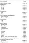
One hundred fifty cases were investigated. Of them, 15 men (10%) whose specimens were negative for both HPV and β-globin were excluded. Three samples negative for HPV DNA were also excluded. Altogether, 132 men were analyzed for the purpose of this study, and their characteristics are presented in Table 1. The ages of these men were between 17 and 68 years (median age, 29 years). Of them, 119 (90.2%) had undergone circumcision. At the time of diagnosis, 57 cases (43.2%) were recurrent. Fifty-four patients (40.9%) had more than five lesions at diagnosis. Genital warts were often located on the penile shaft (65.2% of cases), base of the penis (31.8%), pubic area (12.1%), and coronal sulcus (9.8%). In 49 men (37.1%), lesions were inspected at the sites including the base of the penis or the pubic area, whereas 83 men (62.9%) had genital warts at the remaining sites. On the basis of macroscopic morphology, the lesions were categorized as acuminate or papular/mixed. In 116 men (87.9%), only acuminate lesions were inspected. On the other hand, papular or mixed lesions were seen in 16 men (12.1%).
2. Prevalence of specific genotypes
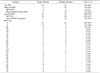
The distribution of HPV types among the 132 cases is shown in Table 2. Low-risk HPV types were detected in 128 cases (97.0%). Of these, 87 cases were infected with a single HPV type, and 41 cases were infected with multiple types. In 101 cases (76.5%), only low-risk HPV types were found. High-risk HPV types were detected in 31 cases (23.5%), and of these, 27 cases (20.5%) contained both high-risk and low-risk HPV types. Four cases were infected with only high-risk types. Regardless of whether it was a single infection, HPV6 (76.5%) was the most common HPV type detected. HPV6 was found as a single-type infection in 67 cases (50.8%) and as a multiple-type infection in 34 cases (25.8%). In 121 cases (91.7%), HPV6 or HPV11 or both were observed. High-risk HPV types detected frequently were HPV16 (6.8%), HPV33 (4.5%), HPV18 (2.3%), and HPV68 (2.3%). In particular, the prevalence of infection with HPV16 or HPV18 or both was 8.3% (11 of 132). Other high-risk HPV types were found in <2% of cases.
3. Determinants of high-risk HPV infection in genital warts of men
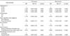
Table 3 shows the association between high-risk HPV prevalence and various characteristics. In the multivariate analysis, there was no statistical association between age and high-risk HPV infection in genital warts of men. Neither the type of genital warts diagnosis nor the number of lesions was significantly associated with high-risk HPV infection. On the other hand, statistically significant associations with high-risk HPV infection were found with the site of lesions, morphology of genital warts, and circumcision status. A higher prevalence of high-risk HPV infection was observed in men with genital warts located at sites including the base of the penis and/or the pubic area (38.8%) than at the other sites (14.5%; OR, 6.92; 95% CI, 2.35-20.41). In men with papular or mixed genital warts, the high-risk HPV prevalence (43.8%) was significantly higher than that in men whose genital warts were only acuminate (20.7%; OR, 6.21; 95% CI, 1.62-23.89). Of the 13 uncircumcised men, 4 men (30.8%) were infected with high-risk HPV types, which was significantly higher than the prevalence (22.7%) of high-risk HPV in circumcised men (OR, 4.67; 95% CI, 1.02-21.34).
DISCUSSION
In the present study, the prevalence of low-risk HPV infection in genital warts of men was 97.0%. The most commonly detected HPV types were HPV6 and HPV11. HPV6 and/or HPV11 was observed in 91.7% of cases. This result is consistent with previous studies, which showed that HPV6 and/or HPV11 was found in a majority (>89%) of genital warts [12,13]. In another study of HPV in a multinational cohort of men, the prevalence of HPV6 and/or HPV11 in genital warts was relatively low; that is, 66.7% of genital warts had HPV positivity [15]. This difference between studies may be due to differences in sample sizes, characteristics of study population, the HPV detection method used, and regional variations in HPV prevalence.
Multiple HPV infection was common in genital warts of men. We observed multiple HPV infection in 41 of 132 cases (31.1%). Consistent with our study, two previous studies of HPV infection in only men with genital warts reported the rate of multiple HPV infection to be 33.8% [12] and 56.7% [13]. Those authors raised the question of whether each HPV type in men with multiple HPV infection is a causative agent of genital warts. In cases of multiple HPV infection in genital warts, however, it is difficult to confirm the etiological role of each infected HPV type. The possible association between genital warts and HPV detected in the warts should not be ignored. Furthermore, in some studies, multiple HPV infection was found to increase the risk of HPV persistence [20].
The prevalence of infection with HPV16 or HPV18 or both, which are known to be responsible for 70% of cervical cancer [21], was 8.3%. This finding is in accordance with the findings of previous studies. In a French study, high-risk HPV prevalence and HPV16 and/or HPV18 prevalence were 24.9% and 6.7% respectively [12]. In men from Hong Kong, the former was 24.6% and the latter was 9.2% [13]. In a multinational prospective study of men residing in the United States, Brazil, and Mexico [15], high-risk HPV prevalence (41.1%) and HPV16 and/or HPV18 prevalence (15.6%) were relatively higher than the results of other studies and ours. It remains uncertain how high-risk HPV types in genital warts of men contribute to the HPV-related disease burden of men and their sexual partners, for example, whether single-type infection with high-risk HPV types causes benign diseases, such as genital warts; how long they persist in men; how much they can be transmitted to women; and how they affect malignancy formation in men and women. However, because of a high prevalence of high-risk HPV types in genital warts of men [12,13,15], the same sexual transmission route of high- and low-risk HPV types [12], and fairly homogeneous transmission rates by oncogenicity [10,11], genital warts in men might be regarded as a clinical risk factor of exposure to high-risk HPV associated with malignant diseases [2,22].
Several studies have reported the association between genital warts of men and increased risk of cancer. Campion et al. [23] demonstrated that 32% of sexual partners of men with penile genital warts had had premalignant cervical lesions confirmed on histology. This result infers that the man with penile genital warts is a carrier of high-risk HPV to his sexual partner. In a case-control study in Denmark that investigated the role of the male factor, a history of genital warts in the male was shown to be one of the most significant risk determinants of cervical neoplasia [24]. A recent Danish cohort study showed that individuals with a diagnosis of genital warts had a long-term increased risk of anogenital cancers and head and neck cancers [22]. To determine the influence of high-risk HPV infection in genital warts of men on themselves and their sexual partners, it is necessary to prospectively evaluate whether the disease burden associated with high-risk HPV would increase in men and women.
According to the results of the multivariate analysis, high-risk HPV infection in genital warts of men was associated with the site of lesions, morphology of lesions, and circumcision status. The most significant risk factor for high-risk HPV detection was the lesions existing at the area including the base of the penis or the pubic area. Why high-risk HPV types seemed to occur more frequently in cases where male genital warts were located at the penile base or the pubic area is not known. We are not aware of other studies that have assessed the association between high-risk HPV infection and the site of genital warts; thus, more studies should be undertaken to verify this finding. The prevalence of high-risk HPV infection (43.8%) was significantly higher in men with papular or mixed genital warts than in men whose genital warts were only acuminate (20.7%). This finding complies with the results reported by Lowhagen et al. [16], who demonstrated that high-risk HPV types had been detected in 8% of acuminate lesions, 24% of papular lesions, and 56% of macular lesions.
In the present study, we found more frequent high-risk HPV detection in uncircumcised men (30.8%) than in circumcised men (22.7%). In several previous longitudinal or cross-sectional studies, there was no consistent association of circumcision with high-risk HPV infection in men [20,25-27]. Two recent randomized controlled trials conducted in Africa demonstrated that male circumcision is associated with a decreased prevalence of high-risk HPV infection in men [28,29]. Furthermore, the reduction in high-risk HPV infection in female partners of circumcised men was reported [30]. Taken together, these findings suggest that male circumcision could reduce the risk of high-risk HPV infection in both men and women regardless of genital warts.
There were several limitations to this study. In cases of multiple warts, we brushed all visible warts with a cytobrush. That is to say, one specimen was taken from all visible genital warts of a man irrespective of the genital warts' site and morphology. For more accurate analyses of the site of lesions and morphology, sampling each lesion according to site and morphology and DNA extraction from those specimens would be preferable. Because the majority of patients (90.2%) had undergone circumcision in our study, the number of uncircumcised men was relatively small. It is probable that this reduced study power.
CONCLUSIONS
Our study found that the prevalence of high-risk HPV infection in genital warts of men was substantial. In particular, site and morphology of lesions were significantly associated with the prevalence of high-risk HPV infection. Circumcision could provide a protective effect against high-risk HPV in men with genital warts. Although many questions exist regarding the impact of high-risk HPV presence in men on themselves and their female sexual partners, it is worth considering that HPV vaccination and circumcision are recommended to men for prevention of high-risk HPV infection. Furthermore, female sexual partners of men with high-risk HPV should be examined aggressively with cytology, colposcopy, and preferably PCR for HPV infection to diagnose cervical neoplasms at an early stage.
 XML Download