

 PDF
PDF ePub
ePub Citation
 Print
Print


INTRODUCTION
It is estimated that the prevalence of urinary incontinence (UI) is 10% to 25% in the female population between the ages of 15 and 64 years [1]. Of women older than 60 years, 37% experience some form of UI [2]. Urinary incontinence has been classified as stress UI when patients complain of involuntary leakage upon effort or physical excretion, sneezing, or coughing; as urge incontinence when the patients complain of involuntary leakage accompanied by or immediately preceded by urgency; and as mixed when there is an association of the previous two types. The primary mechanism of continence in females is derived from urethral support by 2 leaves of the levator ani, endopelvic, and pubocervical fasciae. This hammock supports and compresses the urethra when abdominal pressure increases. If these supporting structures are weakened, urethral hypermobility results and stress urinary incontinence (SUI) occurs [3].
Surgical treatment of SUI aims at improving the ability of the bladder outlet to resist increased abdominal pressure. Surgical correction of this disease is based on many theories. One theory suggested that it is enough to pull the bladder neck and proximal urethra to balance the increase in abdominal pressure [4]. According to the integral theory, however, weakness of urethral support may affect the normal transmission of pelvic muscle strength to the urethra [5]. Accordingly, it was suggested to support the proximal urethra and position the tension-free sling below the midurethra without suspension. Although the sling is tension-free, it creates a strong support that restores muscle strength to the urethra.
Since the first report of a urethral sling by Von Giodano in 1907 [6], various sling techniques and materials have evolved [7]. Ulmsten et al in 1996 introduced the tension-free vaginal tape (TVT) procedure, and this technique has spread worldwide, becoming the gold standard therapy for types I and II SUI [5,8]. The long-term success of pubovaginal slings has made them an increasingly popular method of treating SUI [9]. However, the success rate of midurethral slings was found to be decreased after long-term follow-up. A 3-year follow-up study by Ulmsten et al recorded an 86% TVT cure rate with an 11% improvement rate [10]. Nilsson et al recently reported excellent longterm efficacy with TVT, with an 81.3% cure rate at 7 years [11]. The overall 5-year cure rate for anti-incontinence surgery was reported to be 76.8% [12].
There are many expected mechanisms that account for attrition of long-term success, such as the tape being too loose, so that it becomes slackened with time, or misplacement of the midurethral tape [13-15]. Also, loss of tensile properties and relaxation of the TVT are among the expected causes of the decrease in the success rate at longterm follow-up [16]. In this study, these mechanisms were taken in consideration so that long-term success could be maintained.
MATERIALS AND METHODS
This was a prospective study in which a total of 23 female patients underwent anti-incontinence surgery to correct SUI between August 2006 and January 2008. The patients' mean age was 48.2 years (range, 22-73 years). All cases were primary except two patients had undergone previous anti-incontinence surgery. One patient underwent a retropubic procedure of unknown nature and another one underwent the transobturator tension-free vaginal tape (TOT) procedure. All patients included in the study were diagnosed as having SUI and showed no benefit of anticholinergic therapy. Stress urinary incontinence was defined as involuntary leakage on effort, exertion, sneezing, or coughing. The diagnosis of SUI was based on a positive stress test, and the presence of urethral hypermobility was confirmed by the cotton swab test. Urethral hypermobility was assessed with the Q tip test. The degree of cystocele was assessed according to the Baden-Walker prolapse classification [17]. Cystocele was observed in 12 patients: 2 with grade III, 5 with grade II, and 5 patients with grade I. Patients who had mixed incontinence (8 patients) were subjected to urodynamic evaluation, and any patient who was documented to have urodynamically overactive bladder was excluded. An in situ anterior vaginal wall sling, reinforced with equi-size monofilament polypropylene tape (the size of the mesh was similar to the size of the in situ sling), was used as an anti-incontinence procedure. Preoperatively, all patients were given 1 g of ceftriaxone and an iodine vaginal wash was done. The operation was performed under spinal or general anesthesia.
1. Surgical technique
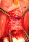
With the patient in the lithotomy position, an 18 F catheter was inserted and the bladder was evacuated. Then placard incision (Fig. 1) at the anterior vaginal wall was performed and dissection of the vaginal flap was carried out to prepare for the midurethral in situ anterior vaginal wall sling (width 1 cm, length 2 cm) (Fig. 2). The length of the midline incision was made according to the degree of cystocele (the transvaginal surgical procedure was used correct cystocele). The dissection at the lateral side of the in situ sling was carried out until the index figure could be felt easily from the suprapubic area. Equi-size monofilament polypropylene mesh (I STOP CL Medical France) was then prepared at the lateral side of the placard incision, 2 suspension sutures of 1/0 polypropylene were inserted in the flap and the mesh (Fig. 3), and using controlled pressure the needle was elevated through the endopelvic fascia, into the space of Retzius, through the rectus muscles, and through the previously created suprapubic skin incision. Two fixation sutures were placed at the lower and upper edges of the in situ sling to keep the mesh over the sling and prevent the dislocation of the mesh (Fig. 3). Before fixation of the suspension sutures to the symphysis pubis periosteum, the bladder was filled with 300 ml isotonic solution and manual pressure was applied to the suprapubic area to test for leakage, thus adjusting the tension of the sutures. Then closure of the placard incision over the in situ sling was done. Intra-operative cystoscopy was done for all patients to rule out urethral or bladder injury. At the end of the operation, a vaginal sponge with Betadine solution and antibacterial cream was placed and left for one night postoperatively. The Foley catheter was removed at 5 to 7 days postoperatively. The mean follow-up period was 30.2 months (range, 24-38 months). Sexual intercourse and carrying heavy weights were avoided for 2 months postoperatively. The absence of a significant post-voiding residual urine (PVR) (50 cc or less was consider insignificant) at the postoperative period was assessed by using real abdominal ultrasound. Postoperative follow-up measures included pelvic physical examination, history of incontinence, stress test, and measurement of PVR. Postoperative cure of SUI was defined as the absence of complaint of leakage and the absence of urine incontinence on stress testing. Improvement was defined as no loss of urine with stress and patient reports of some leakage but overall satisfaction. Persistence of incontinence was considered as failure.
RESULTS
Twenty-two patients (95.65%) benefited from the surgery; 20 patients were cured and 2 patients showed clinical improvements. The ages of these two patients were 53 and 69 years, respectively. The 69-year-old was diabetic and the 53-year-old was morbidly obese (body mass index [BMI] ≥35) and hard- working. Only one patient (4.35%) did not benefit from the surgery (64-year-old, diabetic, BMI=44.98). Urinary retention was observed in one patient (4.34%), which did not resolve after a further 5 days of catheterization. Therefore, the tension of the suspension sutures was reduced under anesthesia, and the patient voided spontaneously after 1 day of catheterization. No intra-operative complications such as bleeding or perforation of the intestine or bladder were observed. No significant postoperative PVR was detected. Vaginal mesh erosion was found in two patients (8.69%) during the gynecological examination in the first month postoperatively; one of them had early sexual intercourse. Both of these patients suffered only from minimal discomfort during sexual intercourse, and the patients refused surgical intervention and a conservative approach (watchful waiting) was applied. Postoperative urgency without urge incontinence was observed in two patients in the early postoperative period, which was resolved after temporary anti-cholinergic therapy for 3 weeks. None of the patients showed suprapubic discomfort or pain due to fixation of the suspension sutures with the symphysis pubis, neither during the physical examination nor during daily physical activities. However, one patient (4.34%) had wound sepsis in the form of suprapubic tenderness and redness in the early postoperative period, which was treated by oral antibiotics and anti-inflammatory drugs for 1 week.
DISCUSSION
Before the era of TVT, surgery for SUI was associated with several complications, including blood loss, pelvic and abdominal organ injury, dyspareunia, urethral erosion, and detrusor instability [4]. Thus, tension-free surgery spread rapidly in the past 15 years as the most common treatment for SUI in females. Unfortunately, TVT had fewer postoperative complications but quite the same rate of intraoperative complications [18-20]. The new TOT approach has resolved some of these problems by passing the obturator fossa to avoid getting in the retropubic space, so that the tape is far enough away from the bladder, vessels, and bowel [21]. However, even this revolutionary approach is associated with some complications, such as thigh pain, hematoma due to significant intraoperative bleeding, and postoperative bladder outlet obstruction [22,23]. In one of the largest studies conducted to date, Kuuva and Nilsson reported that the incidences of postoperative complications after anti-incontinence surgery were 2.3% for urinary retention, 3.8% for bladder perforation, 0.07% for urethral erosion, and 0.8% for wound infection [24]. No significant differences in success rates were found between midurethral sling procedures [25].
In a previous study, we reported encouraging results with the use of a placard technique, using an in situ sling of the anterior vaginal wall for the treatment SUI [26]. Slight modification on the previous technique was adopted by supporting the in situ vaginal sling with equi-size mesh, which aimed to increase the efficacy of the surgical procedure, and the short-term results were encouraging [27]. In the current study, we evaluated the mid-term results of this novel technique. Cost-effectiveness and a low risk of urethral or bladder erosion are the most important advantages of this procedure. The length of the mesh used in our technique is 2 cm, about 15 times less than the length of the same mesh (I -STOP) used for TVT. Urethral erosion is less likely to occur because of the presence of vaginal mucosa between the mesh and the urethra.
Modest long-term success is one of the most disappointing issues facing patients undergoing anti-incontinence surgery. With our present technique, we attempted to solve the mechanism that may lead to reduction in the success rate at the long-term follow-up. In our study group, all patients who were found to be cured at the short-term follow-up [27] showed no deterioration in continence. However, most of the studies that evaluated long-term outcomes after anti-incontinence surgery reported the opposite; Lee et al reported a long-term cure rate of 83% for urinary incontinence after 85.5 months of follow-up and a 93.4% cure rate at 1 year of follow-up [28]. We observed no further complications related to the surgical technique, such as mesh erosion, urge incontinence, or suprapubic pain or tenderness. The probable justifications for the reduction in long-term success in the sling operations are the relaxation of suspension sutures and the mesh, which are less likely to occur in our technique. Lo et al reported that recurrent SUI resulting from slackness of the TVT tape caused by failure to achieve midurethral functional kinking could be rectified by adequate shortening of the excess midurethral sling [29]. Correct midurethral placement of the TVT tape has been regarded as one of the most important technical aspects of the TVT procedure [5,11,13,30]. Therefore, dislocation of the midurethral mesh is also one of the expected mechanisms for failure. In our technique, however, this is not expected because the mesh is fixed with the in situ sling and thus no displacement of the mesh is expected. The patient who did not benefit from the surgery was overly obese (BMI=44.98) and diabetic. Our technique offers the chance to correct the cystocele during the surgery. Cystocele, obesity, and co-morbid diseases are among the factors (e.g., high BMI, urge incontinence, low leak point pressure, high-grade incontinence, severe grade of cystocele) that may have a negative impact on the cure rate after anti-incontinence surgery [12]. Of the two patients who showed only partial benefit from the surgery, both had at least one of these risk factors
No intra-operative complications of the current technique were observed; however, postoperative complications like vaginal erosion and urinary retention were seen. Vaginal erosion occurred in two patients, one of whom had early sexual intercourse, which might cause tape erosion. We believe that as the number of patients increases, the percentage of erosion will be less. Also, we recommend enough dissection at the lateral side of the in situ sling to cover the mesh and in situ sling without tension, thus decreasing the risk of vaginal tape erosion. The urinary retention rate was within reasonable limits compared with that in the literature [24]. This technique seems to be acceptable in secondary cases also. Two patients were undergoing repeat surgeries, and they were cured without any complications. However, performing this technique for secondary cases may carry some difficulties in preparing the in situ sling because of fibrosis resulting from the previous surgery. In these cases, careful dissection is advocated.
CONCLUSIONS
This technique has a good rate of modest long-term success, because relaxation of the suspension sutures and dislocation of the midurethral sling are not expected to occur. The cost-effectiveness and the low risk of urethral erosion, due to the presence of intervening vaginal mucosa, are also important advantages of this technique. In the future, a prospective study recruiting a larger number of patients undergoing this technique with longer term follow-up would be recommended.


 XML Download