

 PDF
PDF ePub
ePub Citation
 Print
Print


Reduction in the size of endourological instruments, improvements in electronic video imaging systems, and advancements in endourological skills have triggered a change in the conventional management regimen for pediatric urolithiasis from open surgery to endoscopic treatment. We present a case of a 10-month-old female infant who had a large upper ureteral stone with bilateral vesicoureteral reflux (VUR). Although it was large, the ureteral stone was removed ureteroscopically, and simultaneously, the contralateral VUR was corrected by Deflux® injection. Here we discuss the possible surgical treatment options that we considered for this infant and pertinent issues when performing ureteroscopic stone removal in children.
CASE REPORT
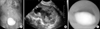
A 10-month-old female infant was referred to our urology department for unresolved febrile urinary tract infection (UTI) despite parenteral antibiotic management. Her first episode of febrile UTI occurred when she was 2 months old. Voiding cystourethrography (VCUG) revealed bilateral VUR (grade 2 in the right, grade 3 in the left), and a 99TcDMSA renal scan demonstrated a focal photon defect on the lateral side of the left kidney. Following this episode, she had another febrile UTI despite prophylactic oral antibiotics when she was 9 months old. The third febrile UTI developed when she was 10 months old, and her fever continued although empirical parenteral antibiotics were given for several days. Ultrasonography and VCUG revealed a newly developed, large (22×10 mm) right ureteral stone at the L4-5 level and consequent moderate hydronephroureterosis containing turbid urine (Fig. 1). She was referred to our urology department at that time for management of the complicated, large upper ureteral stone. A percutaneous nephrostomy tube was inserted into the dilated pelvocaliceal system of the right kidney for drainage of the turbid urine. After this intervention, the patient's fever subsided. After further parenteral antibiotic treatment for 1 week, ureteroscopic surgery for the upper ureteral stone was performed. Deflux® (Oceana Therapeutics, Inc., Edison, USA) injection for correction of the contralateral VUR was performed at the same time.
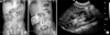
We inspected the bladder with a 9.5 Fr cystoscope. No abnormalities were visualized in the bladder, and the bilateral ureteral orifices were wide open. A 0.035 inch guide wire was placed to the level of the right renal pelvis under direct fluoroscopic and endoscopic guidance. A 6/7.5 Fr self-dilating ureteroscope was advanced over the guide wire just distal to the ureteral stone, and then the working guide wire was removed. A coil guide wire was passed by the ureteral stone and coiled just proximal to the stone for the prevention of upward stone migration. The stone was yellowish and very large but was easily fragmented with an Electro-Medical Systems lithoclast. The fragmented stones were extracted with a stone basket, and the remaining small fragments were flushed down with normal saline irrigation via the nephrostomy tube. After confirmation of complete stone removal from the right ureter, Deflux® injection was performed in the left ureter with the hydrodistention implantation technique. The nephrostomy tube was clamped, but not removed. A postoperative kidney-ureter-bladder (KUB) demonstrated that the stone was completely removed (Fig. 2A). Two days after the surgery, urine leakage developed abruptly around the nephrostomy tube. Antegrade pyelography showed a passage disturbance in the right urinary tract with narrowing of the right distal ureter (Fig. 2B), so the clamped nephrostomy tube was re-opened and the passage disturbance in the right ureter resolved spontaneously. Chemical analysis revealed that the stone was composed of calcium phosphate with carbonate apatite. Ultrasonography performed 2 months after surgery showed no hydronephrosis on the right kidney (Fig. 2C).
DISCUSSION
Because urolithiasis is a rare disease in childhood, the management of pediatric urolithiasis is based on treatment regimens originally developed for adults. However, several clinical situations in the management of pediatric urolithiasis differ from those of urolithiasis in adults [1,2]. In children, there are no clinically insignificant residual stones, and general anesthesia is always required for every procedure. Therefore, careful consideration of the characteristics of pediatric patients should be made when designing treatment strategies for pediatric urolithiasis [3,4].
Extracorporeal shockwave lithotripsy (ESWL), ureteroscopic stone removal (URS), open surgery, or a combination of these modalities could have been applied in our case. We choose ureteroscopic surgery as the surgical treatment option for several reasons. First, it allowed us to remove the entire stone at once. ESWL is currently considered a reasonable treatment option for pediatric urolithiasis and has a success rate of 75% to 90% [5-7]. However, the success rate with a single session of ESWL is only 66% [8]. ESWL monotherapy for large stones often requires multiple sessions under general anesthesia. On the other hand, ureteroscopic surgery offers high success rates after single procedures. Recent studies report that stone clearance after a single ureteroscopic surgery was 100% for stone burdens 10 mm or less and 97% for burdens greater than 10 mm [9]. Although the stone in our case had grown to a large size in only 8 months, there is no definite evidence that pediatric urinary stones grow faster than do stones in an adult.
During URS in young children, the most difficult step is retrograde access of the ureteroscope into the ureter. Our patient was only 10 months old, but was female and had a moderate grade of bilateral vesicoureteral reflux, which allowed for easier ureteroscopic access. Second, the use of URS in this case allowed for simultaneous correction of the contralateral VUR. This infant had a history of two previous febrile UTIs without any evidence of ureteral stones, and the 99TcDMSA renal scan performed during the acute infection period showed a photon defect at the lateral portion of the left kidney. The presence of the photon defect suggested that the left kidney was more vulnerable to infection. Therefore, it was a reasonable choice to simultaneously correct the left VUR in the patient.
Upon reviewing our case, we learned that the ureteral mucosa of young children is very delicate, so ureteroscopic management can induce transient urinary tract obstruction secondary to postoperative edema, which may be clinically significant. The postoperative urine passage disturbance developed abruptly at 2 days after surgery rather than during the immediate postoperative period, however, so we suggest that it was caused by tiny, remnant stone fragments combined with postoperative mucosal edema. If anti-reflux surgery for the ipsilateral ureter had been simultaneously performed, it might have aggravated the disturbance of urine flow and resulted in prolonged urinary tract obstruction. Percutaneous nephrostomy was very useful in our case for the preoperative drainage of retained and turbid urine, for the intraoperative flushing of small stone fragments, and for the postoperative management of the passage disturbance of the urinary tract. However, in our opinion, routine percutaneous nephrostomy is not necessary before URS if the case is not complicated, regardless of stone size.
URS is the first-line surgical treatment for a ureteral stone disease in adults without regard to stone location, but ureteroscopic manipulation in infants is limited because of the ureteroscopic access into the small ureter. In our opinion, if an infant has a moderate degree of VUR on the same side as a ureteral stone, URS is a feasible and moreover a good surgical modality for the ureteral stone regardless of the location or size of the stone. In the case of a female infant, the minimal likelihood of urethral trauma by the ureteroscope is a further indication for choosing ureteroscopic surgery as the initial surgical treatment method.
 XML Download