

 PDF
PDF Citation
 Print
Print


INTRODUCTION
Recent advancements in computer technology have enabled comprehensive analysis of various fields including medicine. The natural history of diseases has been elucidated by detailed assessments of enormous amounts of available data. In the management of cancer treatment, there is an expanding movement that overdiagnosis and over-treatment should be eliminated in order to avoid unnecessary treatment-related invasiveness for the patients and to lower medical costs.
Both active surveillance (AS) and watchful waiting (WW) are accepted policies in several low-risk malignancies, such as prostate cancer [12], small renal masses [34], nonseminomatous germ cell tumors of clinical stage I [5], and papillary thyroid carcinoma [6]. There may be some confusion between AS and WW. According to the NCI Dictionary of Cancer Terms (see the website, http://www.cancer.gov/publications/dictionaries/cancer-terms), AS is defined as 'a treatment plan that involves closely watching a patient's condition but not giving any treatment unless there are changes in test results that show the condition is getting worse.' During AS, tests and exams such as radiographic evaluation, endoscopic exams, and/or blood/urine tests are performed on a regular schedule. On the other hand, WW is defined as 'Closely watching a patient's condition but not giving treatment unless symptoms appear or change. Watchful waiting is sometimes used in conditions that progress slowly.' During WW, patients may be given certain tests and exams.
Clinicians need to consider the adverse effects of surgical intervention and nonsurgical treatment such as radiotherapy. The expense of each intervention should also be considered. The aim of AS is to avoid overtreatment or to delay the need for treatment, while there is still a chance for curative intervention when needed during close follow-up in AS.
Urologists are familiar with the concept of AS for urogenital malignancies such as low-risk prostate cancer [12] and renal cell masses [34]. Although the initial assessment of AS for bladder cancer was reported more than 10 years ago [7], it is still an uncommon option, but it may be ideal in selected patients. The present paper presents the rationale for AS for low-risk primary and recurrent nonmuscle invasive bladder cancer (NMIBC) and reviews the evidence for AS for managing this disease.
EPIDEMIOLOGY AND NATURAL HISTORY OF BLADDER CANCER
Bladder cancer is the sixth most common malignancy in men worldwide, with 297,300 new cases and 112,300 deaths in 2008 [8]. Based on the nationwide data of Japan, estimated new cases and deaths from bladder cancer in 2013 were 17,461 and 7,299 (5,003 men and 2,296 women), respectively [9].
Most visible bladder tumors are diagnosed by visual inspection under cytoscope, and are confirmed of their grade and stage pathologically by transurethral resection (TURBT) specimens and radiographically for initial management.
Approximately 70%–80% of urothelial carcinomas are diagnosed as NMIBC, consisting of Ta (70%), T1 (20%), and isolated Tis (10%) [1011]. The treated natural history of NMIBC is characterized by a favorable prognosis; however, with a high intravesical recurrence rate. NMIBC is known to be a heterogeneous disease with different recommended options for treatment and follow-up regimens, and varying oncologic outcomes. Up to 70% of patients with NMIBCs will experience intravesical recurrences, and 10%–30% will progress to life-threatening muscle-invasive bladder cancer (MIBC, ≥T2) [1213]. However, among NMIBCs, low-risk tumors defined as Ta low-grade solitary papillary tumors are associated with a favorable cancer-specific survival, along with a lower likelihood of stage and grade progression [1415161718]. The 1- and 5-year recurrence rates range from 15%–61% to 31%–78%, respectively, while both the 1- and 5-year progression rates are less than 1% [15].
Kobayashi et al. [18] reported retrospectively the outcomes of newly diagnosed patients with low-grade Ta bladder cancer undergoing initial TURBT. In this retrospective study, worsening progression was defined as recurrence with high-grade Ta, T1, or Tis/concomitant carcinoma in situ (CIS). Among 190 cases, 82 (43%) had recurrence and 21 (11%) had worsening progression during follow-up (median, 101 months). Progression to MIBC was observed in only 2 cases (1%).
Considering the treated natural history of NMIBC, the quality of TURBT and pathologic diagnosis are important factors. The 5-year recurrence-free rate after TURBT monotherapy differed across institutions, ranging from 30% to 70% [1920]. In addition, the residual cancer rate detected by a second TURBT also had a wide range, 20% to 78%, which implies incomplete resection of tumors in initial TURBT is common [21]. The quality of TURBT is not a negligible factor to minimize the risk of recurrence and progression of NMIBC. Discrepancies in pathological findings of TURBT are also key factors depending on examiners because of their experience as well as artifacts due to cauterization. One more issue is the discrepancy on the pathological diagnosis of bladder cancer. Our previous study demonstrated that general pathologists tend to overdiagnose the specimens of bladder tumors both in stage and grade compared to central uropathologists (unpublished data).
FAVORABLE PROGNOSIS OF PAPILLARY BLADDER TUMORS VERIFIED IN ANIMAL MODELS
Experienced urologists have great skills in estimating tumor grade and/or stage by visual appearance on cystoscopy, such as shape, size, and color. A previous report by Herr [22] demonstrated that the histology of low-grade Ta lesions was able to be predicted with an accuracy of 93%, which was increased to 98% when combined with urine cytology. Papillary shaped-bladder tumors have been known to be more indolent than nodular-shaped ones. This concept is supported by animal model investigations regarding urothelial carcinogenesis [232425262728293031].
N-butyl-N-(4-hydroxybutyl) nitrosamine (BBN)-induced bladder cancer is one of the most common bladder cancer models. Bladder tumors in mice exposed to BBN ad libitum in the drinking water progress through hyperplasia, dysplasia, and CIS, and eventually to huge nodular invasive cancers. In contrast, BBN-induced rat bladder cancers are papillary in nature and less aggressive. Bladder urothelia in rats treated with BBN develop papillary/nodular hyperplasia after 4 weeks and eventually form papillary tumors through papilloma. Papillary tumors are multifocal but nonmuscle invasive. BBN-induced bladder cancer in rats rarely progresses to muscle invasive lesions.
BBN-induced urothelial carcinogenesis of the bladder in dogs (beagle dogs and mongrel dogs) has been investigated in detail by our colleagues, Okajima et al. [2627282930]. When 2 beagles were treated daily with 160 mg of BBN in oral capsules, multifocal papillary G1/G2 tumors were seen 137 weeks after the initiation of BBN treatment. At the time of tumor detection, BBN administration was discontinued and the two beagle dogs were observed without any treatment for over 11 years [30]. Pathology revealed papillary NMIBC with increased number and size, and small foci of concomitant CIS at autopsy. In addition, no distant metastasis was observed.
In the bladder cancer models of beagles and rats, urothelial carcinogenesis was initiated by papillary growing tumors. The results indicate that these tumors remained papillary NMIBC over a long period of time, strongly suggested that the majority of papillary bladder tumors inherently possess little potential to progress to life-threating MIBC. This clearly suggests that low-risk papillary bladder cancer is a candidate for an AS policy.
INVASIVENESS OF BLADDER PRESERVATION TREATMENT
Needless to say, the fundamental goal of treatment of NMIBC is the bladder preservation. It consists of TURBT with or without intravesical treatment with chemotherapeutic reagents and/or Bacille de Calmette et Guérin. There are several possible complications, comorbidities and substantial risks associated with these procedures. Intraoperative and postoperative complications of TURBT have been reported previously [3132]. In a series of 2,821 patients with 65 years old (range, 16–94 years) of average age 145 complications (5.1%) were observed. Most common ones were bleeding in 78 patients (2.8%) and bladder perforation in 36 (1.3%) requiring open surgery in 4 patients (11%). Bladder perforation occurs more frequently in elderly or female patients who tend to have a thin bladder wall [33]. The true incidence of perforation may be higher, with small perforations that are unrecognized and treated with routine indwelling catheterization without incident. Prospective studies have revealed that leakage of contrast media during cystography was detected in more than half of patients undergoing TURBT [3435]. Blood transfusion was performed in 2.3%–3.4% [3132]. Collado et al. [31] reported the blood coagulation system disorders (<1%) such as lower-extremity deep vein thrombosis, pulmonary embolism, and acute myocardial infarction, which are known to be potentially life threatening. The authors demonstrated that the incidence of complications correlated with the size and number of tumors, but did not with tumor stage, grade or location.
Other potential demerit of TURBT comprises lower urinary tract symptoms due to the resection and urethral catheterization, which make the bladder susceptible to infection. The repeated extensive resection using electric cautery may cause bladder scarring [35]. In addition, routine cystoscopic examination is also a big issue in the follow-up protocol. The sensitivity of urine cytology is low for the detection of recurrence in low-risk bladder cancers, while cystoscopy is able to detect small recurrent lesions. Cystoscopy (even with flexible scope) is a discomfort and invasive examination, and has substantial psychological impact to the patients. It potentially causes urethral trauma and subsequent urethral stricture during the long-term follow-up. As to the medical cost, cystoscopy coupled with urine cytology is expensive at outpatient clinic, approximately $150 in Japan with refer to the exchange rate against US dollar value as of March 2016.
EVIDENCE ABOUT ACTIVE SURVEILLANCE FOR NMIBC
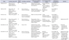
Based on the clinical data of the favorable prognosis associated with low-grade Ta bladder cancer, there may be no need for immediate TURBT or fulguration for primary or recurrent disease. It has been over a decade since Soloway et al. [7] presented the initial series of expectant management and reported the feasibility and safety of this concept in patients with low-risk bladder cancer in 2003. To date, several reports have shown AS (sometimes referred to as expectant management) for this disease is a reasonable option [73637383940]. Table 1 shows the study design and outcomes of AS for low-risk bladder cancer. It is still not a common option that urologists provide to their patients. One of the issues may be a lack of optical follow-up protocols regarding the interval between surveillance cystoscopy and the timing of treatment interventions.
The initial report of AS was a retrospective review of 32 patients with one or more tumors that were visually small, papillary, or low-grade appearing [33]. All patients had a history of pathologically diagnosed Ta or T1 cancers. Surveillance cystoscopy was performed in the office every 3 to 6 months. Criteria for TURBT were tumor growth or any changes in tumor appearance implying grade and/or stage progression or gross hematuria. During follow-up (mean, 38 months), the mean number of tumor recurrences was 3.8 per patient. Of 45 observed tumors that were preceded by TaG1–2, only three (6.7%) resected by TURBT revealed progression to high-grade lesions: high-grade Ta in one and T1 in two, while no tumor progressed to MIBC. Gofrit et al. [36] reported the first homogenous series of low-risk bladder cancer defined by strict inclusion criteria. Two years later, the authors updated their series with 31 patients and 43 observation periods at a mean observation follow-up time of 16.1 months [38]. Of 4 observation periods, 35 were terminated because of the appearance of additional tumors (n=15), excessive tumor growth (n=12), patients' request (n=7), and the need for prostatectomy (n=1). No patients progressed to MIBC and none died from bladder cancer.
Hernández et al. [3940] reported two consecutive studies evaluating AS for low-risk NMIBC in 2009 and 2015. Patients were allocated to AS with the criteria similar to the studies by Gofrit et al. [3638] and low-grade T1 tumors were included. In the largest and most recent study, Hernandez et al. [40] evaluated 186 patients (252 observation periods) including 161 men and 25 women with a mean age of 66±12 years. During active follow-up including urine cytology plus cystoscopy (first 2 years) or urine cytology plus ultrasound test (thereafter), intervention treatments were performed in 203 (80.2%) out of 252 observation periods. The most frequent reason for termination of AS was an increment of lesion number/size, accounting for 178 out of the total 252 observation periods (70.6%). As a result, 17.1% (43 patients) remained under AS for more than 2 years and 8.7% (22 patients) have been under AS for more than 3 years. One of the most important issues of AS is oncological safety representing no progression in stage and grade. Across the surveillance periods, the stage progression-free rate and grade progression-free rate were 86.4% and 79.3%, respectively. Progression to T2 was seen in only four patients; all of them originally had T1G2 tumors.
CRITERIA AND FOLLOW-UP PROTOCOL FOR ACTIVE SURVEILLANCE OF BLADDER CANCER
Inclusion criteria for AS in patients with recurrent low-risk NMIBC are refined by Gofrit et al. [36] as follows: (1) previous resection of low-grade (G1–2) Ta tumors; (2) no history of previous high-grade (G3) tumors; (3) asymptomatic, small (<10 mm) papillary tumor found on routine cystoscopy; (4) negative urinary cytology before and during surveillance; and (5) patient willingness to participate in the surveillance protocol. However, it is still controversial whether criteria can include T1 tumors or not. The largest study reported by Hernández et al. [40] included the past history of T1 tumors in their criteria. Of 252 observation periods, T1G1 tumors and T1G2 tumors account for 25 (10%) and 42 (17%), respectively. Although there was no progression to MIBC in 25 T1G1 tumor, but four cases (10%) progressed to MIBC in 42 T1G2 tumors. In addition, European Association of Urology (EAU) guidelines of NMIBC categorize T1 tumors into the high-risk group [41]. These findings imply that a history of T1G2 tumor may not meet the criteria for AS.
The pathological information including tumor grade, stage and lymphovascular involvement is not available unless TURBT or biopsy are performed. Recently there are several novel supplementary imaging techniques have been applied in the clinical setting. There is increasing evidence on the usefulness of the enhanced photodynamic imaging [42], narrow band imaging [43], optical coherence tomography [44] and confocal laser endomicroscopy [45], which might be promising aids for cancer detection, disease monitoring and risk stratification. Nakai et al. [46] reported the data suggesting high grade and high stage bladder tumors show significantly higher fluorescent intensity compared to low grade and low stage ones. This result implies imaging data or information highly reflect tumor aggressiveness and biology. To date, there has been no report showing the clinical potential of these novel imaging techniques in the patient selection and monitoring during AS.
Another issue in AS for bladder cancer is that the follow-up protocol has not been established yet. Many previous studies included cystoscopy and urine cytology every 3 months for the first 2 years, every 6 months for the next 3 years, and annually thereafter (Table 1, summary of standard follow-up protocols), which complies with the NMIBC guidelines of EAU, American Urological Association, and the National Comprehensive Cancer Network for patients treated with TURBT. These follow-up protocols may be too strict in the routine clinical practice for low-risk NMIBC. In contrast, the consensus of the Société Internationale d'Urologie experts indicted that cystoscopy can be skipped for up to 12 months if the first cystoscopy at three months after TUR does not detect recurrent tumors [41]. We have conducted a couple of clinical trials to examine the natural history of NMIBC including TaG1–2 and T1G2 [1947] treated with TURBT alone or combined with prophylactic intravesical chemotherapy. In a subanalysis regarding the recurrence detection rate by follow-up cystoscope on the standard protocol, 41 recurrences were observed out of 784 cystoscopic examinations in the TUR alone group, and 20 recurrences out of 881 cystoscopic examinations in the epirubicin-treated group during the follow-up period (Table 2). It turned out that number of cystoscope to detect a recurrence was 19.1 times in the control group and 44.1 times in the epirubicin-treated group. As mentioned above, urologists should also bear in mind that cystoscopy is a discomfort, invasive and expensive examination, and have substantial psychological impact to the patients.
Although papillary low-risk NMIBC is characterized by frequent intravesical recurrence and a low potential for disease progression, the intervals between cystoscopy could be delayed or even skipped. In order to advocate AS as a standard option in selected patients, a specific follow-up protocol including intervals of cystoscopy, urine cytology, urine markers, and other radiographic exams needs to be validated on the basis of a high level of evidence, so that unnecessary cytoscopic examinations can be avoided.
Additional parameters estimating the tumor biological behavior are expected in the development of tailor-made follow-up strategies. A recent report assessed the approved urinary tests such as Urinary cytology, ImmunoCyt, BTA Stat, hemoglobin dipstick, and NMP22 BladderChek as potential predictors of recurrence and progression of NMIBC [48]. Among these five, only NMP22 had an independent prognostic impact on intravesical recurrence and disease progression. Because NMP22 is an approved test that is easy to access, this result may be applicable to the clinic [4849]. Several studies have been carried out to confirm the clinical usefulness of molecular biomarkers for predicting risks [13505152]. Biomarkers such as the Ki-67 labeling index [5354], gene mutation of the fibroblast growth factor receptor 3 (FGFR3) [515253555657], p53 status [525859] and chromosomal abnormalities detected by fluorescence in situ hybridization [60] have been reported to be promising molecular tests; however, there is still a need for them to be validated prior to application to clinical practice.
The activated missense mutation of FGFR3 is one of the most frequent gene aberrations in human bladder cancer. Among low-grade NMIBCs, 50%–70% harbor the mutation, whereas it is detected in only 10%–20% of high-grade MIBCs and CIS [13555657]. These findings support the idea that activated FGFR3 plays critical roles in both carcinogenesis and the cell growth of low-grade NMIBC. Hernandez et al. [55] reported the largest prospective study to date to investigate the potential of FGFR3 mutation as a prognostic factor for NMIBC. The results showed that mutated FGFR3 was associated with a higher risk of intravesical recurrence, but was also associated with a lower risk of disease progression and cancer-specific mortality. An FGFR3 mutation test can be supplementary to determining the optimal follow-up schedule. MicroRNA and genetic alterations detected in the exfoliated urine cells should also be regarded as important auxiliary markers to monitor patients during AS. Influence of cell growth stimulators such as growth factors and cytokines in urine or tissue are not negligible as urine-based markers [61]. In the meantime, urinary cytology using photodynamic diagnosis is likely to become a novel tool of the AS follow-up program [62].
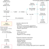
Algorithm of AS for NMIBC is shown in Fig. 1. When peduncular and papillary tumor is found on cystoscopy, AS can be recommended to the patient as a one of treatment options. In case of the patient with history of NMIBC, it is mandatory that the patients have no past history of high grade tumors, CIS or MIBC. Tumor size, tumor number, and negative urine cytology were included in the criteria for AS in most of the previous reports [736383940]. However, specific size and number have not been determined yet. Thus, in the inclusion criteria of the algorithm, tumor size, tumor number and negative urine cytology were included as optional conditions [4849]. However, patients for whom urologists hesitate to perform TURBT due to high age and severe concomitant diseases would be allocated to WW rather than AS. After the patient is allocated to the AS program, he or she is followed up at the regular interval with cystoscopy, urine tests and radiographic examinations. Interval of cystoscopy depends on the tumor behavior such as tumor size, tumor number and FGFR3 mutation status if available. Changes in tumor appearance, occurrence of tumor-related symptom and patient's request are absolute indications for treatment intervention. Tumor growth, increased tumor number, urine cytology and NMP-22 urine test are not included, because they have not been validated and optimized yet. Even if patients have a history of allocation to an AS program, they can be reallocated repeatedly. As treatment intervention, TURBT as a whole resection seems to be smarter than TURBT at every recurrence.
CONCLUSIONS
Urologists are intuitively aware that immediate removal is not required in all recurrent tumors such as low-grade Ta bladder cancer with an indolent behavior. In spite of considerable evidence supporting AS for bladder cancer, follow-up protocols and intervention modalities have not yet been validated at a high level of evidence. However, accumulating data suggest the feasibility, clinical significance, and economic impact of this option. Large-scale randomized clinical trials are required to prove the real benefit of AS for well-selected patients. Similarly, we need to make maximal efforts to obtain significantly reliable biomarkers for AS in the urine or sera and to establish monitoring systems.

 XML Download