

 PDF
PDF ePub
ePub Citation
 Print
Print


Abbreviations
AE
adverse event
CTLA-4
cytotoxic T-lymphocyte antigen 4
DC
dendritic cell
EGF
epidermal growth factor
EGFR
epidermal growth factor receptor
FDA
Food and Drug Administration
IFN-γ
interferon gamma
MAGE
melanoma-associated antigen
MM
multiple myeloma
NK
natural killer
NSCLC
non-small-cell lung cancer
ORR
overall response rate
OS
overall survival
PD-1
programmed cell death 1
PD-L1
program death-ligand 1
PFS
progression-free survival
PI3K
phosphatidylinositol 3-kinase
SCLC
small-cell lung cancer
TAA
tumor-associated antigen
TC
tumor cell
Treg
regulatory T cell
INTRODUCTION
Lung cancer is one of the primary causes of death worldwide (1). The high mortality rate of the disease may be associated with the obscurity of the symptoms that remain undiagnosed for a significant period of time. According to studies, approximately 57% of all lung cancers are metastatic in nature and can affect various tissues and organs (2). There are multiple subtypes, but lung cancer can be categorized into following 2 major subtypes: non-small-cell lung cancer (NSCLC) and small-cell lung cancer (SCLC) (3). According to studies, NSCLC is the most prevalent type of lung cancer and constitutes 80%–85% of all lung cancer cases (1). Generally, NSCLC is known as a slow persistent tumor that eventually becomes metastatic and spreads to the nearby tissues and cells (4). Whereas, SCLC spreads rapidly during the initial stage and is more common in smokers than non-smokers (5).
Together, NSCLC and SCLC account for approximately 1.5 million deaths each year, however, most studies have been conducted on NSCLC because of its diverse and metastatic nature. As statistic reports, only 5% of patients with NSCLC can survive after stage IV (6). Clinical options for cancer therapies, such as surgery, chemotherapy, irradiation, etc., have been used to treat such type of cancers, however, still require better efficacy to cure of cancer especially to treat the advanced stages of cancer (7). The prognosis of NSCLC is difficult because of unidentified symptoms. Most patients do not feel any pain or exhibit symptoms during early stages of NSCLC (6). Due to this, it is challenging to diagnose the disease in initial stages. In addition, the tumor cells (TCs) have the ability to hijack host's immune system and use it like normal cells, resulting in immune editing. Immunosuppression is one of the main characteristics of NSCLC. Hence, the drugs that can counteract this action can be beneficial to treat the disease.
In the past few years, immunotherapy has been used to treat various types of cancers, however, the therapy was considered unsuitable for lung cancer. It was believed that lung cancer is a non-immunogenic disease and does not provoke profound immune responses (8). Nevertheless, the main reason behind such myths is the ability of lung tumors to escape all the checkpoints and suppress overall immune response by altering T-cell mediated cytotoxicity. The recent technological advancement has helped in determining the immunogenic nature of NSCLC and since then, various types of immunotherapies, such as monoclonal antibodies, cytokines, vaccines, etc., have been used to treat these cancers. However, each method has its own advantages and disadvantages, and therefore, it is postulated that agglomeration of multiple therapies can be more beneficial than employing a single method (9).
In the last decade, scientists discovered 2 immune checkpoint inhibitors that target programmed cell death 1 (PD-1) and program death-ligand 1 (PD-L1). These inhibitors are found to be effective even against advanced NSCLC. Therefore, the TCs that express a high level of PD-1 and PD-L1 can easily be identified and targeted to treat the patients. The current mini review will discuss the benefits and significance of these checkpoint inhibitors, cytotoxic T-lymphocyte antigen 4 (CTLA-4) blockades and neoantigen-specific vaccines in the treatment of NSCLC. Nevertheless, this review also acknowledges that identification of potential neoantigens and biomarkers is a challenging task that requires vigorous clinical testing to ascertain safety and efficacy.
SIGNIFICANCE OF IMMUNOTHERAPY
Most NSCLC cannot be detected until the advanced stages of the disease. Unfortunately, at this stage, NSCLC shows minimal response to chemotherapy and becomes fatal. In general, chemotherapy can only cure 15%–30% of all lung cancer cases (10). In most case, patients in early stages have to undergo surgery to remove tumor, however, the symptoms may recur shortly due to metastatic nature of the disease (11). Hence, there is an emergent need to identify alternative methods to cure NSCLC. Immunotherapy is one of such methods that has become successful in treating different types of tumors with manageable side effects (211). Research shows that immunostimulants can be a useful class of drugs that can counteract cancer cells by enhancing the level of tumor specific antibodies, cytotoxic-T cells, natural killer (NK) cells, dendritic cells (DCs), macrophages, and cytokines in blood plasma (2). Among all these immune cells, T-cells play the most crucial role in identifying the antigen on TCs. The tumor-specific CD8+ T cells are activated by the release of IL-12 by antigen presenting DCs. These activated CD8+ T cells can kill TCs directly.
In addition, one subset of CD4+ T helper cells 1 also promotes the activation of CD8+ T cells through their secreted interferon gamma (IFN-γ), while the other subset of CD4+ T helper cells 2 (Th2 CD4+) stimulates an antibody-mediated immune response and activates B cells by releasing IL-4. Both CD8+ and CD4+ T cells secrete IFN-γ and a T helper 1 (Th1) cytokine, which can sensitize TCs to CD8+ T cells and activates other immune cells, thereby favoring tumor destruction.
IMMUNOEDITING
Although immune system can fight various foreign substances in the body, the cancer cells are smart enough to escape the trap. Cancer cells alter host's immune system for its own benefits and expansion. This immunoediting (immune escaping) by TCs is usually achieved in 3 common steps: elimination, equilibrium, and escape (111213) (Fig. 1). In the first phase (elimination), immune cells identify and destroy most cells with abnormal growth by instigating apoptosis. However, some of the TCs survived this stage and proceeded to next stage. Equilibrium stage is a stagnant stage for cancer cells (1415). By this stage, the overall immunity of the body diminishes significantly. Hence, the primary role of immune system remains confined to impede the further growth of TCs. In the final stage, TCs overpower host's immune system by suppressing active immune responses, decreasing regulatory action and escaping apoptosis (1415). In other words, TCs gain the ability to circumvent immune responses and emerge as malignant cells (Fig. 1). These cells disguise themselves as normal cells and mimic their actions in various ways.
Figure 1
Immunoediting (immune escaping) mechanism.
TGF-β, transforming growth factor-β; VEGF, vascular endothelial growth factor; IDO, indoleamine-pyrrole 2,3-dioxygenase; MDSC, myeloid-derived suppressor cell.
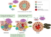
Mechanisms that are used by cancer cells to evade the host immune response are valuable targets for immunotherapy. The loss of antigen expression and resistance to cytotoxicity are the 2 ways by which TCs overcome immunity as they can no longer be recognized by T-cells or phagocytes. Cancer cells can also change their microenvironment to an immunosuppressive state via cytokine expression of transforming growth factor- β (TGF-β), vascular endothelial growth factor (VEGF), or indoleamine-pyrrole 2,3-dioxygenase (IDO) (Fig. 1). Additionally, TCs can actively induce immunosuppression through the recruitment of regulatory T cells (Tregs; a subtype of helper T-cells) and myeloid-derived suppressor cell (MDSC). Recent advancement in cancer biology has focused on reshaping the tumor microenvironment. Several immunotherapeutic strategies that inhibit tumor-induced immunosuppression have been developed in the past few years. Specifically, the use of mAbs directed at PD-1, PD-L1, and CTLA-4 has shown promising results.
IMMUNE CHECKPOINT MOLECULES
PD-1 and CTLA-4
PD-1 and CTLA-4 immune checkpoints are negative regulators of T-cell immune function (16). These targets lead to high activation of the immune system, and their inhibition has led to new immunotherapies for melanoma, NSCLC, and other cancers (17). Ligation of both PD-1 and CTLA-4 block CD3/CD28-mediated upregulation of glucose metabolism and Akt activity, although each accomplishes this regulation using different mechanisms (18). CTLA-4-mediated inhibition of Akt phosphorylation is sensitive to okadaic acid, giving direct proof that protein phosphatase 2A (PP2A) plays a role in mediating CTLA-4 suppression of T-cell activation (19). In comparison, PD-1 signaling inhibits Akt phosphorylation by inhibiting CD28-mediated activation of phosphatidylinositol 3-kinase (PI3K) (19). The capability of PD-1 to suppress PI3K/Akt activation depends on the immunoreceptory tyrosine-based switch motif locating its cytoplasmic tail, adding more significance to this domain in mediating PD-1 signal transmission (19). PD-1 litigation is more efficient in suppressing CD3/CD28-induced changes in the T-cell transcriptional profile, implying that differential regulation of PI3K activation by CTLA-4 and PD-1 ligation leads to varying cellular phenotypes (20). In combination, CTLA-4 and PD-1 inhibit T-cell activation via separate and potentially synergistic mechanisms. The functions of PD-1 and CTLA-4 in inhibiting immune responses such as antitumor responses are fairly different. While PD-1 suppresses T-cells later in an immune response, mainly in lymph peripheral tissues, CTLA-4 is believed to regulate T-cell proliferation early in an immune response (16). Hence, the clinical profiles of immuno-oncology agents prohibiting these 2 checkpoints may vary according to their functional differences.
PD-L1 and PD-L2
PD-L1 and PD-L2 are ligands for PD-1, a costimulatory molecule, which plays an inhibitory function in regulating T-cell activation in the periphery (21). This ligation is crucial for immunotherapy based on PD-1 blockade. IFN-γ-JAK1/JAK2-STAT1/STAT2/STAT3-IRF1 axis mainly regulates PD-L1 expression, with IRF1 binding to its promoter (22). Similarly, PD-L2 responds to IFN-β and IFN-γ, and is regulated via both IRF1 and STAT3 that bind to the PD-L2 promoter (22). PD-L1 is extremely rampant in multiple myeloma (MM), myelodysplastic syndrome (MDS), and acute myelogenous leukemia (AML), with considerable expression by non-tumor hematopoietic cells, mainly CD8+ T-cells (23). PD-L2 expression is considerably absent in myeloid diseases but detectable in MM (24). Interestingly, PD-L1 expression is most common on TCs in MM and on non-tumor hematopoietic cells in MDS, while expression on non-tumor and TCs in AML was comparable (25). PD-L1 up-regulation relies on TLR4 and STAT1, while PD-L2 relies on IL-4Rα and STAT1 in dermal fibroblasts from alopecia areata (26). Th1/T helper 2 (Th2) cells also differentially up-regulate PD-L1 and PD-L2 expression on inflammatory macrophages (2728). According to our study, IFN-γ treatment induces PD-L1 up-regulation and this up-regulation is further augmented by epigenetic priming, azacitidin which is DNA methyl tranferase inhibitor in lung cancer cells (data not shown).
IMMUNOTHERAPY FOR NSCLC
Several clinical trials have been conducted to evaluate the efficacy of immunotherapy, however only 20%–25% of patients have shown positive response towards this relatively new approach (161718). Nevertheless, immunotherapy can be an effective approach to treating lung cancer as it acts through boosting host's own immune system and is more specific and patient-centric than other treatment. Recently, many studies have been conducted to evaluate the efficacy of immune checkpoint inhibitors (e.g., PD-1/PD-L1 inhibitors and CTLA-4 inhibitors) and vaccine therapy (e.g., neoantigen-specific vaccines) to attenuate the severity of NSCLC and enhance the overall life expectancy (2).
Studies show that success of immunotherapy depends upon various factors, including the type of therapy, expression level of targeting agents, immunotherapy strategies, tumor-stage, and duration of therapy (9). The patients, who express a low level of PD-1 and PD-L1 cannot be diagnosed easily during the test. Such patients might express different types of molecules or might have modified immune response that suppresses the level of these biomarkers (2).
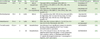
In last 5 years, monoclonal antibodies, such as nivolumab, pembrolizumab, and atezolizumab have been approved to treat NSCLC (2). Nivolumab and pembrolizumab target PD-1, while atezolizumab targets PD-L1 (29303132). Additionally, ipilimumab and tremelimumab have been designed to block CTLA-4 receptors (33). Immune checkpoint inhibitors have been found to exert some adverse events (AEs); however, incidence and severity of such reactions are lesser than the other contemporary approaches, such as chemo- or radiotherapy. Due to their advantages, immunotherapy has become one of the preferred drug choices and is acclaimed as effective second-line therapy for NSCLC. Table 1 summarizes the results of phase III clinical trials of anti-PD-1 and anti-PD-L1, and phase II clinical trials of anti-CTLA-4. Such trials are essential to evaluate the true competency of immunotherapy in the treatment of lung cancer. Numeral clinical trials are ongoing and the outcomes are encouraging so far (3435).
Table 1
Phase III clinical trials for anti-PD-1/PD-L1 and phase II for anti-CTLA-4 in lung cancer
| Immunotherapy | Target | Phase | No. | Cancer type | Results | Trials or ClinicalTrials.gov # | Reference |
|---|---|---|---|---|---|---|---|
| Nivolumab | PD-1 | III | 272 | NSCLC (squamous) | Two-year ORR vs. docetaxel: 23% (95% CI, 16%–30%) vs. 8% (95% CI, 4%–13%), AEs: 10% (grade 3–4) | CheckMate 017 | (36) |
| NCT01642004 | |||||||
| III | 582 | NSCLC (non-squamous) | Two-year ORR vs. docetaxel: 29% (95% CI, 24%–34%) vs. 16% (95% CI, 12%–20%), AEs: 10% (grade 3–4) | CheckMate 057 | (36) | ||
| NCT01673867 | |||||||
| Pembrolizumab | PD-1 | III | 1,034 | NSCLC | PD-L1 positive only, ORR: 19% for low dose and 20% for high dose, MS: 14.9 mon for low dose, 17.3 mon for high dose, AEs: 13% for low, 16 for high dose (grade 3–5) | Keynote 010 | (31) |
| NCT01905657 | |||||||
| Atezolizumab | PD-L1 | III | 1,225 | NSCLC | ORR: 14%, MS: 13.8 mon (95% CI, 11.8–15.7) vs. 9.6 mon (8.6–11.2) of docetaxel, AEs: 15% | OAK | (37) |
| NCT02008227 | |||||||
| Tremelimumab | CTLA-4 | II | 382 | Mesothelioma | Monotherapy is not significant compare to placebo for the primary endpoint of OS 7.7 vs. 7.3 mon (tremelimumab vs. placebo) | NCT01843374 | (38) |
| II | 29 | Mesothelioma | CR: 0, PR: 2 (6 and 18 mon), OS: 10.7 mon, 93% patients had at least one grade 1–2 AEs (cutaneous rash, prutirus, colitis, or diarrhea) and 14% had 3–4 grade AEs (2 gastrointestinal, 1 neurological, 2 hepatics, and 1 pancreatic) | NCT01649024 | (39) |
PD-1/PD-L1 CHECKPOINT INHIBITORS
Recently, Food and Drug Administration (FDA) approved nivolumab and pembrolizumab as PD-1 checkpoint inhibitor (40). These drugs block PD-1 receptors, found on T-cells, B-cells, monocytes, DCs, or NK cells. PD-1 can impede cytokine production by binding with either PD-L1 or PD-L2. PD-1/PD-L1 complex actuates the death of tumor-specific T-cells (40) and directs transformation of CD4 T cells into Tregs. Increased level of Tregs may further lead to immune tolerance and resistance towards tumors.
The ligands PD-L1 and PD-L2 are usually expressed on TCs, and therefore, their increased level can be used a potential biomarker for NSCLC. These ligands bind with PD-1 receptors, presented on activated T-cells and negatively regulates its action. Consequently, the production and proliferation of tumor-specific T-cells decrease significantly (25). Moreover, the cytokine production also diminishes and may result in a reduced level of active immune cells in the blood and impair overall immunity of the patient (25). The drugs, like nivolumab and pembrolizumab, can compete with PD-L1 or PD-L2 to bind with PD-1 receptors. These drugs are IgG4 monoclonal antibodies that facilitate the death of TCs by blocking the receptor (41).
The phase III trials on nivolumab were conducted on 272 participants, with the advanced level of squamous NSCLC (36). These patients expressed a high level of PD-L1 and were prescribed various doses to estimate the immune response and relative risk. The drug showed promising results with an overall response rate (ORR) of 23%. Another Phase III trial included 582 patients with non-squamous NSCLC and focused on overall survival (OS) rate and AEs associated with the drug (36). There was a mark reduction of 27% in mortality rate with a significant improvement in patients' condition. The ORR was reported to be nearly 29% in this trial. Nivolumab did not induce severe AEs in most patients. The most common side effects were skin allergy and pulmonary or gastrointestinal toxicity. Some of these symptoms were the result of escalated autoimmunity and can be treated with anti-inflammatory drugs. Nivolumab was approved by FDA in March 2015 for NSCLC patients (4243).
Pembrolizumab is another PD-1 blocker that was approved in 2014 by FDA and is first immune checkpoint blockade drug as first-line treatment in May 2017 for metastatic NSCLC with expression of PD-L1 on at least 50% of TCs (44). The phase I trial included 495 patients with significant symptoms and high level of PD-L1 (44). According to study, approximately 53% patients reported some sort of AEs after the therapy organs (2). However, none of them were serious and were restricted to rashes and allergies. Phase II and phase III trials collectively included 1,034 participants (31). The drug showed favorable results with remarkable improvement in progression-free survival (PFS) and minimal AEs. The ORR was approximately 19%–20% with 13%–16% of noticeable AEs (31). Both nivolumab and pembrolizumab were tested on patients who had simultaneously undergone surgery, chemotherapy or radiation therapy. Consequently, it was difficult to evaluate the precise effects of these mAbs on patients. However, agglomeration of immunotherapy with traditional approaches improves the symptoms significantly.
PD-L1 inhibitors are another important class of drugs that stimulate the immune system to combat cancer cells. Unlike PD-1 blockers, these drugs inhibit the interaction between PD-1 and PD-L1 and thus, prevent the death of tumor-specific T-cells. Atezolizumab and durvalumab are the most common drugs of this class. These drugs are completely humanized and consist of IgG1 Abs (45). The drugs have undergone several clinical trials and showed favorable outcomes with minimal AEs (45). Atezolizumab has been tested on 1,225 patients with advanced NSCLC for its safety and efficacy (37). The drug showed significant outcomes with 14% ORR and 15% AEs (37). The AEs were limited to fatigue, dehydration, and nausea. Some patients left the treatment due to these discomforts, however, no lethal effects have been noticed. In another study, atezolizumab was reported to improve ORR by 63% organs (2). Nonetheless, the study was conducted on a small number of population (n=37), and the drug was given in combination with other therapeutic agents, such as epigenetic, chemotherapy, radiation therapy, etc. (1846474849).
CTLA-4 BLOCKADES
The activation of T-cells is regulated by co-stimulatory and co-inhibitory mechanisms. T-cells become activated when they bind to the major histocompatibility complexes to destroy the foreign antigen. However, overstimulation of T-cells can result in autoimmune disease in which the body can develop less tolerance for even self-antigen. These stimulatory and inhibitory pathways are regulated by CD28 and CTLA-4, respectively (50). The CTLA-4 is overexpressed in NSCLC patients and competes with CD-28 for B7 ligands on antigen-presenting cells (5152). Binding of CTLA-4 to the ligand inhibits activation of T-cells, production of interleukin-2 and stimulation of other immune cells (5152). Ipilimumab and tremelimumab are the examples of monoclonal antibodies that can counteract the action of CTLA-4 and may help in restoring immune response by promoting T-cell activation (5354). Ipilimumab is still undergoing phase III trials and showed promising results when prescribed with chemotherapeutic agents. Ipilimumab increased immune-related PFS, however, no significant improvement has been reported in OS rate (5354). The phase II trials for tremelimumab were conducted on patients with mesothelioma. Two studies included 382 and 29 patients, respectively, and showed an OS of 7.7 to 10.7 months (3839). However, vigourous clinical studies are required to estimate the actual benefits.
NEOANTIGEN-SPECIFIC VACCINES
One of the most significant breakthroughs in NSCLC is the identification of neoantigens. Neoantigens are tumor-specific antigens (TSAs) that are expressed only on TCs (55). These antigens are altered protein sequences that were produced due to the accumulation of mutation in TCs (56). The most common types of mutations found in NSCLC are missense mutation, frameshift, translocation, and mRNA splicing variants that alter post-translational processes (57). These mutations alter the normal protein sequences in the host cells and give rise to new protein sequences which are commonly called as neoantigens. The type and the total number of neoantigens may vary from person to person. It is postulated that a high number of neoantigens can provide more biomarkers to design new therapies.
Most neoantigens are immunogenic and hence, are critical to exploit the actual benefits of checkpoint inhibitors and other immunotherapies (55). The activated T-cells identify and bind to these specific antigens to destroy TCs. In the traditional approach, cancer vaccines are prepared using tumor-associated antigens (TAAs) (55). These antigens are not unique to TCs and can also be expressed on normal cells. Thus, the TAAs-containing vaccines can cause severe autoimmune disorder due to lack of differentiation from ‘self’ to ‘non-self.’ Neoantigens can solve this problem due to their high specificity and can be used to design personalized-vaccines (58). Currently, the majority of research is focused on producing TSA vaccine, however, identifying correct antigen is still not a feasible approach yet. It is evident that more technological advancement is needed to detect potential neoantigens.
Melanoma-associated antigen (MAGE)-A3 is considered as tumor-specific protein as it is primarily expressed in approximately 39.2% NSCLC cases (5960). Nonetheless, some scientists put it in the class of TAAs as it is also expressed on normal cells of testis and placenta (61). MAGE-A3 containing vaccines had undergone various trials. The vaccine did not exert any severe effects and was well-tolerated among a large number of patients, but the therapy was terminated as it could not improve the OS rate or PFS significantly (62).
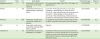
Epidermal growth factor (EGF) vaccine is another important vaccine that contains epidermal growth factor receptor (EGFR) oncogene. The level of EGFR is very high in NSCLC patients. CIMAvax-EGF vaccine is a human recombinant vaccine that is targeted to treat advanced NSCLC (6364). The vaccine is, currently, undergoing phase III trials and showed significant improvement in OS rate (median OS, 11.7 vs. 5.33 months). Talactoferrin, TG4010, belagenpumatucel-L, etc. are other antigen-specific vaccines that have been developed to combat advanced NSCLC (6566676869). These vaccines are still under scrutiny, and their overall impact on survival rate has been summarized in Table 2.
Table 2
Antigen-specific vaccines for NSCLC
| Vaccines | Phase | No. | Study design | Results | ClinicalTrials.gov # | Reference |
|---|---|---|---|---|---|---|
| Belagenpumatucel-L | II | 75 | Randomized, 3 dose cohorts (1.25, 2.5, or 5×10⁷ cells/injection) | OS improves with higher dose (581 vs. 252 days). No significant AEs | NCT01058785 | (65) |
| TG4010 | II | 148 | Combination with first-line chemotherapy in advanced NSCLC | 6 mon PFS: 43.2% (32 of 74; 95% CI, 33.4–53.5) in TG4010 + chemotherapy vs. 35.1% (26 of 74; 25.9–45.3) in the chemotherapy, AEs: more common in TG4010 + chemotherapy vs. 35.1% (26 of 74; 25.9–45.3) in the chemotherapy. TG4010 enhances the effect of chemotherapy in advanced NSCLC alone | NCT00415818 | (69) |
| MAGE-A3 | III | 2,312 | Multicenter, double blind, randomized, MAGE-A3 vs. placebo | No significant improvement in disease-free survival compare to placebo with MAGE-A3-positive NSCLC | NCT00480025 | (62) |
| EGF | III | - | Open-label, multicenter, randomized wild type EGFR NSCLC patients | Ongoing | NCT02187367 | - |
| Talactoferrin | II | 110 | Randomized, double blind, talactoferrin vs. placebo | Combination with C/P, demonstrated improvement in PFS (4.2 vs. 7.0 mon), and OS (8.5 vs. 10.4 mon) vs. placebo in patients with previously untreated stage IIIB/IV NSCLC. Enhance activity without significant additional toxicity | NCT00706862 | (70) |
In a recent study, personalized RNA mutanome vaccines have been developed to determine the T cell response against wide spectrum of mutations (71). The vaccine was successful in increasing PFS rate in patients suffering from melanoma. In another study, vaccine-induced T-cell response was evaluated for 20 neoantigens (72). The therapy not only enhanced survival rate but also reduced the probability of recurrence. Both studies indicate the benefits of personalized immunotherapy in the management of wide-variety of cancers. However, these studies have been conducted on a small number of patients, and vigorous clinical trials are needed to determine the efficacy and safety of personalized-vaccines.
MAJOR OBSTACLES IN IMMUNOTHERAPY
Immunotherapy is an effective way of treating NSCLC with mild to moderate side effects. The therapy has achieved success in increasing OS rate and preventing the progression of the disease. Nevertheless, it is not devoid of challenges, and more research is required to derive the best possible outcome out of this therapy. Recent development of immunotherapy is dependent on expression of PD-1/PD-L1 biomarkers. Therefore, the patients who do not produce sufficient level of PD-L1 cannot be diagnosed using these biomarkers and hence, remain untreated for a long period. Also, some patients have a relatively weaker immune system and do not produce ample level of T-cells to destroy the tumor. Blocking of CTLA-4 receptors is susceptible to cause overexpression of immune cells. Such reactions may lead to severe autoimmune disorder and can be lethal if not treated timely.
Another major challenge is the detections of immunogenic neoantigens. NSCLC is a heterogenous disease, and it is still impossible to identify all mutated genes associated with the disease (2). Not only more than one mutation is responsible for such tumors, but also their expression varies from person to person (7374). Hence, there can be several unidentified neoantigens that may contribute to enhancing the severity of the disease. TCs often disguise by constantly evolving and adapting to changing microenvironment (5575). These cells can express a wide variety of neo-antigens that cannot be identified with existing diagnostic tests and mass spectrometry. Also, some antigens are of small size and do not produce sufficient immunogenic response (75). Therefore, it is difficult to isolate these antigens for further analysis. Sometimes, the antigens change their conformation with the progression of the disease (75). In this case, routine biopsies are required to produce most potential antigens, but the process is both uncomfortable and risky and therefore, many patients choose not to continue with the therapy (5575).
CONCLUSION
Immunotherapy is considered as an effective new method to treat advanced NSCLC. Recently, a large number of studies has been conducted to evaluate the relative efficacy and safety of various immunotherapies. There is no study in which immunotherapy was used as an only treatment method to cure NSCLC. Hence, it is difficult to predict the true efficacy and safety of this approach. Although, development of immune checkpoint inhibitors and neoantigen specific vaccines can be a promising step to cure lung cancer, however, there is a long way to go to attain desirable success in this field. The future research should focus on the development of advanced mass spectrometry and sequencing methods to determine the potential biomarkers and neoantigens and overcome the limitations of existing strategies. Additionally, detailed clinical studies should be conducted to estimate and compare the overall efficacy and safety of immunotherapy and other therapeutic approaches.
 XML Download