

 PDF
PDF ePub
ePub Citation
 Print
Print


INTRODUCTION
Pro-inflammatory cytokine interleukin-12 (IL-12) was originally identified as a NK cell stimulatory factor (1) and a cytotoxic T lymphocyte (CTL) maturation factor (2,3). IL-12 induces Th1 responses (4,5) enhances the generation of CTL and lymphokine activated killer cells (6,7). IL-12 also stimulates T cells to produce cytokines including IFN-γ, GM-CSF and TNF-α (8).
IL-12 is a heterodimer formed by p40 and p35 subunits. The p40 subunit is mainly produced in excess of p35 subunit in dendritic cells and macrophages, and these cells also produce p40 homodimers (9). In addition, both monomers and homodimers of p40 are produced in transfected cells (10) and in mice (11). The biological function of p40 homodimer is controversial yet; acts as a receptor antagonist (12), induces production of TNF-α in macrophages (13,14), and promotes the migration of dendritic cells and macrophages (15,16). IL-12 is also found as a cell surface-associated form in a human monocytes and a mouse macrophage cell line, and is up-regulated in response to IFN-γ and LPS stimulation (17). In addition, an alternative membrane-associated form of the p35 molecule may be produced (18). However, the biological function of membrane-associated forms of the IL-12 and the p35 subunit has not been determined yet.
Administration of recombinant IL-12 exerts anti-tumor effect in experimental tumor models (19,20), but toxicities associated with systemic administration hamper clinical extension (21-23). To avoid the toxic problem of recombinant cytokines, the cytokine gene transfer method was adopted to achieve local cytokine production. Alternatively, expression of cytokine as a membrane-bound form further confined the functional range of the cytokines and lowered toxicity of various cytokines (24); TNF-α (25,26), GM-CSF (12,27-29), IFN-γ (27), IL-2 (30-34), IL-4 (35,36), and IL-12 (32,33,35,37,38). We hypothesized that a tumor cell vaccine may avoid such side effect if it stimulates selectively the TAA-specific CD8+ T cells, without involving CD4+ T helper cells and antigen presenting cells like dendritic cells (39).
We generated MethA tumor clones expressing membrane-bound form of IL-12p35 (mbIL-12p35) or IL-12p40 (mbIL-12p40) and investigated if macrophages are activated by the mbIL-12 expressing tumor clones. Moreover, we examined the tumor growth of mbIL-12p35 expressing tumor clone in CD4+ or CD8+ T cell-depleted mice to understand which T cell subpopulation functions mainly in the immune responses to the tumor clone cells.
MATERIALS AND METHODS
Cells and mice
The methylcholanthrene-induced fibrosarcoma MethA (BALB/c origin) was maintained in RPMI-1640 medium (Gibco-BRL, Rockville, MD), supplemented with 10% heat-inactivated fetal bovine serum (FBS; Gibco-BRL), 2 mM L-glutamine (Sigma, St. Louis, MO), 100 U/ml penicillin (Sigma, St. Louis, MO), 100µg/ml streptomycin (Sigma, St. Louis, MO), and at 37℃ under 5% CO2. Female BALB/c mice were obtained from Daehan Biolink (Eumseong, Korea), and all mice were used between 6~8 weeks of age. All animal procedures were approved and guided by the committee for experimental animal care and use at Chungnam National University.
Antibodies and reagents
Control rat-IgG, PE-anti-IL-12p40, PE-anti-CD4, PE-anti-CD8, purified anti-Ld, PE-conjugated goat anti-rat IgG, PE-conjugated rat anti-mouse IgG, and recombinant mouse IL-2 and IL-12 were purchased from BD PharMingen (San Diego, CA). Purified anti-IL-12p35 mAb was purchased from Santa Cruz Biotechnology (Santa Cruz, CA). Enzyme-linked immunosorbent assay (ELISA) kit for mouse TNF-α was purchased from R&D Systems (Menneapolis, MN). G418 were purchased from Sigma (St. Louis, MO).
Vector construction, transfection, and drug selection
The construction of pcDNA3.1(+) based chimeric cDNAs expression vectors of mbIL-12p35 and mbIL-12p40 was previously described (38). The 240 bp TNF-α cDNA fragment encoding transmembrane region, cytoplasmic region, and part of the extracellular region was followed by the cDNA fragments of IL-12p35 (encoding 215 amino acids) or IL-12p40 (encoding 335 amino acids). MethA cells were transfected with mbIL-12p35, mbIL-12p40, or mock vector alone using the Lipofectamine 2000 (Invitrogen). After 24 hr, cells were plated in 96 well plates (0.5 cell/well) in G418 (1 mg/ml)-containing medium. Viable colonies were usually visible 2~3 weeks after transfection.
Isolation of mouse peritoneal macrophages
Mice were injected with 0.8 ml of 4% thioglycollate (DIFCO, Detroit, USA) intraperitoneally, and 48 hr later, macrophages were obtained by peritoneal lavage with sterile RPMI-1640 medium containing 1% FBS. Cells were washed three times with RPMI-1640 at 4℃ and were maintained in 5% CO2 at 37℃, and plated at a concentration of 1×10⁶ cells/ml (in a volume of 0.5 ml/well) in a 24 well plates. After 1 hr, non-adherent cells were removed by washing, and MMC (mito mycine-C)-inactivated tumor clones (0.5 ml/well) were mixed for co-culture. As a control, LPS (2µg/ml) were added to the adherent cells.
FACS analysis
To analyze the expression of mbIL-12p35 or mbIL-12p40 on cell surface, mbIL-12p35 or mbIL-12p40 clones were incubated for 30 min at 4℃ with fluorochrome-conjugated IL-12 p35 or p40-specific antibody appropriately diluted in staining buffer (1×PBS containing 0.02% sodium azide, and 2% FBS), respectively. Cells were then washed with staining buffer three times. Stained cells were analyzed on a FACSCalibur flow cytometer with CELLQuest (BD, San Jose, CA).
ELISA
To test of whether mbIL-12p40 and mbIL-12p35 proteins released on tumor clones, 5×10⁵ cells of wild type, mock vector transfected, mbIL-12p35 and mbIL-12p40 clones were cultured for 48 hr in 2 ml of normal medium, respectively. To investigate whether production of TNF-α on macrophages by mbIL-12p35 or mbIL-12p40 tumor clones, 1×10⁶ cells of mouse peritoneal macrophages were co-cultured with 5×10⁵ cells of MMC-treated wild type, mock vector transfected, mbIL-12p35 and mbIL-12p40 expressing tumor clones for 24 hr, respectively. All culture supernatants were harvested and measured for TNF-α and IL-12 levels by specific ELISA kit of corresponding cytokines following the manufacturer's instructions. As a positive controls for IL-12 and TNF-α, culture supernatant from the LPS (2µg/ml)-treated mouse peritoneal macrophages was used.
Tumor growth in CD4+ T cell or CD8+ T cell-depleted mice
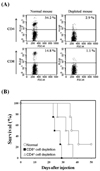
To deplete CD4+ T cells or CD8+ T cells in vivo, mice were injected intraperitoneally with antibodies specific to CD4 (GK1.5, 200µg/time) or CD8 (53-6.72, 200µg/time), respectively, on days -3, 0, 3, 7. Depletion of the target cells was confirmed by FACS analysis using CD4 and CD8-specific antibodies. After injection of mbIL-12p35 expressing tumor clone (5×10⁴ cells) on day 0, survival was monitored.
RESULTS
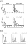
The mbIL-12 subunit molecules are stably expressed on tumor cell surface stably
The MethA cells were transfected with mbIL-12p35 or mbIL-12p40 expression vectors based on pcDNA3.1(+) vector. After transfection, cells were selection with G418-containing medium, and the drug-resistant clones were isolated and cultured in mass. The drug-resistant clones were then analyzed for the expression of mbIL-12p35 or mbIL-12p40. To analyze the effect of mbIL-12 on the expression of MHC class I molecules, the expression level of MHC class I (Ld) on the tumor clones was analyzed by FACS analysis. The expression of mbIL-12 p35 or p40 molecules was stable for more than three months in vitro culture (Fig. 1A), and they were expressed equivalent levels of MHC class I (Ld) (Fig. 1B), suggesting that the expression of the mbIL-12 subunits do not affect severely the expression of MHC class I.
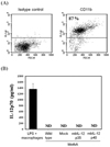
The mbIL-12p35 and the mbIL-12p40 molecules are not released from the tumor clones
To analyze whether the mbIL-12p35 or the mbIL-12p40 molecules are cleaved and released from the mbIL-12p35 or the mbIL-12p40 expressing tumor clones, culture supernatants of wild type MethA cells, mock vector transfected, mbIL-12p35 and mbIL-12p40 expressing clones were analyzed by ELISA for the presence of IL-12 or its subunits. As a positive control for IL-12, culture supernatant from LPS (2µg/ml)-treated peritoneal macrophages was used (Fig. 2A). The purity of isolated macrophages was measured by FACS analysis using anti-CD11 antibody. After 48 hr, the culture supernatants were harvested and measured for IL-12 by ELISA. The mbIL-12p35 or mbIL-12p40 was not detected in any culture supernatant but LPS-activated macrophages (Fig. 2B). This result indicates that the membrane-bound form of p35 and p40 molecules are not released from the tumor clones, thus, only cells in physical contact with the mbIL-12p35 or the mbIL-12p40 tumor clone would be affected.
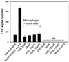
The mbIL-12p35 or the mbIL-12p40 expressing tumor clones fails to induce TNF-α production in macrophages
Soluble form of the IL-12p40 monomer or homodimer induces production of TNF-α on primary macrophages were reported (13). To investigate whether the mbIL-12p35 or the mbIL-12p40 clone induces production of TNF-α on macrophages, isolated peritoneal macrophages from the normal mouse were co-cultured with MMC-inactivated wild type MethA cells, mock vector transfectant clone, mbIL-12p35 or mbIL-12p40 expressing tumor clone, respectively. In contrast to the LPS-treated macrophage group, TNF-α levels of the other groups were similar to the group cultured macrophages only (Fig. 3), indicating that the mbIL-12p40 or the mbIL-12p35 expressing tumor clones do not activate macrophages to produce TNF-α.
Depletion of CD8+ T cells accelerates IL-12p35 expressing MethA tumor cell growth
In previous study, we evaluated tumorigenicity of mbIL-12 subunit expressing tumor clones, and found that the IL-12p35 expressing tumor clone was less tumorigenic than mbIL-12p40 expressing tumor clone (38). The results suggest that the mbIL-12p35 expressing tumor clone is more effective than mbIL-12p40 expressing tumor clone to activate anti-tumor immune responses. To analyze which T cell subpopulation plays a critical role in the anti-tumor immune responses, mice were depleted CD4+ or CD8+ T cells by injecting specific monoclonal antibodies (Fig. 4A). The depleted mice were then injected with mbIL-12p35 expressing tumor clone subcutaneously, and survival was monitored. As shown in Fig. 4B, the group depleted with CD8+ T cell subpopulation was most susceptible to the mbIL-12p35 expressing tumor clone, suggesting that the CD8+ CTLs are responsible for the immune response to mbIL-12p35 expressing tumor clone.
Taken together, the expression of membrane-bound form of cytokine, p35 and p40 subunit of IL-12, was stable for extended period of time, and the molecules were not released into culture supernatants. Moreover, both the mbIL-12p35 and the mbIL-12p40 expressing tumor clones did not activate macrophages in vitro, but the mbIL-12p35 expressing tumor clone lost tumorigenicity by involving mainly CD8+ T cells.
DISCUSSION
We reported previously that the mbIL-12p35 expressing MethA tumor clone was highly immunogenic in vivo, so that the tumor clone failed to form tumor in BALB/c syngeneic mice (38). In this study we investigated more about the expression characteristics of membrane-bound form of IL-12 subunits and their effects on macrophages in vitro. The expression of membrane-bound form of IL-12 p35 or p40 subunits was stable for more than 3 months, and the molecules were not released into culture supernatants. The peritoneal macrophages were not activated by the mbIL-12p35 expressing tumor clones in vitro, but growth of the mbIL-12p35 expressing tumor clone in CD8+ T cell-depleted mice was accelerated than in CD4+ T cell-depleted mice and normal mice. These results suggest that the mbIL-12p35 expressing tumor clone cells may stimulate CD8+ T cells by direct priming, without involving antigen presenting cells or CD4+ T helper cells.
The main concern in the clinical application of IL-12 in tumor therapy is its systemic side effects. Various toxicities of recombinant IL-12 were recorded in mice and human; elevated transaminases, leukopenia, and liver dysfunction (21-23,40). Tumor cells genetically engineered to express IL-12 also showed side effects (41,42). In our laboratory, IL-2 or IL-4 was expressed on MethA tumor cells and their anti-tumor effects were analyzed (30,34,36). As expected the membrane-bound form of cytokine chimeric molecules with TNF-α were stably expressed for a long time. The chimeric cytokine molecules were not detected in culture supernatant, suggesting that the membrane-bound form may not be shed so much.
With anti-tumor effect of tumor clone expressing the mbIL-12p35 molecule, we have been interested in elucidating which cell populations are critical to display such anti-tumor effects. As an indirect way we analyzed stimulatory effect with the tumor clones expressing mbIL-12p35 or p40 on peritoneal macrophages. Clearly the tumor clones were not effective to stimulate the macrophages. These results do not reconcile with the positive effect of soluble p40 monomer or homodimer to induce TNF-α in macrophages (13). We speculate that the membrane-bound form of p40 on tumor cells may require proper orientation to interact with specific IL-12 receptors. The membrane-bound form may have limited flexibility or totally different orientation. Interestingly, the mbIL-12p35 expressing tumor clone showed anti-tumor effect (38), but the tumor clone failed to activate macrophages to produce TNF-α in vitro. These results suggest that the antigen presenting cells may not be critical to induce anti-tumor effect by the mbIL-12p35 expressing tumor clone, at least in vitro condition. Further study is required to prove whether antigen presenting cells are dispensable for the anti-tumor effects. Consistently, the growth in vivo of the mbIL-12p35 expressing tumor clone was accelerated in CD8+ T cell-depleted mice than CD4+ T cell-depleted mice as previously reported. We also reported previously that NK cells were not critical in the induction of anti-tumor immunity induced by mbIL-12p35 expressing tumor clone (38).
To develop a tumor cell vaccine that stimulates TAA-specific CD8+ T cells selectively, without involving CD4+ T helper cells and antigen presenting cells like dendritic cells or macrophages, tumor cells should be equipped with the ability to provide signal 1 and costimulatory signals to fully activate CTLs. Practically, we could induce anti-tumor effect by genetically modifying tumor cells to express membrane-bound form of p35 subunit of IL-12.
 XML Download