

 PDF
PDF ePub
ePub Citation
 Print
Print


INTRODUCTION
Allogeneic hematopoietic stem cell transplantation (HSCT) after total body irradiation is a common treatment for nonmalignant and malignant hematologic disorders. Graft-versus-host disease (GVHD) is a life threatening complication following the treatment and a great impediment of success of the HSCT. GVHD is considered to progress through three distinct phases (1,2): inflammation induced by irradiation, activation of allo-reactive T cells, and then tissue damage in target organs, such as spleen, liver, lung and intestine, which is caused by the activated T cells. Donor-originated T cells recognize allo-antigens displayed by host tissues and initiate allo-responses in the GVHD hosts. Especially, T cell responses specific for peptides originated from allogeneic minor histocompatibility antigens play major roles in induction of GVHD upon HSCT between MHC-matched individuals.
Pre-conditioning of the recipients of HSCT, total body irradiation before the transplantation, is necessary for destroying malignant cells residing in the recipients, and for creating space in the recipient's bone marrow in order to facilitate the engraftment of injected bone marrow cells (3). Intensity of the total body irradiation, however, correlates with increase in risk of GVHD (4). Irradiation with high dose increases systemic inflammatory cytokine and endotoxin levels and drives the interaction between DC and donor T cells, leading to severe GVHD (5,6).
Inflammatory cytokines such as IL-1, TNF-α, and IL-6 have been known to be related to increase in GVHD severity (7,8). Cytokine profiles of T cells, IFN-γ and IL-4 have also been demonstrated to be important in the pathogenesis of GVHD (9). IL-17 is another inflammatory cytokine and production of IL-17 by CD4 T cells play a critical role in triggering inflammation and tissue injury in autoimmune diseases (10,11). Most of the studies regarding IL-17 in relation to GVHD have been focused on the role of CD4 T cells producing IL-17 (Th17) with controversies over suppressive role or contribution to local damages (12-14). A human study demonstrated that IL-17 producing T cells contributed to acute GVHD (15). Although the relationship between the increase in inflammatory cytokine levels, especially cytokine secretion by T cells, and the severity of GVHD has been well known, the role of myeloid cells in GVHD and the production of cytokines by those cells have been obscure.
Myeloid cells participate in diverse immune function, supporting or regulating immune responses by producing cytokines, nitric oxide, and reactive oxygen species, etc. And the myeloid lineage cells consist of diverse subpopulations: Gr-1+ and Mac-1+ cells represent granulocytes and macrophages, respectively, and Gr-1+ Mac-1+ cells, named myelid-derivved suppressor cells (MDSC), have recently been identified to be a population that can suppress T cell function (16). Since GVHD has been considered as T cell-mediated immunopathology in which myeloid cells have rarely been considered as important factors, detailed studies on myeloid cells related to GVHD, for example the kinetic study or contribution of myeloid cells during GVHD development, have rarely been done.
In our previous GVHD study using a MHC-matched mouse model (B6 → BALB.B, intravenous injection of bone marrow (BM) cells and splenocytes from B6 mice into irradiated BALB.B mice), expansion of CD8 T cells with specificity for minor H antigens haves been detected in peripheral blood and target tissues (17). And immunodominance phenomenon has been observed during the course of GVHD: that is, among the minor H antigen-specific CD8 T cells, CD8 T cells for H60 dominated over those for H13 and HY during the GHVD progression. Since then, we were interested in the role of myeloid cells in GVHD progression, because we observed that proportions of Gr-1+ cells and Mac-1+ cells in the peripheral blood were high at early time points before CD8 T cell expansion period and late time points before morbid phase of GVHD hosts. And we presumed that the myeloid cells would produce inflammatory cytokines at the early time point and contribute to development of GVHD by helping CD8 T cell expansion. As the first step to test this presumption, we chose irradiation for pre-conditioning as an inflammation inducing factor and studied the influences of irradiation doses on GVHD development. Two different irradiation doses were selected, which could generate full chimerism (myeloablative conditioning) vs mixed chimerism (non-myeloablative conditioning) of leukocytes in the B6 → BALB.B GVHD hosts and influence GVHD survival. Using the two different doses, we compared immune kinetics in blood and target organs of GVHD hosts, and production of IL-17 by the myeloid cell population. The results show that the effects of different doses of irradiation are more noticeable in the leukocytes infiltrating target organs, and IL-17 production by myeloid cells is higher in the GVHD hosts that received high dose irradiation.
MATERIALS AND METHODS
Mice
Male C.B10-H2b/LiMcdJ (BALB.B) and female C57BL/6 (B6) mice were obtained from the Jackson Laboratory (Bar Harbor, ME, USA) and used as recipients and donor of bone marrow (BM) and spleen cells, respectively. Mice were housed under specific pathogen-free conditions at the center for animal resource development of Seoul National University College of Medicine in Korea. All the experiments were performed under approval from The Seoul National University Institutional Animal Care and Use Committee (IACUC).
GVHD induction
Bone marrow (BM) cells were prepared from female B6 mice by flushing femurs and tibias with 1×PBS (Phosphate buffered saline). Spleen cells from female B6 mice were prepared by grinding spleens through steel mesh. Recipient male BALB.B mice were irradiated with split does from a 137Cs source with a 5 hr interval. The recipient mice pre-conditioned either with 900 cGy (myeloablative conditioning) or 400 cGy (non-myeloablative conditioning) were injected with a mixture of B6 BM (5×10⁶) and splenocytes (2×10⁷) in a volume of 300 µL into a lateral tail vein after 5 hours after the second irradiation.
Preparation of tissue infiltrating leukocytes
BALB.B GVHD hosts were perfused with PBS/heparin (75 U/ml; Sigmal, St Louis, MO, USA) before sacrifice to deplete circulating peripheral blood leukocytes (PBLs). The preparation of leukocytes infiltrating livers and lungs was described in detail previously (17). In brief, livers were pressed through stainless steel mesh and suspended in 5% fetal calf serum (FCS)-PBS. The cell suspensions were admixed with 33% Percoll containing 100 U/ml heparin and were then centriguged 200 rpm for 15 min at room temperature. The cell suspensions were applied to Percoll gradients and RBC lysis, and then resuspended in 5% FCS-PBS. Lungs were cut into small pieces and treated with collagenase at 37℃ for 30 minutes after washing with 1×Hanks balanced salt solution (HBSS)/HEPES(N-2-hydroxyethylpiperzine-N'-2-ethaaaanesulfonic acid: 10 mM). Then, after washing twice with 5% FCS-PBS, the cells were applied to Percoll gradients and resuspended in 5% FCS-PBS after washing with 1×PBS.
Cell staining and flow cytometry
Fresh PBLs, splenocytes, or lymphocytes infiltrating livers and lungs from the GVHD hosts were incubated at 4℃ for 30 minutes in FACs buffer (1×PBS with 0.1% bovine calf serum and 0.05% sodium azide) containing PE-labeled H60/H-Kb tetramer and saturating amount of FITC-conjugated anti-CD11a (2D7, eBiosicence, San Diego, CA) and APC-conjugated anti-CD8 (eBioscience) monoclonal antibodies. Anti-β2 microglobulin B (anti-β2mb; S19.8, Santa Cruz Biotechnology) antibody was used to distinguish donor (B6:β2mb)-originated cells from recipient (BALB.B: β2ma)-originated ones. Other monoclonal antibodies used were PE or APC-conjugated anti-Mac-1 (M1/70, eBioscience), FITC-conjugated anti-Gr-1 (RB6-8C5, eBioscience), PE-conjugated anti-CD4 (GK1.5, eBioscience). For intracytoplsmic staining for IFN-γ and IL-17 production, cells were stained with anti-Mac-1 or anti-Gr-1 antibodies, washed, and then fixed with 4% paraformaldehyde at room temperate for 20 minutes. After washing with 1×PBS, the cells were permeabilized with saporine and stained with APC-conjugated anti-IFN-γ (XMG1.2, eBioscience) or PE-conjugated IL-17 (IL-17A; TC11-18H10, BD Bioscience, San Diego, CA) antibodies at 4℃ for 2 hrs. The stained cells were analyzed using a FACSCalibur equipped with CellQeust software (BD Bioscience).
RESULTS
Irradiation doses corresponding to myeloablative and non-myeloablative conditioning in GVHD hosts
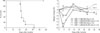
To select two different irradiation doses that correspond to myeloablative high intensity vs non-myeloablative mild intensity doses in the B6 → BALB.B GVHD system, 900 cGy, 600 cGy, and 400 cGy of irradiation doses were tested for chimerism formation in the PBLs of BALB.B host mice. BALB.B mice were irradiated with the three different doses, transplanted with BM and splenocytes from B6 mice, and then eye-bled on days 4, 7, and 14 post-transplantation. Pooled PBLs prepared from three BALB.B mice of each group were stained with antibodies for marker antigens, Gr-1, Mac-1, CD4 and CD8, in combination with anti-β2mb antibody to distinguish the cell origins, donor (β2mb) or recipient (β2ma), and then analyzed via flow cytometry. Flow cytometry results showed that, on day 4 after the transplantation with 900 cGy irradiation dose, Gr-1+, Mac-1+, CD4+ T cell and CD8+ T cells in PBL of BALB.B hosts showed mixed chimerism, mixed with cells of donor and recipient origins. However, from day 7, they showed full chimerism with all the cell populations replaced with the cells of B6 origin (Fig. 1). On the contrary, host-originated Gr-1+ and Mac-1+ populations were still detected in of the PBLs of the BALB.B GVHD hosts with irradiation dose of 400 cGy, showing mixed chimerism status, until the day 14 after the transplantation, while most of CD4 T and CD8 T cells were of B6 origin, probably due to expansion of allo-reactive CD4 and CD8 T cells of B6 origin in the BALB.B hosts. With 600 cGy of irradiation dose, overall pattern of PBL chimerism was similar to that seen in BALB.B recipients with 900 cGy irradiation (data not shown). Based upon these results, we selected 900 cGy and 400 cGy irradiation doses as myeloablative and non-myeloablative conditioning doses, leading to full and mixed-chiemerism in the PBLs of the BALB GVHD hosts, respectively.
Correlation between pre-conditioning intensity and GVHD severity
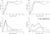
Next, we examined the effect of the selected myeloablative (900 cGy) and non-myeloablative (400 cGy) irradiation doses on GVHD severity in our allogeneic B6 → BALB.B model. BALB.B mice which were irradiated under the two different conditions and transplanted with allogeneic B6 BM and spleen cells were observed for their survival for >50 days and regularly checked for their weight changes for 35 days after transplantation with BM and splencotyes from B6 mice. Syngeneic control transplantation, B6 → B6, was performed under the same irradiation conditions. Myeloablative high dose pre-conditioning and subsequent allogeneic transplantation led to death of BALB.B hosts with drastic weight loss and other GVHD symptoms, such as diarrhea and hunched posture (Fig. 2). With non-myeloablative low dose pre-conditioning, allogeneic BALB.B hosts survived longer than 50 days. None of the syngeneic B6 hosts developed GVHD under any circumstatnces. Even though the B6 hosts under the myeloablative condition went though weight loss early after transplantation, they recovered soon afterward and survived longer. This result demonstrated that the selected irradiation does of 900 cGy (termed myeloablative) and 400 cGy (non-myeloablative) resulted in different outcome of GVHD induction, not only different PBL chimerism in B6 → BALB.B system, implying that they are suitable doses for investigating the irradiation effects on immune kinetics during GVHD development.
Kinetics of peripheral blood leukocytes in GVHD hosts with myeloablative vs non-myeloablative irradiation
Then, we compared PBL profiles along the time course after GVHD induction between the BALB.B hosts conditioned with myeloabltive vs non-myeloablative irradiation, wondering whether PBL profiles would be different depending upon the GVHD severity, so that they could be used to distinguish and predict GVHD outcomes. Bloods from the GVHD survivors of each pre-conditioning group were collected periodically and pooled for PBL preparation. The PBLs were typed for profiling proportions of Gr-1+, Mac-1+, CD4+ T and CD8+ T cells in the blood via staining with antibodies specific for corresponding antigens and subsequent flow cytometric analysis, and the proportions of each cell type were plotted as kinetics graphs. On the contrary to our expectation, there was no big difference in the percentages of Gr-1+ or Mac-1+ cells present in bloods between the myeloabaltively vs non-myeloablatively conditioned BALB.B hosts. Their fractions reached peak, about 90% of PBL, on day 7 after the GVHD induction and continued to be high afterward (Fig. 3). The kinetics of CD4+ T cells in the blood was not significantly different between the two groups, either. The only significant difference was found in CD8 T cell population: the proportion of CD8 T cells in bloods of GVHD hosts was higher under the myeloablative (~12%) condition than the non-myeloabative (4~7%) condition (Fig. 3). The results from the PBL profile study demonstrated that there was no big difference in myeloid cell proportions between the GVHD hosts with myeloablative vs non-myeloablative irradiation, even though the GVHD hosts with the two different pre-conditioning doses experienced different degrees of the disease severity.
Comparison between the profiles of leukocytes that infiltrate target organs of GVHS hosts under myeloablative vs non-myeloablative conditions
Since the PBLs profiles of GVHD hosts with different doses of irradiation were not significantly different, especially considering the proportions of myeloid lineage cells, we explored the leukocytes that infiltrated into target organs during GVHD course, presuming that the infiltration of myeloid cells into target tissues might have been influenced by the irradiation doses. Leukocytes infiltrating spleens, livers and lungs were periodically prepared from three BALB.B mice under each condition, pooled for staining and subsequent flow cytometry to profile Gr-1+, Mac-1+, CD4+ T and CD8+ T cells in the organs along the GVHD course.
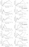
Overall kinetics of the four types of leukocytes infiltrating target tissues was significantly different, depending upon the pre-conditioning doses (Fig. 4). Proportions of Gr-1+ cells continuously increased in the spleens (Fig. 4A), livers (Fig. 4B), and lungs (Fig. 4C) of BALB.B hosts with high intensity irradiation and accounted for more than 60% of filtrating leukocytes at late time points (later than day 21 post-transplantation) in the livers and lungs. With low intensity irradiation, the kinetics of Gr-1+ infiltrating cells showed patterns of early increase followed by decrease in the spleens, livers, and lungs of the BALB.B hosts. The kinetics of Mac-1+ cells was very similar to that of Gr-1+ cells residing in all the three organs in both myeloabltively and non-myeloabltively conditioned hosts.
Percentages of CD4+ T cells reached peak on day 4 (17%), day 7 (45%), and day 14 (32%) in the spleens, livers, and lungs of the BALB.B hosts with high intensity irradiation, respectively, and then waned afterward (Fig. 4A-C). This initial up and then down pattern of kinetics was observed in the case of CD4 T cells infiltrating the tissues of the BALB.B hosts with low intensity irradiation, but the peak values were comparatively low (14% in spleens, 30 % in livers, and 10% in lungs on day 4 post-transplantation).
Patterns of CD8 T cell kinetics was similar to those of CD4 T cell in the tissues of the GVHD hosts with myeloablative pre-conditioning with peak values of 13% (day 4 post-transplantation), 35% (day 14), and 42% (day 14) in the spleens, livers, and lungs, respectively. In the GVHD hosts with non-myeloablative pre-conditioning, proportion CD8 T cells gradually increased in the spleens with peak values of 12% on day 21 but, in the lungs and livers, they reached peak on day 14 post-transplantation.
Altogether the results obtained from the profiling of leukocytes infiltrating target organs of GVHD, it could be concluded that immune kinetics in the target organs is influenced by the pre-conditioning intensity. Among the three organs, liver and lung showed very similar kinetics, while spleen showed its own kinetics pattern for each cell population, possibly due to its role as secondary lymphoid organ as well as a GVHD target organ. However, on the contrary to our expectation, proportion of myeloid cells infiltrating spleens and lungs cells at early time point (on day 4 post-transplantation) was lower in the GVHD hosts with myeloabaltive irradiation than non-myeloablative irradiation, for example, 12 % vs 15 % of Gr-1+ cells in the spleens and 17.3% vs 44% of Gr-1+ cells in the lungs of myeloabaltively conditioned vs non-myeloabaltively conditioned hosts, respectively.
IL-17 production by Gr-1+ or Mac-1+ cells that infiltrate target organs of GVHD hosts
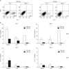
Even though proportions of myeloid populations in target tissues on day 4 post-transplantation did not correlate with irradiation intensities, we wondered whether proportion of cells producing inflammatory cytokine in the infiltrating myeloid cells would correlate with the irradiation dose. To test this, we prepared infiltrating leukocytes from the spleen, liver, and lung on day 4 after GVHD induction and analyzed for the production of IL-17 by Gr-1+ or Mac-1+ cells. Intracytoplasmic staining of the cells with anti-IL-17 and anti-IFN-γ antibodies and subsequence flow cytometry analysis revealed that proportions of IL-17 producing cells in Gr-1+ and Mac-1+ cells present in the spleen were higher when the hosts received myeloabaltively high dose irradiation than those with non-myeloabaltively low dose irradiation (Fig. 5A). They did not produce IFN-γ. Proportion of IL-17 producing Gr-1+ cells was higher in the spleens (18.7% on average) than in the livers (7.63% on average) or lungs (10.4% on average) of the GVHD hosts with myeloablative irradiation (Fig. 5B). In the case of Mac-1+ cells, proportions of IL-17 producing cells were higher in the lungs (16.7% on average) than those in the spleens (5.85% on average) and the livers (5.13% on average) of the myeloablatively conditioned hosts. In all the three organs, proportions of IL-17 producing cells were lower in non-myeloablatively conditioned hosts than in the myeloabltively conditioned ones. On day 7 after the GVHD induction, IL-17 production by the myeloid cells that infiltrated target organs subsided to very low levels (Fig. 5B). Altogether, these results demonstrate that, even though proportions of Gr-1+ or Mac-1+ cells was little lower in the target organs of myeloablatively conditioned hosts than those of non-myeloabaltively conditioned ones at early time points after GVHD induction, proportions of myeloid cells producing inflammatory cytokine IL-17 in the infiltrating cells were higher in the former than the latter.
DISCUSSION
In this study, we investigated the effects of irradiation pre-conditioning dose on the kinetics of leukocytes residing in blood and tissues of GVHD hosts, and production of an inflammation cytokine, IL-17, by myeloid cells. And the results showed that different irradiation doses affected kinetics of leukocytes infiltrating GVHD target organs and IL-17 production by the myeloid cells.
B6 → BALB.B, used in this study, is a mouse model for GVHD occurring after BM transplantation between MHC-matched unrelated individuals (17). In terms of pathogenesis of GVHD, allogeneic T cell activity, especially allo-minor H antigen specific CD4 T and CD8 T cells have been considered to major risk factor, and the minor H antigen-reactive CD8 T cells have been detected in the blood and tissues of BALB.B GVHD hosts (17). Even though it has been known that alloreactive CD8 T cells execute tissue damage in the target organs, the connection between myeloid cells and the activation of allo-reactive T cells has been obscure. Here, our data showed that myeloid cells actually produce cytokine, IL-17, in the GVHD target tissues, in response to irradiation inflammatory stimulus. Therefore, it could be suggested that even though proportions of myeloid cells in blood and target organs at early time point after GVHD themselves did not correlate with irradiation intensity, fractions of IL-17 producing cells in the tissue infiltrating myeloid cells reflected intensity of pre-conditioning. Based on this result, it could be suggested that such difference in the activity of tissue infiltrating myeloid cells to produce IL-17 would influence the adaptive immunity in the target tissues following the inflammation phase during the GVHD course, resulting in the difference in the magnitudes of expansion of allo-reactive CD4 T and CD8 T cells in the target organs between the GVHD hosts with different irradiation doses.
IL17 has been known to be involved in development of some autoimmune diseases. Regarding the role of IL-17 in GVHD, focus has been onto the role of Th17 in GVHD and Th17 has been reported to be involved in cutaneous and pulmonary GVHD in a mouse model (12) and the increase of Th17 cells or Tc17 cells correlated with a higher incidence of acute GVHD in human study (18). Our result demonstrates that IL-17 can be produced by myeloid cells as well. However, Gr-1+ granlocytes and Mac-1+ macrophages may contribute to GVHD progression in various ways, not just simply through Il-17 production. They could produce other cytokines, IL-1, IL-16, and TNF, and IFN-γ in responding to irradiation stimulation. Moreover, the myeloid cells are composed of various cell types, including recently identified myeloid suppressive cells (Gr-1+Mac-1+). Therefore, elucidating the exact role of myeloid cells to GVHD progression in B6 → BALB.B system should await further detailed study including dissection of myeloid cells and investigation on their cytokine profiles and migration along the GVHD course.
The results from PBL profiling study demonstrate that just leukocyte typing of PBL does not reflect or predict the GVHD severity: there is no big difference in proportions of each leukocyte population between the GVHD hosts with high and low disease severity, except for CD8 T cell population. Rather, significant difference in the leukocyte profiles between the GVHD hosts with high and low doses irradiation was detected in the leukocytes infiltrate target organs of GVHD, suggesting a close relationship between irradiation doses and leukocyte infiltration. In clinic, it is often observed that tumors relapse in the GVHD patients with mild symptoms, who have received non-myeloid conditioning, and the relapse occurs in the extra bone marrow tissues (19). Based upon our data, this is supposed to be due to reduced infiltration of inflammatory cytokine producing myeloid cells at early time points post-irradiation and hence reduced activity of allo-reactive CD8 T cells afterwards, which can help to eradicate tumors in the tissues. Therefore, for double purpose of success of HSCT and complete remission of tumors, a tool to control inflammation to help tissue infiltration of leukocytes to eradicate tumors but avoid adverse GVHD development should be developed.
In summary, our results demonstrate that pre-conditioning dose influence leukocyte infiltration into target organs, resulting in different kinetics of leukocyte profiles in GVHD hosts, different degrees of IL-17 production by myeloid cells, and finally different outcome of GVHD. Investigation on the immune kinetics in detail, including cytokine profiling produced in each different target organ and detailed phenotyping of infiltrating cells, will enhance the understanding of pathophysiology of GVHD. The results from this study will be helpful for better understanding of pathophysiology of GVHD and development of proper method to control GVHD and tumor relapse in clinic.

 XML Download