

 PDF
PDF ePub
ePub Citation
 Print
Print


Abstract
Chronic Recurrent Multifocal Osteomyelitis (CRMO) is a non-bacterial inflammatory disorder of unknown cause occurring in children and adolescents. It is characterized by the insidious onset of pain and swelling to multifocal involved bones, recurring over months to years. Non-steroid anti-inflammatory drugs (NSAIDs) and steroids are the first choice for the initial and relapse treatment. However, multifocal and frequent relapses might require more intensive anti-inflammatory treatment. Here, we report that an adolescent with CRMO refractory to antibiotics, NSAIDs and steroids over a two-year responded well to bisphosphonate. To our knowledge, this is the first case using bisphosphonate in adolescent refractory CRMO in Korea.
REFERENCES
1. Giedion A, Holthusen W, Masel LF, Vischer D. Subacute and chronic “symmetrical” osteomyelitis. Ann Radiol (Paris). 1972; 15:329–42.
2. Van Howe RS, Starshak RJ, Chusid MJ. Chronic, recurrent multifocal osteomyelitis. Case report and review of the literature. Clin Pediatr (Phila). 1989; 28:54–9.
3. Bj0rkstén B, Boquist L. Histopathological aspects of chronic recurrent multifocal osteomyelitis. J Bone Joint Surg Br. 1980; 62:376–80.
4. Chun CS. Chronic recurrent multifocal osteomyelitis of the spine and mandible: case report and review of the literature. Pediatrics. 2004; 113:e380–4.

5. Khanna G, Sato TS, Ferguson P. Imaging of chronic recurrent multifocal osteomyelitis. Radiographics. 2009; 29:1159–77.
6. El-Shanti HI, Ferguson PJ. Chronic recurrent multifocal osteomyelitis: a concise review and genetic update. Clin Orthop Relat Res. 2007; 462:11–9.
7. Miettunen PM, Wei X, Kaura D, Reslan WA, Aguirre AN, Kellner JD. Dramatic pain relief and resolution of bone inflammation following pamidronate in 9 pediatric patients with persistent chronic recurrent multifocal osteomyelitis (CRMO). Pediatr Rheumatol Online J. 2009; 7:2.
8. Mandell GA, Contreras SJ, Conard K, Harcke HT, Maas KW. Bone scintigraphy in the detection of chronic recurrent multifocal osteomyelitis. J Nucl Med. 1998; 39:1778–83.
9. Fritz J, Tzaribatchev N, Claussen CD, Carrino JA, Horger MS. Chronic recurrent multifocal osteomyelitis: comparison of whole-body MR imaging with radiography and correlation with clinical and laboratory data. Radiolog. 2009; 252:842–51.
10. King SM, Laxer RM, Manson D, Gold R. Chronic recurrent multifocal osteomyelitis: a noninfectious inflammatory process. Pediatr Infect Dis J. 1987; 6:907–11.
11. Girschick HJ, Huppertz HI, Harmsen D, Krauspe R, Müller-Hermelink HK, Papadopoulos T. Chronic recurrent multifocal osteomyelitis in children: diagnostic value of histopathology and microbial testing. Hum Pathol. 1999; 30:59–65.
12. Carpenter E, Jackson MA, Friesen CA, Scarbrough M, Roberts CC. Crohn's-associated chronic recurrent multifocal osteomyelitis responsive to infliximab. J Pediatr. 2004; 144:541–4.
13. Girschick HJ, Raab P, Surbaum S, Trusen A, Kirschner S, Schneider P, et al. Chronic non-bacterial osteomyelitis in children. Ann Rheum Dis. 2005; 64:279–85.
14. Simm PJ, Allen RC, Zacharin MR. Bisphosphonate treatment in chronic recurrent multifocal osteomyelitis. J Pediatr. 2008; 152:571–5.
15. Duffy CM, Lam PY, Ditchfield M, Allen R, Graham HK. Chronic recurrent multifocal osteomyelitis: review of orthopaedic complications at maturity. J Pediatr Orthop. 2002; 22:501–5.
Figure 1.
(A) Bony tissue of left arm shows marrow fibrosis with chronic nonspecific inflammation. (B) Cortical bone and periosteum of right arm reveals osteocartilaginous tissue without histologic abnormality (A, B: H&E, ×40)
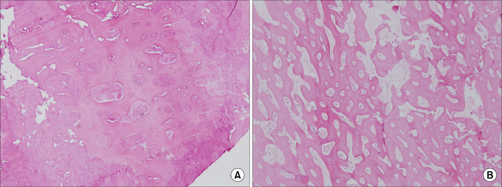
Figure 2.
(A) Plain radiography of the right femur shows osteolytic lesion and periosteal reaction in midshaft of right femur with overlying soft-tissue swelling, (B) plain radiography of the left humerus shows small lytic lesion in the mid humerus with mild associated periosteal reaction, after 13 months of bisphosphonate treatment, plain radiography of the right femur (C) and left humerus (D) shows improvement in inflammation and osteolytic lesion.
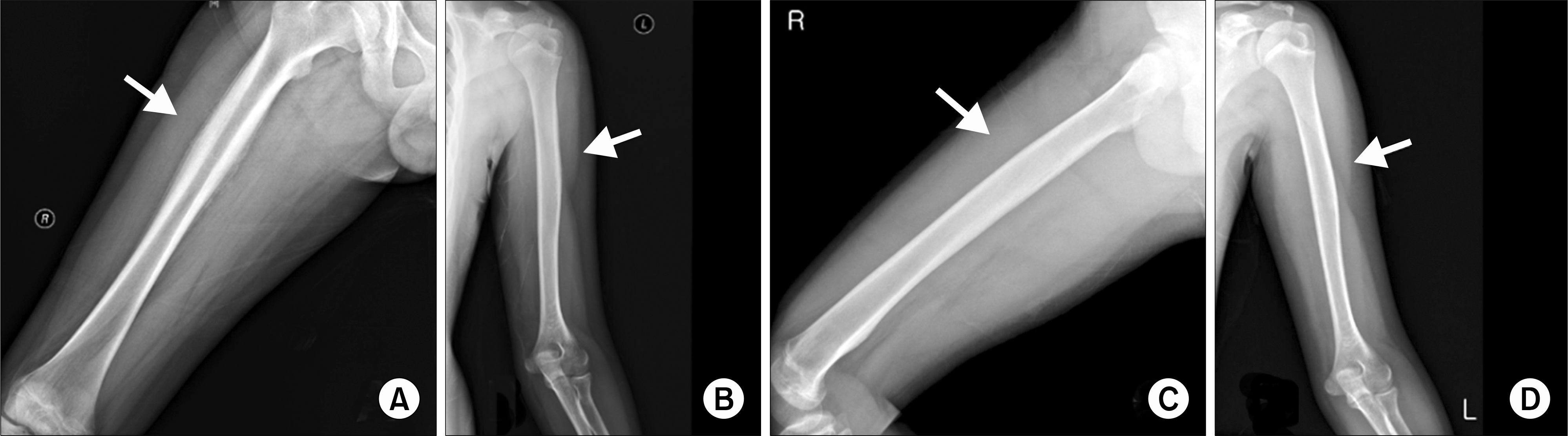
Figure 3.
Coronal T2-weighted magnetic resonance image shows newly developed fracture on the right supracondylar femur and increased extent of bone marrow edema on right distal femur with diffuse periosteal reaction.
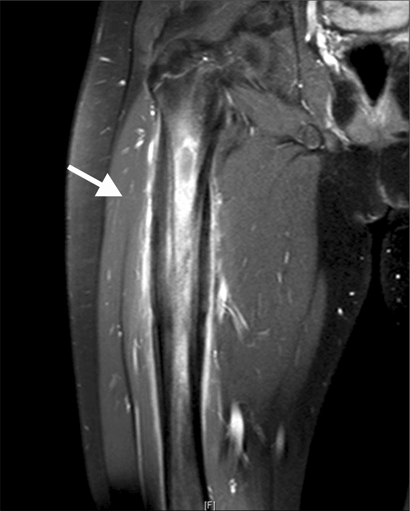
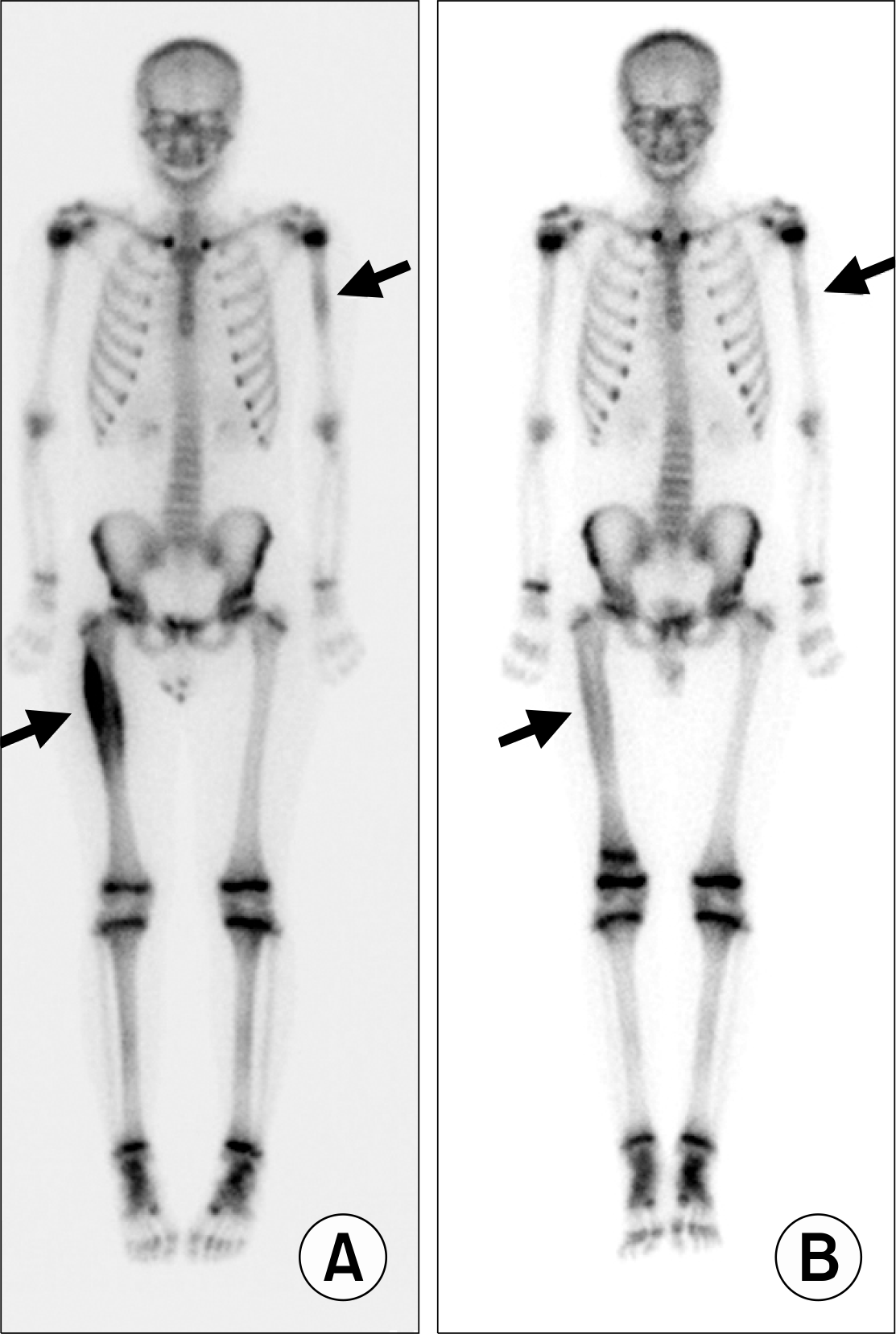
Figure 4.
(A) Technetium 99m bone scan shows diffusely increased multifocal radiotracer uptake in the right femur shaft and left proximal humerus. After 13 months of bisphosphonate treatment, Technetium 99m bone scan (B) reveals further decrease in intensity of radiotracer uptake in the right femur shaft and left proximal humerus, probably associated with healing state of osteomyelitis.
 XML Download