

 PDF
PDF ePub
ePub Citation
 Print
Print


Introduction
In humans, right cardiac catheterization was first performed via the antecubital fossa vein by using the cut-down technique in 1929.1) Right cardiac catheterization has subsequently been regarded as the standard procedure for obtaining a precise diagnosis in patients with many cardiac diseases, such as congenital heart disease. Currently, right cardiac catheterization is generally performed through the femoral vein because it allows an easy approach to the right heart. Nonetheless, right cardiac catheterization with a femoral vein approach involves several potential problems, including bleeding, hematoma, prolonged bed rest, and a risk of infection.
Left cardiac catheterization through the radial artery including coronary angiography and percutaneous coronary artery intervention has become widespread because it is more advantageous for the patient than the femoral artery approach. Patients who undergo left cardiac catheterization through the radial artery achieve early ambulation and have a reduced duration of hospital stay. Further, it is associated with fewer bleeding complications than the femoral or brachial artery approaches.2)3) Simultaneous right and left cardiac catheterizations are required in some patients. In such cases, it is difficult to choose the access sites for the right cardiac catheterization when considering the radial artery approach for the left cardiac catheterization. As catheter technology has developed (including the production of smaller catheters), it is reportedly feasible to perform right cardiac catheterization through the superficial vein of the forearm, and furthermore, the approach is associated with fewer complications.4)5)6)7)8) However, Asian patients often have a smaller body size than western patients, leading to some concerns about the feasibility and safety of this procedure in Asians. To date, there are no studies of right cardiac catheterization using the forearm vein in Korean patients. Herein, we reported our experiences with right heart catheterization using the antecubital fossa vein, including evaluations of its feasibility and safety in Korean patients.
Subjects and Methods
Patients
Between January 2003 and December 2014, we performed right cardiac catheterizations through the femoral or antecubital fossa veins of 132 patients with various clinical indications. The decision to use the femoral or antecubital fossa vein approach depended on the operator's discretion, based on the presence of a visible or palpable vein. The usefulness of the antecubital fossa vein was evaluated by application of a tourniquet to the upper arm by a nurse; if 1 of 2 veins (cephalic or basilic vein) was prominent (visible or palpable), the antecubital fossa vein approach was considered possible. If the patient required simultaneous left cardiac catheterization, such as in coronary angiography, we were able to perform the left cardiac catheterization via the radial artery and the right cardiac catheterization via the antecubital fossa vein.
We reviewed the patients' medical records retrospectively for demographic data (age, sex, weight, height, and body mass index), indications for right cardiac catheterization, and procedural and outcome data (initial success rate, procedure time, compression to ambulation time, and complications).
All patient blood tests were performed prior to cardiac catheterization. We included only diagnostic right heart catheterizations and excluded all cases of right cardiac catheterizations with simultaneous left cardiac catheterization, cardiac biopsy, an electrophysiologic study, or pericardiocentesis.
The antecubital fossa vein denotes either the cephalic or basilic vein. The initial success rate was defined as the rate of successful completion of the procedure, without a change from the access site that had initially been used. The procedure time was defined as the time between entering and exiting the cardiac catheterization laboratory. The compression to ambulation time was defined as the time between removal of the sheath and ambulation. Complications were defined as bleeding, hematoma, dissection of a vessel, infection, delayed hemorrhage, arteriovenous fistula, aneurysm of a vessel, or cardiac injury. Each patient was additionally checked for any procedure-related complications during the follow-up period.
We comparatively analyzed patient characteristics, indications, and outcomes between the antecubital fossa vein group and the femoral vein group.
Study protocol
Femoral vein approach (using a 6-French vein sheath). We used traditional methods when performing right cardiac catheterization via the femoral vein. The patients were required to fast for 12 hours and provided written informed consent for the procedure. A 6-French vein sheath was inserted in the femoral vein by the standard Seldinger technique under local anesthesia with 2% lidocaine infiltration. A 6-French, balloon-tipped, 2-lumen catheter, 110 cm catheter (Balloon Wedge Pressure Catheter, Arrow International, Reading, PA, USA) was passed through the sheath and into the right heart for hemodynamic evaluation. After this procedure, the vein sheath was removed and direct manual compression was applied to the puncture site for 10 min. Subsequently, a sandbag was applied for 2-4 hours to achieve venous hemostasis. During this time, the patients were required to have bed rest.
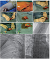
Antecubital fossa vein approach (using a 6-French vein sheath). The patients fasted for 12 hours and provided written informed consent for the procedure. Evaluation of the usefulness of the antecubital fossa vein was by tourniquet application to the upper arm by a nurse; if 1 of the 2 veins (either the cephalic or basilic vein) was prominent, the antecubital fossa area was cleaned with antiseptic solution (povidone-iodine). The nurse punctured the antecubital fossa vein with a 20-gauge Instyle intravenous cannula (IV Catheter, Becton Dickinson Medical, Singapore) (Fig. 1A and B). Subsequently, the tourniquet was removed aseptically. The Instyle intravenous cannula was exchanged for a 6-French vein sheath under local anesthesia with 2% lidocaine infiltration (Fig. 1C to H). A 110 cm, 6-French, balloon-tipped, 2-lumen catheter (Balloon Wedge Pressure Catheter, Arrow International, Reading, PA, USA) was passed via the sheath into the superior vena cava (SVC) without balloon inflation. After the SVC, the hemodynamics of the right heart were evaluated with the inflated balloon catheter (Fig. 1I to K). Although it was sometimes difficult to pass the catheter into the SVC or the inferior vena cava (IVC), these difficulties were easily overcome by using a 0.035-inch hydrophilic wire (Terumo Medical Corporation, Somerset, NJ, USA). We removed the sheath, manually compressed the puncture site, and subsequently dressed the puncture site with gauze and elastic plaster. The patients were permitted to ambulate immediately after the procedure.
Statistical analysis
Continuous variables were summarized as means±standard deviations, while categorical variables were summarized as absolute numbers and percentages (%). We used the Chi-square test, Fisher's exact test, or Student's t-test for all comparisons between the 2 groups. In the interpretation of the statistical test results, p<0.05 was considered significant. The statistical analysis was performed using SPSS version 18.0 (SPSS, Inc., Chicago, IL, USA).
Results
Right cardiac catheterization was performed in 132 patients via the antecubital fossa vein and the femoral vein in 37 (28%) and 95 (72%) patients, respectively. The baseline characteristics of the patients in the 2 groups were shown in Table 1. No significant differences in any of the baseline characteristics, including age, sex, weight, height, and body mass index were observed between the 2 groups.
In both groups, evaluation of the intracardiac shunt was the most frequent indication for right cardiac catheterization (antecubital fossa vein group: n=23, 62%; femoral vein group: n=63, 66%; p=0.65). Evaluation of dyspnea was also a common indication in both groups (antecubital fossa vein group: n=10, 27%; femoral vein group, n=14, 15%; p=0.10). Other indications were shown in Table 2.
All right cardiac catheterization procedures were successful and there were no conversions of the access site between the 2 groups. The procedural time was not significantly different between the antecubital fossa and femoral vein groups (21.6±16.8 min vs. 25.6±12.6 min, p=0.14). The compression to ambulation time was significantly shorter in the antecubital fossa vein group than in the femoral vein group (0.0 min vs. 201.2±48.1 min, p<0.01).
During the follow-up period, there were no procedure-related complications in either group, including bleeding, hematoma, vessel dissection, infection, delayed hemorrhage, arteriovenous fistula, vessel aneurysm, or cardiac injury. The procedural and outcome data were shown in Table 3.
Discussion
The results of this study indicated that right cardiac catheterization through the antecubital fossa vein may be as useful as the femoral vein approach. Previous studies have shown that procedures performed via the antecubital fossa vein have many advantages, such as early ambulation, reduced bleeding with easy hemostasis, and greater convenience for simultaneous left cardiac catheterization via the radial artery.4)5)6)7)8) However, some of the previous studies were conducted in Western populations5)6)8) and only 2 of the previous studies4)7) were performed in Taiwan. In the present study of a Korean population, we likewise showed that right cardiac catheterization through the antecubital fossa vein may be feasible in select patients.
Gilchrist et al.9) have previously reported the feasibility and safety of this technique, as well as the methods for performing right cardiac catheterization through the forearm vein. They suggested that the access site would be considered from the antecubital fossa veins to the wrist vein near the radial artery. However, Lo et al.6) suggested that, to increase the success rate of percutaneous venous cannulation, antecubital fossa veins might be preferable because they are less technically demanding.
In the present study, the success rate of percutaneous venous cannulation was 100% in the antecubital fossa vein group. The high success rate was due to selection of patients with prominent antecubital fossa veins on tourniquet assessment. However, despite the use of a tourniquet, it was still necessary to use the femoral vein in 6 patients because the antecubital fossa veins were not visible or palpable. These 6 patients were elderly or obese, and comprised 13.9% of the patients in whom we initially tried to use the antecubital fossa vein.
Our results also showed similar procedural time between the 2 groups. A previous study showed a significantly shorter procedural time in the forearm group than in the femoral group.7) Another study showed a shorter procedural duration in the percutaneous femoral approach group than in the percutaneous arm approach group.6) The shortened procedural time in the forearm group in the former study may have been related to the anatomical structure that followed a natural vascular curvature; whereas, in the latter study, the prolonged procedural time in the forearm group may have been related to the identification and puncture of a suitable antecubital fossa vein, as well as the operators' lack of experience with the procedure during the early stages of its introduction. In our experience, the antecubital fossa vein was easier to puncture than the femoral vein because the operator or nurse was able to confirm the cephalic or basilic vein by identifying the antecubital fossa. However, it was generally difficult to find the antecubital fossa veins in elderly and obese patients. Additionally, in the early stages of our study on the antecubital fossa vein approach, we found that it was difficult to pass the catheter into the SVC or IVC. Nonetheless, when using a 0.035-inch hydrophilic wire, it was easy for us to overcome these obstacles and reduce the procedural time. A recent study showed that fluoroscopy time was shorter in patients receiving the antecubital vein approach than in patients receiving the proximal vein approach.8) This result indicated that the use of the antecubital fossa vein for right cardiac catheterization may reduce radiation exposure times for patients and operators.
In the present study, the compression to ambulation time was significantly shorter in the antecubital fossa vein approach group than in the femoral vein approach group. These results were easily explained, since the patients in the antecubital fossa vein approach group were able to walk immediately after the procedure and did not require bed rest for hemostasis. In contrast, the patients in the femoral vein approach group needed bed rest with sandbags for hemostasis. Moreover, 7 of the patients in this group (7.3%) complained of dysuria during bed rest. Considering both the time that was required to completely stop bleeding and the discomfort that was associated with several hours of bed rest, the antecubital fossa vein approach may be more advantageous for patients than the femoral vein approach. Moreover, right cardiac catheterization through the antecubital fossa vein would be useful for invasive evaluation of exercise hemodynamics via supine bicycle exercises, because the patients are able to move both legs freely during examination.10)
There were no complications in the present study. Similarly, Lo et al.6) reported that there were no complications (such as bleeding) among 28 patients who received anticoagulation treatment and underwent left and right heart catheterizations via the percutaneous arm approach. These results suggest that the antecubital fossa vein approach and the femoral vein approach are equally safe. However Shah et al.8) reported some complications, such as arterial puncture and hematoma, in a proximal venous access group. Although we did not observe any complications, these kinds of complications could occur at any time because of the anatomic relationship of the femoral artery and vein.
Our study had some limitations. First, it had the usual limitations of single-center, retrospective, observational studies that include relatively small numbers of patients. Second, we excluded patients who had invisible and impalpable antecubital fossa veins from the antecubital fossa vein approach group. The choice of the access site for cardiac catheterization was made at the discretion of each operator. Additionally, procedural times and other procedural results depended on the operator's technique and experience, which could have introduced bias into the analysis. Finally, we did not assess pain, radiation exposure, fluoroscopy time, or the times required for each stage of the procedure (such as the vascular access stage and hemodynamics evaluation stage).
Conclusions
This single-center, retrospective, observational study showed that the right cardiac catheterization via the antecubital fossa vein could be a feasible method in Korean patients. The excellent success rate of this access approach was accompanied by a low complication rate. Additionally, this method could provide patients with the convenience of early ambulation. Therefore, the antecubital fossa vein could be considered as an alternative to the femoral vein when determining the vascular access site for right cardiac catheterization in selected patients.


 XML Download