Journal List > Korean Circ J > v.46(1) > 1101463

Marquez, Lee, Kim, and Han: Post-Translational Modifications of Cardiac Mitochondrial Proteins in Cardiovascular Disease: Not Lost in Translation

Abstract

Protein post-translational modifications (PTMs) are crucial in regulating cellular biology by playing key roles in processes such as the rapid on and off switching of signaling network and the regulation of enzymatic activities without affecting gene expressions. PTMs lead to conformational changes in the tertiary structure of protein and resultant regulation of protein function such as activation, inhibition, or signaling roles. PTMs such as phosphorylation, acetylation, and S-nitrosylation of specific sites in proteins have key roles in regulation of mitochondrial functions, thereby contributing to the progression to heart failure. Despite the extensive study of PTMs in mitochondrial proteins much remains unclear. Further research is yet to be undertaken to elucidate how changes in the proteins may lead to cardiovascular and metabolic disease progression in particular. We aimed to summarize the various types of PTMs that occur in mitochondrial proteins, which might be associated with heart failure. This study will increase the understanding of cardiovascular diseases through PTM.

Introduction

Post-translational modifications (PTMs) are changes or alterations in a protein occurring after the completion of the translational process by ribosomes that are catalyzed by numerous enzymes. PTMs occur either when a functional group is covalently added to a protein, or during the proteolytic processing and folding processes. Protein PTMs play a key role in several physiological and cellular processes including cellular differentiation,1) protein degradation,2) signaling and regulatory processes,3) regulation of gene expression,4)5) and protein-protein interactions.6)7) PTMs act as a mechanism for the specification of proteins, through conformational changes that either minutely8) or largely9)10) change the overall tertiary structure of a protein.11) These modifications increase the variety and difference of proteins.12) Protein PTM dysfunction via external stimuli13)14)15) or aberrant signaling16)17) eventually leads to disease progression either through aberrant signaling or impaired PTM crosstalk.18)19) Non-native protein PTM leads to either biochemical dysfunction20) or a structural modification21) in the amino acid due to 'crosstalk.'12)22)
Mitochondria encompass 90% of the energy produced mainly by oxidative phosphorylation via electron transfer and adenosine triphosphate (ATP) synthase complexes.23) Subtle regulation of mitochondrial functions is mediated PTMs24) such as phosphorylation,25) acetylation,26) succinylation,27) and O-GlcNAcylation of mitochondrial proteins.28)29) Furthermore, since mitochondria are the central hubs of energy production, these networks are involved in various human pathological phenotypes; furthermore, mitochondrial proteomic dysfunction is directly associated with heart diseases.30)31) The availability of high-tech mass spectrometric techniques combined with advanced modified proteins/peptides by affinity chromatography methods has resulted in breakthroughs regarding the role of PTMs in cellular protein, particularly in mitochondrial proteins. Enzymes and stimuli involved in changes of PTMs are equally important; however, these issues are beyond the scope of this review. A better understanding of PTMs helps clinicians and researchers alike, but also facilitates development of targeted strategies for disease intervention.

Mitochondria, Heart Failure, and Post-Translational Modifications

According to the latest report by the World Health Organization, cardiovascular disease (CVD) is the leading cause of mortality in developed countries. Despite much progress in the advancement in the prevention, diagnosis, and management of CVD in the past decades,32)33)34)35) heart failure (HF) continues to be prevalent since current trends do not target direct cure in HF patients (except those with congenital heart diseases), but only decreases in the mortality rates.36)
Heart failure is a multifactorial clinical condition that is characterized by a dysfunction in the contractility of the myocardium, which results to the inability of the heart to provide enough blood for the metabolic needs of surrounding tissues.37) Increased preload and afterload, neurohormonal dysregulation, cardiac ischemia, and intrinsic abnormalities of the myocardium are common etiologic factors of HF. The gradual development of CVD to HF is a multicomponent and a multistep process, wherein cumulative acute cardiovascular injuries like myocardial ischemia/reperfusion (I/R) result in a chronic dysfunction.38)
HF progresses partly via changes in major signal transduction pathways,39) dysfunction in calcium homeostasis and energy fluxes,40) and alterations of the contractile apparatus in the heart.41) The underlying principles of bioenergetics is a key in further understanding its effects on CVD and HF (i.e., synthesized myocardial ATP transfer is required to maintain the excitationcontraction coupling, continuously supporting the optimal functioning of systolic and diastolic periods in the heart). Since cellular energetics and metabolism is heavily influenced and regulated by the mitochondria, there is undoubtedly a link between the heart and mitochondria function. Dysfunction in mitochondria such as oxidative damage,42) respiration impairment,43) and substrate utilization alterations have been reported in HF.44)
A frail heart undergoes complex changes in energy metabolism and substrate utilization due to mitochondrial dysfunction that are still unclear. These metabolic changes or remodeling occur when genes of interest trigger structural, functional, and electrical changes, which result in decreased cardiac function. One mechanism of this remodeling involves PTMs, which break down misfolded and damaged proteins in addition to proteins involved in contractile apparatus and hypertrophic gene expression. Likewise, proteins in respiratory chain and fat/glucose oxidation, which possess at least 1 reversible acetylation mark in complexes I, II, and V, and pyruvate dehydrogenase (PDH), and numerous acyl-CoA dehydrogenases, are involved.45)46) Regulation of metabolic enzymes within the mitochondria by acetylation implies that altered acetylation states within the mitochondria could play a role in the pathophysiology of heart failure.47)
Protein post-translational modifications are important in the study and analysis of disease progression such as those involving CVD, since the interplay between regulatory PTMs and the induced changes of the organelle dysfunction including mitochondria are potentially important factors in CVD progression.48) Several studies on the relationship of mitochondrial PTM and heart failure are reported.49)50) O'Rourke et al.48) in a study on heart mitochondria isolated from HF reported that cAMP-activated protein kinase might be involved in the increased protein phosphorylation during HF. Oxidative phosphorylation brought about by subunit-specific phosphorylation of complex IV regulates the incorporation or destabilization of the supercomplexes.51) In addition, growing evidence shows mitochondrial protein acetylation as a common mechanism in response to cardiac stress. Moreover, decreased nicotinamide adenine nucleotide (NAD+)/nicotinamide adenine nucleotide (reduced) (NADH) ratio and increased mitochondrial protein acetylation with increased sensitivity of mitochondrial permeability transition pore (mPTP) are associated during calcium sensitization. However, whether hyper-acetylation of a single protein or a select group of protein targets is the main contributor to increased sensitivity to cardiac stress remains to be determined.52)
The mitochondria plays a significant role in the regulation of cellular and physiological processes, hence it is important to study the associated proteins. We review the most common PTMs occurring in select mitochondrial proteins, as well as minor PTMs and the complex relationship with other PTMs that contribute to the progression of heart disease.

Post-Translational Modifications of Mitochondrial Proteins

Phosphorylation

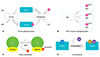
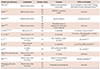
Signaling cascades are important in the study of acute cardiac pathologies, particularly in cases of I/R.12) Kinase activators and inhibitors are important mediators in pathological diseases.53) Phosphorylation is one of the well-studied PTMs that are responsible for altering a target protein's conformation that leads to either activation or inactivation of mitochondrial functions (Table 1 and Fig. 1A). Protein phosphorylation is a reversible PTM regulated by kinases and phosphatases, which is responsible for the phosphorylation and dephosphorylation of substrates, respectively.54) Protein kinases are key enzymes in modulating phosphate group transfer to serine, tyrosine, and threonine residues of the targeted proteins and thereby generate negatively charged side chains, which can either attract or repel target proteins in an external stimulus-dependent manner.55) Protein phosphatases on the other hand are signal transducing enzymes that dephosphorylate phosphoproteins.56)
Phosphorylation regulates the catalysis of numerous mitochondrial enzyme complexes by specific kinases associated with these complexes, making the process more efficient. One of the earliest studies regarding phosphorylation focuses on protein kinase capable of phosphorylating proteins in the rat liver mitochondria.53) Table 1 shows selected mitochondrial proteins from the Pagliarini and Dixon57) review on phosphoproteins found in the matrix, inner membrane, intermembrane space and outer membrane.
Pyruvate dehydrogenase complex (PDC), a kinase/phosphatasedependent regulatory cascade, is composed of 3 principle subunits: pyruvate dehydrogenase E1, dihydrolipoamide acetyltransferase E2, and dihydrolipoamide dehydrogenase E3. PDC links glycolysis to tricarboxylic acid cycle (TCA) cycle,58) catalyzing pyruvate conversion to acetyl CoA and is regulated by allosteric effectors and by reversible phosphorylation.59) E1 subunit complex phosphorylation at sites Ser264, 271, and 204 by pyruvate dehydrogenase kinase (PDK) leads to its inactivation,60)61) while dephosphorylation, via pyruvate dehydrogenase phosphatase results in its activation. Inhibition or phosphorylated state is brought about by high concentrations of immediate products such as acetyl CoA and NADH, and terminal products such as ATP levels. Thus, phosphorylation of PDC E1 by a phosphatase switches off the activity of the complex, thereby deactivating it (Fig. 1B).
Pyruvate dehydrogenase kinase (PDHK) exists mostly in organisms of eukaryotic lineages,62) and is notably absent only in organisms with reduced or absent mitochondria.61) The sequence of PDK bears significant similarity with histidine kinases,63)64) which are widely distributed sensory transducers in prokaryotes.65) PDHK is made up of 2 different subunits i.e., PDHK α-subunit with kinase activity on selective proteolytic cleavage and PDHK β-subunit, a regulatory subunit.66) Like phosphatase, kinase activities are activated by increased ratios of acetyl-CoA/CoA and NADH/NAD+67)68) and inhibited by elevated adenosine diphosphate (ADP) levels69) and by dichloroacetate70) in the mitochondrial matrix.
The inner mitochondrial membrane protein branched-chain α-ketoacid dehydrogenase (BCKD) complex, responsible for the oxidative decarboxylation of 3 branched amino acids (valine, leucine, and isoleucine), possesses a thiamine pyrophosphate-dependent branched-chain α-ketoacid decarboxylase (E1). Inactivation of this complex occurs via serine 193 phosphorylation through BCKD kinase (BCKDK).71) Branched chain amino acid levels are maintained during protein starvation by BCKDK-mediated phosphorylation and complex inhibition, a pathological characteristic of maple syrup urine disease that presents with severe neurological dysfunction.72)
End-stage heart failure may be partly due to reduced 3',5'-cyclic adenosine monophospate (cAMP)-dependent phosphorylation in various oxidative phosphorylation (OXPHOS) subunits.73) cAMPdependent phosphorylation regulates OXPHOS activity by elevating cAMP levels, which leads to increased nicotinamide adenine dinucleotide dehydrogenase (ubiquinone) Fe-S protein 4 (NDUFS4) phosphorylation complex I (CI) activity by twofold.74) In line with this, CI reportedly undergoes cAMP-responsive phosphorylation at the 10 and 18-kDa subunits.75) Mass spectrometry technique also shows that purified CI possesses phosphorylation sites at the 42-kDa subunit in addition to the B16.6, B14.5a, and B14.5b subunits of CI.76) Complex IV (CIV) subunit phosphorylation is considered not only beneficial, but critical for CIV activity in healthy and pathological cardiac mitochondria. Hypoxia and ischemia increase protein kinase A-dependent phosphorylation of IVi1 and Vb subunits of CIV,77) which are associated with lower CIV activity and increased reactive oxygen species (ROS) production.78)
Currently, phosphorylation is known to regulate proteins in the different mitochondrial compartments in a very specific manner, and its regulation leads to alterations that either directly or indirectly cause heart failure. However, the kinases and phosphatases involved during mitochondrial protein phosphorylation, including its subunits are not fully understood. In addition, the phosphorylation events regulating mitochondrial functions or dysfunctions in vivo remain to be confirmed.

Acetylation

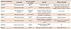
Acetylation occurs when an acetyl group is introduced into a compound, wherein the hydrogen atom of a hydroxyl group is exchanged with an acetyl group yielding acetate.79) Studies have suggested that non-histone-acetylases and deacetylases are both involved in cardiac remodeling (Table 2 and Fig. 1C).
Sirtuins (SIRTs) are NAD+-dependent protein deacetylases that play key roles in regulation of mammalian metabolism. Cells have 7 different SIRTs, 3 of which are localized in the mitochondria: SIRT3, SIRT4, and SIRT5.80) When SIRT3 is abolished, mitochondrial proteins become hyperacetylated and exhibit altered function that eventually leads to mitochondrial dysfunction.81) SIRT3 targets many enzymes, which is suggestive of its role in heart failure. For example, mitochondrial protein acetyl-CoA synthetase 2 (AceCS2) activities are directly regulated by SIRT3 in an NAD+-dependent manner.82)83) SIRT3 in neonatal rat ventricular myocytes (NRVMs) deacetylates nuclear protein Ku70, preventing mitochondrial translocation of Bax and enhancing H2O2 tolerance.84) SIRT3 also deacetylates FOXO3a responsible for increasing manganesedependent superoxide dismutase (MnSOD, SOD2) expression, which in turn diminishes mitochondrial superoxide.85) A new study has shown the role of SIRT3 in fatty acid oxidation (FAO), wherein it deacetylates Lys42 of the 8 acetylation site on long-chain acyl-CoA dehydrogenase (LCAD) leading to FAO pathway activation.86) Aside from AceCS2 and LCAD, acetylation also regulates other mitochondrial components such as malate dehydrogenase87) and isocitrate dehydrogenase88) in the TCA cycle. Mitochondrial malate dehydrogenase (MDH2) becomes acetylated at Lys185, Lys301, Lys307 and Lys314 and Lys314, resulting in increased activity (rapid reduction of oxaloacetate to malate when coupled with high NADH) during the hyperacetylated MDH2 state,89) thereby increasing gluconeogenesis and hyperglycemia risks.87) On the other hand, SIRT4 and SIRT5-deficient mice do not show any significant changes in their acetylation status,90) and thus both SIRT4 and SIRT5 have limited deacetylase activities.91)

S-nitrosylation

S-nitrosylation is the specific attachment of nitric oxide (NO)-related species to a thiol group to form S-nitrosothiol, which also serves as a cellular signal due to its similar nature with phosphorylation.92) The actions of denitroylases are thought to be similar with those of the phosphatases in kinase signaling; in addition, they are considered as major regulators of the cytosolic and mitochondrial thioredoxin reductases.93)94)
In mitochondria, regulation of oxidative phosphorylation and glycolysis by NO-mediated protein S-nitrosylation is important in both physiologic and pathophysiologic conditions (Fig. 1D). S-nitrosylation of mitochondrial aldehyde dehydrogenase 2 family, involved in NAD+-dependent oxidation of the different aldehydes produced during intermediary metabolism, leads to reversible inhibition.95) Creatine kinase, responsible for conversion of creatine, uses ATP to produce phosphocreatine and ADP; it is inhibited by S-nitrosoglutathione (GSNO) dose-dependently via transnitrosylation96) and reversibly regulated through S-nitrosylation of Cys283 in adult rat ventricular myocytes.97) Another study using isolated rat heart mitochondria as a model shows inhibition of complex I by the S-nitrosylation of the 75-kDa subunit through the exogenous addition of GSNO. In an endothelial cell model, the inhibition of mitochondrial complex IV/cytochrome c oxidase possibly occurs by S-nitrosylation at Cys196 and Cys200, which are both active residues.98) However, the exact mechanism of this PTM in vivo, briefly described as the addition of NO to reactive cysteines, still remains largely unknown.99)100)

Cross-talk with various protein post-translational modifications

The respective actions of each PTM are clearly essential modulators of protein structure-function relationships. The numerous PTMs are potentially related, forming networks, as evident in different biological systems.101) Three criteria are suggested in crosstalk of PTMs: 1) similar site competition; 2) modification that facilitates conformational change in the second site accessibility that allows another PTM to occur, and; 3) direct alteration of the modifying enzyme of the other PTM.102) One of the earliest studies concerning potential PTM crosstalk focuses on the potential sites of O-linked β-N-acetylglucosamine (O-GlcNAc), which is modification of serine or threonine hydroxyl moieties by β-N-acetylglucosamine, or phosphorylation modification, which both target serine and threonine dynamically and transiently in nature.28) However, a recent study shows that the location of the O-GlcNAcylation machinery within the cell partially dictates its function. The regulation of O-GlcNAcylation through subcellular redistribution of OGT/OGA and functional consequences that have immediate therapeutic potential to improve cardiac contractility posits a new concept in PTM cross-talk.103) Furthermore, PTM sumoylation is the covalent protein modification by addition of ubiquitin-like polypeptides. Crosstalk between sumoylation and phosphorylation is also suggested, since the small ubiquitin-like modifier (SUMO) attachment lysine site is located 4 sites from a phosphorylated serine in numerous sumoylated proteins.104)105)
Phosphorylation and lysine acetylation are likewise involved in crosstalk.23) For example, adenosine monophosphate-activated protein kinase and the SIRT family. A study shows that there is an approximately 80% overlap between the interacting sites of mitochondrial lysine acetylation and succinylation; for instance, approximately 25% of known SIRT5 target sites were also modified by SIRT3.106) These findings indicate the possible cooperation between proteins in order to maintain balance in the mitochondria.
Crosstalk also signals degradation, as shown in I/R injury where myosin light chain 2 is reduced between a deamidated asparagine and a phosphorylated serine, exhibiting 3 PTMs occurring within 2 amino acids. Similarly, there is a complicated relationship between S-glutathionylation and major PTMs. S-glutathionylation, in which protein cysteine residues are modified during glutathione addition, has 2 main mitochondrial roles i.e., oxidant stress defense and redox signaling. During the oxidation of the mitochondrial glutathione, the cysteine-rich 75-kDa subunit of complex I becomes the main target.107) Glutathionylation/oxidation acts as a buffer against ROS under these conditions, keeping protein thiols away from the gradual oxidation to sulfinic acid and sulfonic acid, which might lead to irreversible protein dysfunction (Fig. 1).108)109) Another study108) shows that glutathionylation of Complex I (CI) due to diamide-induced glutathion depletion inhibits CI activity; however, ROS levels remain unchanged during nitrosylation on the same subunit. CI undergoes nitrosylation and glutathionylation, hence, mitochondrial complex II (CII) is a protein that persistently undergoes glutathionylation. In an I/R model induced by coronary ligation, the 70-kDa subunit of CII undergoes markedly reduced glutathionylation that is related with the loss of electron transfer activity. Thus, glutathionylation likely plays a key role in the maintenance of CII function.110)

Limitations of mitochondrial protein post-translational modification studies

With the technological advancements of recent years, hundreds of mitochondrial proteins are identified through mass spectrometry-based proteomics. This has led to a completely novel way of understanding CVD. Extensive validation of PTM sites on mitochondrial proteins and the correlation with protein function remains to be done. A full understanding of the intricate web of post-translational signaling and regulation in the mitochondria, as well as the identification of target proteins, should be the next step before further development of strategies to combat CVD. However, integrative approaches including computational biology, protein arrays, and biochemical analyses will quickly advance the progress of studies. Hofer and Wenz111) raised several questions whether PTMs are regulatory in nature given the small percentage of target proteins that are modified during the process; and if so, the regulatory mechanisms involved need to be elucidated. In addition, it is not clear whether all regulatory PTMs are beneficial to the continued functioning of the system, or detrimental. Moreover, questions regarding tissue-specificity and time-dependence should be considered during experimentation. Addressing these issues will greatly aid in understanding disease mechanisms and suggest targeted strategies for disease intervention.

A Clinical Perspective of Mitochondrial Protein Post-Translational Modification for Combating Cardiovascular Disease

Current therapies do not directly address the treatment of CVD, but rather, are aimed at slowing its progress to HF. For example, statins that slow the formation of atherosclerotic lesions,112) antianginals targeting ischemic tissues,113) and antiplatelet or anticoagulation agents which hinder the formation of a clot.113) Cardiac ischemia can at times be predicted, such as in the cases of cardiac surgery or balloon inflation employed in percutaneous coronary intervention.114) More recently, preconditioning (PC) of the heart has emerged in clinical practice,115) wherein the heart is subjected to short intermittent I/R cycles before index ischemia that reduces the infarct size and improves postischemic function.116) Targeting the MPTP is a new trend with more promising effect.
MPTP opening is a key factor that drives necrotic cell death in I/R injury.117) The key components of MPTP are still unclear, but transgenic animal models show that adenine nucleotide translocase as the phosphate carrier can regulate the pore opening.118) Cyclophilin D119) and voltage-dependent anion channel (VDAC)120) are associated with MPTP but their roles remain unclear. Other proteins related with MPTP include hexokinase II,121) which serves as a connection between the pore and cellular metabolism and mitochondrial translocator protein, which communicates with VDAC.122)
PC can act on MPTP either directly on the MPTP to inhibit its opening123) and/or by reducing calcium or ROS levels that trigger MPTP.117) Considering that cardioprotective signaling pathways are activated through direct MPTP inhibition, these signaling pathways are capable of modifying MPTP components. Thus, we can expect that cardioprotective signaling leads to mitochondrial protein PTM.
The importance of epigenetics in gene regulatory mechanisms leading to cardiovascular complications have also been widely studied (particularly histone and DNA modifications) as a therapeutic target for CVD. Factors such as diet, environmental changes, and activity affect gene expression in an entity and its offspring via epigenetics, without affecting the genomic sequence. A clinical study124) conducted by the Diabetes Control and Complications Trial/Epidemiology of Diabetes Interventions and Complication (DCCT/EDIC), show that patients on constant intensive therapy have lower cardiovascular complications, as compared to those who are first on conventional therapy and then switched to intensive therapy. This supports the evidence that a history of hyperglycemia possibly leads to long-lasting molecular changes that ultimately puts the patients on the fast-track to the development and progression of CVD. Thus, PTM-mediated regulation of the cardiac proteome can serve as a foundation laid early in life or transmitted from the parental units.125)
HF is a multifactorial syndrome that progresses largely due to myocardial dysfunction brought about by mitochondrial modifications. Therefore, it is important to understand mitochondrial cytopathy in HF as a potential foundation for therapeutic strategies to maintain mitochondrial integrity, enhance substrate metabolism, protect and reduce oxidative stress in the environment, and improve the myocardial contractility.126) These changes are undoubtedly related to the modification of mitochondrial proteins through various PTMs. Some novel mitochondrial targets of phosphorylation, S-nitrosylation, and acetylation are elucidated, most of which are linked to single protein activity or total mitochondrial function. Collectively, the understanding of mitochondrial PTMs will provide an insight into controlling mitochondria-related conditions such as HF, but more systemic and long-term research is needed.

Figures and Tables

Fig. 1

Several types of post-translational modifications. (A) Reversible protein phosphorylation. Protein kinase transfers a phosphate group (P) from ATP (ADPP) to the target protein. A protein phosphatase is responsible for removing the phosphate group via hydrolysis. (B) Protein kinases mediate phosphorylation at various amino acid sites of serine, threonine and tyrosine side chains. In the example shown, PDH is a phosphorylated protein found in the mitochondrial matrix. PDH is a large complex made up of several units (E1, E2, and E3), which is responsible for catalyzing oxidative decarboxylation of pyruvate, to form acetyl-CoA. PDH is phosphprylated at Ser264, 271, and 204. (C) Reversible protein acetylation. In the given example, Sirtuin 3 (SIRT3) deacetylases the acetylated form of NDUFA9. (D) Redox-mediated S-nitrosylation occurs through the covalent reaction of nitric oxide (NO)-related species with a cysteine thiol group on the target protein. ATP: adenosine triphosphate, ADP: adenosine diphosphate, PDH: pyruvate dehydrogenase, Ser: serine, NDUFA9: NADH dehydrogenase subcomplex A9, NO: nitric oxide, CI: complex I, SH: thiol subunit, S: sulfur.

kcj-46-1-g001
Table 1

Brief outline of phosphorylation in mitochondrial proteins

kcj-46-1-i001
Protein and references Localization Phospho site(s) Function Effect on function
PDC E1α127) Mitochondrial matrix Ser Oxidative decarboxylation of pyruvate Acute modulation of active PDC,128) delays reactivation through dephosphorylation128)
PDHK129)130)131) Mitochondrial matrix Ser
Thr
Tyr
OXPHOS to glycolysis switch Unknown
BCKAD25)132) Mitochondrial matrix Ser
Thr
Tyr
Leucine oxidation ↓Complex activity71)
MDH133)134) Mitochondrial matrix Ser
Thr
Tyr
Reversible malate to oxaloacetate conversion, malate-aspartate Unknown
CI: ESSS133)135)136) Matrix arm of CI Ser CI assembly ↑CI activity in bovine hearts74)
CI: 10 kDa135) Intermembrane of CI Ser CI assembly ↑CI activity, ↓ ROS production in bovine hearts137)
CIV I135) Mitochondrial inner membrane Tyr Cyt c oxidation, oxygen reduction ↓CIV activity138)
CIV Vb139) Intermembrane side of CIV Ser CIV regulation ↓CIV activity, ↑ROS production78)
CV β140) CV matrix (F1) Thr ATP synthesis from ADP and Pi ↓CIV activity141)
NAD (P) transhydrogenase 142)143)144) Mitochondrial inner membrane Tyr H+ pump Unknown
ANT142)145) Mitochondrial inner membrane Tyr Adenine transmembrane transporter ↑Cellular respiration146)
Phosphate carrier protein132)142)147) Mitochondrial inner membrane Ser
Thr
Tyr
Phosphate group transporter Unknown

Proteins presented are adapted from the Table in a 2006 review by Pagliarini and Dixon57) with more relevant and recent sources, including a cross-reference to PhosphoSite Plus studies and a more specific function. General phosphorylation sites are described due to space limitations. Up and down arrows indicate increase and decrease, respectively. PDC: pyruvate dehydrogenase complex, Ser: serine, PDHK: pyruvate dehydrogenase complex kinase, OXPHOS: oxidative phosphorylation, Thr: threonine, Tyr: tyrosine, BCKAD: branched-chain alpha-keto acid dehydrogenase, MDH: malate dehydrogenase, CI: complex I, ESSS: ESSS subunit of NADH:Ubiquinone oxidoreductase (complex I), kDa: kilodalton, ROS: reactive oxygen species, CIV: complex IV, Cyt c: cytochrome C oxidase, CV: complex V, ATP: adenosine triphosphate, F1: fraction 1, ADP: adenosine diphosphate, NAD: nicotinamide adenine dinucleotide transhydrogenase, P: phosphate, ANT: adenine nucleotide translocase

Table 2

Brief outline of acetylation in mitochondrial proteins

kcj-46-1-i002
Protein and references Localization Lysine (K) acetylation site(s) Function Effect on function
AceCS283) Mitochondrial matrix K64283) Acetate/CoA ligation Deacetylation ↑acetate conversion
ALDH2148) Mitochondrial matrix K377*149) Acetaldehyde metabolism Deacetylation by SIRT3 allows NAPQI binding to ALDH2, ↓ activity
ATP5A1150) Mitochondrial inner membrane K427, K531, K539150) Produces ATP from ADP in the presence of a proton gradient ↑ETC activity
IDH2151) Mitochondrial matrix K155, K180152) Oxidative decarboxylation of isocitrate to 2-oxoglutarate ↓Catalytic activity
LCAD153) Mitochondrial matrix K254154) Mitochondrial fatty acid oxidation ↑Lipid processing
MDH2153) Mitochondrial matrix K16587) Reversible malate to oxaloacetate conversion, malate-aspartate shuttle ↑Malate export, ↑Gluconeogenesis and hyperglycemia
NDUF9A52) Mitochondrial matrix K89*155) Accessory subunit of NADH dehydrogenase CI ↑CI
SOD2156) Mitochondrial matrix K68*156) Destroys superoxide anion radicals ↓Enzymatic activity

The examples are adapted from the Table in a 2014 review by Papanicolaou, et al.26) The Table is updated with the addition of acetylation/deacetylation on function. Common acetylation sites among rat, mouse, and guinea pig samples are chosen as representative acetylation site(s) due to space limitations. *Indicates acetylation in liver. Up and down arrows indicate increase and decrease, respectively. K: lysine, AceCS2: acetyl-CoA synthetase 2, ALDH2: aldehyde dehydrogenase 2, SIRT: sirtuins, NAPQI: N-acetyl-p-benzoquinone imine, ATP5A1: ATP synthase subunit 5A alpha subunit isoform 1, ATP: adenosine triphosphate, ETC: electron transport chain, ADP: adenosine diphosphate, IDH2: isocitrate dehydrogenase 2, LCAD: long-chain acyl-CoA dehydrogenase, MDH: malate dehydrogenase, NDUF9: NADH dehydrogenase subcomplex A9, CI: complex I, NADH: nicotinamide adenine dinucleotide (reduced), SOD2: superoxide dismutase 2

Acknowledgments

This work was supported by the Priority Research Centers Program (2010-0020224) and the Basic Science Research Program (2011-0028925 and 2012R1A2A1A03007595) through the National Research Foundation of Korea (NRF), funded by the Ministry of Education, Science, and Technology.

Notes

The authors have no financial conflicts of interest.

References

1. Grotenbreg G, Ploegh H. Chemical biology: dressed-up proteins. Nature. 2007; 446:993–995.
2. Geiss-Friedlander R, Melchior F. Concepts in sumoylation: a decade on. Nat Rev Mol Cell Biol. 2007; 8:947–956.
3. Morrison RS, Kinoshita Y, Johnson MD, et al. Proteomic analysis in the neurosciences. Mol Cell Proteomics. 2002; 1:553–560.
4. Fukuda H, Sano N, Muto S, Horikoshi M. Simple histone acetylation plays a complex role in the regulation of gene expression. Brief Funct Genomic Proteomic. 2006; 5:190–208.
5. Chu S, Ferro TJ. Sp1: regulation of gene expression by phosphorylation. Gene. 2005; 348:1–11.
6. Li X, Foley EA, Kawashima SA, et al. Examining post-translational modification-mediated protein-protein interactions using a chemical proteomics approach. Protein Sci. 2013; 22:287–295.
7. Chavez JD, Weisbrod CR, Zheng C, Eng JK, Bruce JE. Protein interactions, post-translational modifications and topologies in human cells. Mol Cell Proteomics. 2013; 12:1451–1467.
8. Wang S, Ionescu R, Peekhaus N, Leung JY, Ha S, Vlasak J. Separation of post-translational modifications in monoclonal antibodies by exploiting subtle conformational changes under mildly acidic conditions. J Chromatogr A. 2010; 1217:6496–6502.
9. Slade DJ, Subramanian V, Fuhrmann J, Thompson PR. Chemical and biological methods to detect post-translational modifications of arginine. Biopolymers. 2014; 101:133–143.
10. Warnecke A, Sandalova T, Achour A, Harris RA. PyTMs: a useful PyMOL plugin for modeling common post-translational modifications. BMC Bioinformatics. 2014; 15:370.
11. Prabakaran S, Lippens G, Steen H, Gunawardena J. Post-translational modification: nature's escape from genetic imprisonment and the basis for dynamic information encoding. Wiley Interdiscip Rev Syst Biol Med. 2012; 4:565–583.
12. Liddy KA, White MY, Cordwell SJ. Functional decorations: posttranslational modifications and heart disease delineated by targeted proteomics. Genome Med. 2013; 5:20.
13. Han SJ, Lonard DM, O'Malley BW. Multi-modulation of nuclear receptor coactivators through posttranslational modifications. Trends Endocrinol Metab. 2009; 20:8–15.
14. Lonard DM, O'Malley BW. Nuclear receptor coregulators: judges, juries, and executioners of cellular regulation. Mol Cell. 2007; 27:691–700.
15. Rosenfeld MG, Lunyak VV, Glass CK. Sensors and signals: a coactivator/corepressor/epigenetic code for integrating signaldependent programs of transcriptional response. Genes Dev. 2006; 20:1405–1428.
16. Ren RJ, Dammer EB, Wang G, Seyfried NT, Levey AI. Proteomics of protein post-translational modifications implicated in neurodegeneration. Transl Neurodegener. 2014; 3:23.
17. Kemper JK. Regulation of FXR transcriptional activity in health and disease: Emerging roles of FXR cofactors and post-translational modifications. Biochim Biophys Acta. 2011; 1812:842–850.
18. Butkinaree C, Park K, Hart GW. O-linked beta-N-acetylglucosamine (O-GlcNAc): extensive crosstalk with phosphorylation to regulate signaling and transcription in response to nutrients and stress. Biochim Biophys Acta. 2010; 1800:96–106.
19. Ito K. Impact of post-translational modifications of proteins on the inflammatory process. Biochem Soc Trans. 2007; 35:281–283.
20. Moreno-Gonzalo O, Villarroya-Beltri C, Sánchez-Madrid F. Post-translational modifications of exosomal proteins. Front Immunol. 2014; 5:383.
21. Pejaver V, Hsu WL, Xin F, Dunker AK, Uversky VN, Radivojac P. The structural and functional signatures of proteins that undergo multiple events of post-translational modification. Protein Sci. 2014; 23:1077–1093.
22. Peng M, Scholten A, Heck AJ, van Breukelen B. Identification of enriched PTM crosstalk motifs from large-scale experimental data sets. J Proteome Res. 2014; 13:249–259.
23. Koc EC, Koc H. Regulation of mammalian mitochondrial translation by post-translational modifications. Biochim Biophys Acta. 2012; 1819:1055–1066.
24. Zhang J, Lin A, Powers J, et al. Perspectives on: SGP symposium on mitochondrial physiology and medicine: mitochondrial proteome design: from molecular identity to pathophysiological regulation. J Gen Physiol. 2012; 139:395–406.
25. Deng N, Zhang J, Zong C, et al. Phosphoproteome analysis reveals regulatory sites in major pathways of cardiac mitochondria. Mol Cell Proteomics. 2011; 10:M110.000117.
26. Papanicolaou KN, O'Rourke B, Foster DB. Metabolism leaves its mark on the powerhouse: recent progress in post-translational modifications of lysine in mitochondria. Front Physiol. 2014; 5:301.
27. Zhang Z, Tan M, Xie Z, Dai L, Chen Y, Zhao Y. Identification of lysine succinylation as a new post-translational modification. Nat Chem Biol. 2011; 7:58–63.
28. Hart GW, Slawson C, Ramirez-Correa G, Lagerlof O. Cross talk between O-GlcNAcylation and phosphorylation: roles in signaling, transcription, and chronic disease. Annu Rev Biochem. 2011; 80:825–858.
29. Gucek M, Murphy E. What can we learn about cardioprotection from the cardiac mitochondrial proteome? Cardiovasc Res. 2010; 88:211–218.
30. Narayan N, Lee IH, Borenstein R, et al. The NAD-dependent deacetylase SIRT2 is required for programmed necrosis. Nature. 2012; 492:199–204.
31. Hollander JM, Baseler WA, Dabkowski ER. Proteomic remodeling of mitochondria in heart failure. Congest Heart Fail. 2011; 17:262–268.
32. Nabel EG, Braunwald E. A tale of coronary artery disease and myocardial infarction. N Engl J Med. 2012; 366:54–63.
33. Chen J, Normand SL, Wang Y, Krumholz HM. National and regional trends in heart failure hospitalization and mortality rates for Medicare beneficiaries, 1998-2008. JAMA. 2011; 306:1669–1678.
34. Jhund PS, Macintyre K, Simpson CR, et al. Long-term trends in first hospitalization for heart failure and subsequent survival between 1986 and 2003: a population study of 5.1 million people. Circulation. 2009; 119:515–523.
35. Lee HA, Park H. Trends in ischemic heart disease mortality in Korea, 1985-2009: an age-period-cohort analysis. J Prev Med Public Health. 2012; 45:323–328.
36. Park JJ, Choi DJ. Treatment of heart failure with reduced ejection fraction: current update. Korean J Med. 2015; 88:127–134.
37. Rosca MG, Hoppel CL. Mitochondrial dysfunction in heart failure. Heart Fail Rev. 2013; 18:607–622.
38. White MY, Edwards AV, Cordwell SJ, Van Eyk JE. Mitochondria: a mirror into cellular dysfunction in heart disease. Proteomics Clin Appl. 2008; 2:845–861.
39. Haq S, Choukroun G, Lim H, et al. Differential activation of signal transduction pathways in human hearts with hypertrophy versus advanced heart failure. Circulation. 2001; 103:670–677.
40. De Sousa E, Veksler V, Minajeva A, et al. Subcellular creatine kinase alterations. Implications in heart failure. Circ Res. 1999; 85:68–76.
41. van der Velden J, Papp Z, Zaremba R, et al. Increased Ca2+-sensitivity of the contractile apparatus in end-stage human heart failure results from altered phosphorylation of contractile proteins. Cardiovasc Res. 2003; 57:37–47.
42. Dai DF, Rabinovitch PS, Ungvari Z. Mitochondria and cardiovascular aging. Circ Res. 2012; 110:1109–1124.
43. Boudina S, Laclau MN, Tariosse L, et al. Alteration of mitochondrial function in a model of chronic ischemia in vivo in rat heart. Am J Physiol Heart Circ Physiol. 2002; 282:H821–H831.
44. Sack MN, Rader TA, Park S, Bastin J, McCune SA, Kelly DP. Fatty acid oxidation enzyme gene expression is downregulated in the failing heart. Circulation. 1996; 94:2837–2842.
45. Kim SC, Sprung R, Chen Y, et al. Substrate and functional diversity of lysine acetylation revealed by a proteomics survey. Mol Cell. 2006; 23:607–618.
46. Choudhary C, Kumar C, Gnad F, et al. Lysine acetylation targets protein complexes and co-regulates major cellular functions. Science. 2009; 325:834–840.
47. Wagner GR, Payne RM. Mitochondrial acetylation and diseases of aging. J Aging Res. 2011; 2011:234875.
48. O'Rourke B, Van Eyk JE, Foster DB. Mitochondrial protein phosphorylation as a regulatory modality: implications for mitochondrial dysfunction in heart failure. Congest Heart Fail. 2011; 17:269–282.
49. Bayeva M, Gheorghiade M, Ardehali H. Mitochondria as a therapeutic target in heart failure. J Am Coll Cardiol. 2013; 61:599–610.
50. Kurdi M, Booz GW. Focus on mitochondria dysfunction and dysregulation in heart failure: towards new therapeutic strategies to improve heart function. Congest Heart Fail. 2011; 17:255–256.
51. Rosca M, Minkler P, Hoppel CL. Cardiac mitochondria in heart failure: normal cardiolipin profile and increased threonine phosphorylation of complex IV. Biochim Biophys Acta. 2011; 1807:1373–1382.
52. Karamanlidis G, Lee CF, Garcia-Menendez L, et al. Mitochondrial complex I deficiency increases protein acetylation and accelerates heart failure. Cell Metab. 2013; 18:239–250.
53. Hughes WA, Halestrap AP. The regulation of branched-chain 2-oxo acid dehydrogenase of liver, kidney and heart by phosphorylation. Biochem J. 1981; 196:459–469.
54. Johnson LN. The regulation of protein phosphorylation. Biochem Soc Trans. 2009; 37(Pt 4):627–641.
55. Hanks SK, Hunter T. Protein kinases 6. The eukaryotic protein kinase superfamily: kinase (catalytic) domain structure and classification. FASEB J. 1995; 9:576–596.
56. Barford D. Protein phosphatases. Curr Opin Struct Biol. 1995; 5:728–734.
57. Pagliarini DJ, Dixon JE. Mitochondrial modulation: reversible phosphorylation takes center stage? Trends Biochem Sci. 2006; 31:26–34.
58. Sun W, Liu Q, Leng J, Zheng Y, Li J. The role of Pyruvate Dehydrogenase Complex in cardiovascular diseases. Life Sci. 2015; 121:97–103.
59. Strumilo S. Short-term regulation of the mammalian pyruvate dehydrogenase complex. Acta Biochim Pol. 2005; 52:759–764.
60. Patel MS, Korotchkina LG. Regulation of the pyruvate dehydrogenase complex. Biochem Soc Trans. 2006; 34(Pt 2):217–222.
61. Gudi R, Bowker-Kinley MM, Kedishvili NY, Zhao Y, Popov KM. Diversity of the pyruvate dehydrogenase kinase gene family in humans. J Biol Chem. 1995; 270:28989–28994.
62. Patel MS, Korotchkina LG. Regulation of mammalian pyruvate dehydrogenase complex by phosphorylation: complexity of multiple phosphorylation sites and kinases. Exp Mol Med. 2001; 33:191–197.
63. Popov KM, Kedishvili NY, Zhao Y, Shimomura Y, Crabb DW, Harris RA. Primary structure of pyruvate dehydrogenase kinase establishes a new family of eukaryotic protein kinases. J Biol Chem. 1993; 268:26602–26606.
64. Kennelly PJ, Potts M. Fancy meeting you here! A fresh look at "prokaryotic" protein phosphorylation. J Bacteriol. 1996; 178:4759–4764.
65. Wolanin PM, Thomason PA, Stock JB. Histidine protein kinases: key signal transducers outside the animal kingdom. Genome Biol. 2002; 3:REVIEWS3013.1–REVIEWS3013.8.
66. Baker JC, Yan X, Peng T, Kasten S, Roche TE. Marked differences between two isoforms of human pyruvate dehydrogenase kinase. J Biol Chem. 2000; 275:15773–15781.
67. Bowker-Kinley MM, Davis WI, Wu P, Harris RA, Popov KM. Evidence for existence of tissue-specific regulation of the mammalian pyruvate dehydrogenase complex. Biochem J. 1998; 329(Pt 1):191–196.
68. Sugden MC, Holness MJ. Recent advances in mechanisms regulating glucose oxidation at the level of the pyruvate dehydrogenase complex by PDKs. Am J Physiol Endocrinol Metab. 2003; 284:E855–E862.
69. Roche TE, Hiromasa Y, Turkan A, et al. Essential roles of lipoyl domains in the activated function and control of pyruvate dehydrogenase kinases and phosphatase isoform 1. Eur J Biochem. 2003; 270:1050–1056.
70. Whitehouse S, Cooper RH, Randle PJ. Mechanism of activation of pyruvate dehydrogenase by dichloroacetate and other halogenated carboxylic acids. Biochem J. 1974; 141:761–774.
71. Xu M, Nagasaki M, Obayashi M, Sato Y, Tamura T, Shimomura Y. Mechanism of activation of branched-chain alpha-keto acid dehydrogenase complex by exercise. Biochem Biophys Res Commun. 2001; 287:752–756.
72. Zhang B, Zhao Y, Harris RA, Crabb DW. Molecular defects in the E1 alpha subunit of the branched-chain alpha-ketoacid dehydrogenase complex that cause maple syrup urine disease. Mol Biol Med. 1991; 8:39–47.
73. Schmidt U, Hajjar RJ, Kim CS, Lebeche D, Doye AA, Gwathmey JK. Human heart failure: cAMP stimulation of SR Ca(2+)-ATPase activity and phosphorylation level of phospholamban. Am J Physiol. 1999; 277(2 Pt 2):H474–H480.
74. Scacco S, Vergari R, Scarpulla RC, et al. cAMP-dependent phosphorylation of the nuclear encoded 18-kDa (IP) subunit of respiratory complex I and activation of the complex in serum-starved mouse fibroblast cultures. J Biol Chem. 2000; 275:17578–17582.
75. Papa S, De Rasmo D, Scacco S, et al. Mammalian complex I: a regulable and vulnerable pacemaker in mitochondrial respiratory function. Biochim Biophys Acta. 2008; 1777:719–728.
76. Palmisano G, Sardanelli AM, Signorile A, Papa S, Larsen MR. The phosphorylation pattern of bovine heart complex I subunits. Proteomics. 2007; 7:1575–1583.
77. Fang JK, Prabu SK, Sepuri NB, et al. Site specific phosphorylation of cytochrome c oxidase subunits I, IVi1 and Vb in rabbit hearts subjected to ischemia/reperfusion. FEBS Lett. 2007; 581:1302–1310.
78. Prabu SK, Anandatheerthavarada HK, Raza H, Srinivasan S, Spear JF, Avadhani NG. Protein kinase A-mediated phosphorylation modulates cytochrome c oxidase function and augments hypoxia and myocardial ischemia-related injury. J Biol Chem. 2006; 281:2061–2070.
79. Lee S. Post-translational modification of proteins in toxicological research: focus on lysine acylation. Toxicol Res. 2013; 29:81–86.
80. Sack MN, Finkel T. Mitochondrial metabolism, sirtuins, and aging. Cold Spring Harb Perspect Biol. 2012; (4):pii: a013102.
81. Anderson KA, Hirschey MD. Mitochondrial protein acetylation regulates metabolism. Essays Biochem. 2012; 52:23–35.
82. Hallows WC, Lee S, Denu JM. Sirtuins deacetylate and activate mammalian acetyl-CoA synthetases. Proc Natl Acad Sci U S A. 2006; 103:10230–10235.
83. Schwer B, Bunkenborg J, Verdin RO, Andersen JS, Verdin E. Reversible lysine acetylation controls the activity of the mitochondrial enzyme acetyl-CoA synthetase 2. Proc Natl Acad Sci U S A. 2006; 103:10224–10229.
84. Sundaresan NR, Samant SA, Pillai VB, Rajamohan SB, Gupta MP. SIRT3 is a stress-responsive deacetylase in cardiomyocytes that protects cells from stress-mediated cell death by deacetylation of Ku70. Mol Cell Biol. 2008; 28:6384–6401.
85. Sundaresan NR, Gupta M, Kim G, Rajamohan SB, Isbatan A, Gupta MP. Sirt3 blocks the cardiac hypertrophic response by augmenting Foxo3a-dependent antioxidant defense mechanisms in mice. J Clin Invest. 2009; 119:2758–2771.
86. Hirschey MD, Shimazu T, Goetzman E, et al. SIRT3 regulates mitochondrial fatty-acid oxidation by reversible enzyme deacetylation. Nature. 2010; 464:121–125.
87. Zhao S, Xu W, Jiang W, et al. Regulation of cellular metabolism by protein lysine acetylation. Science. 2010; 327:1000–1004.
88. Schlicker C, Gertz M, Papatheodorou P, Kachholz B, Becker CF, Steegborn C. Substrates and regulation mechanisms for the human mitochondrial sirtuins Sirt3 and Sirt5. J Mol Biol. 2008; 382:790–801.
89. Ghanta S, Grossmann RE, Brenner C. Mitochondrial protein acetylation as a cell-intrinsic, evolutionary driver of fat storage: chemical and metabolic logic of acetyl-lysine modifications. Crit Rev Biochem Mol Biol. 2013; 48:561–574.
90. Lombard DB, Alt FW, Cheng HL, et al. Mammalian Sir2 homolog SIRT3 regulates global mitochondrial lysine acetylation. Mol Cell Biol. 2007; 27:8807–8814.
91. Mathias RA, Greco TM, Oberstein A, et al. Sirtuin 4 is a lipoamidase regulating pyruvate dehydrogenase complex activity. Cell. 2014; 159:1615–1625.
92. Hess DT, Matsumoto A, Kim SO, Marshall HE, Stamler JS. Protein S-nitrosylation: purview and parameters. Nat Rev Mol Cell Biol. 2005; 6:150–166.
93. Benhar M, Forrester MT, Hess DT, Stamler JS. Regulated protein denitrosylation by cytosolic and mitochondrial thioredoxins. Science. 2008; 320:1050–1054.
94. Sengupta R, Ryter SW, Zuckerbraun BS, Tzeng E, Billiar TR, Stoyanovsky DA. Thioredoxin catalyzes the denitrosation of low-molecular mass and protein S-nitrosothiols. Biochemistry. 2007; 46:8472–8483.
95. Moon KH, Kim BJ, Song BJ. Inhibition of mitochondrial aldehyde dehydrogenase by nitric oxide-mediated S-nitrosylation. FEBS Lett. 2005; 579:6115–6120.
96. Wolosker H, Panizzutti R, Engelender S. Inhibition of creatine kinase by S-nitrosoglutathione. FEBS Lett. 1996; 392:274–276.
97. Arstall MA, Bailey C, Gross WL, Bak M, Balligand JL, Kelly RA. Reversible S-nitrosation of creatine kinase by nitric oxide in adult rat ventricular myocytes. J Mol Cell Cardiol. 1998; 30:979–988.
98. Zhang J, Jin B, Li L, Block ER, Patel JM. Nitric oxide-induced persistent inhibition and nitrosylation of active site cysteine residues of mitochondrial cytochrome-c oxidase in lung endothelial cells. Am J Physiol Cell Physiol. 2005; 288:C840–C849.
99. Costa NJ, Dahm CC, Hurrell F, Taylor ER, Murphy MP. Interactions of mitochondrial thiols with nitric oxide. Antioxid Redox Signal. 2003; 5:291–305.
100. Zhang Y, Hogg N. S-Nitrosothiols: cellular formation and transport. Free Radic Biol Med. 2005; 38:831–838.
101. Lee JS, Smith E, Shilatifard A. The language of histone crosstalk. Cell. 2010; 142:682–685.
102. Seo J, Lee KJ. Post-translational modifications and their biological functions: proteomic analysis and systematic approaches. J Biochem Mol Biol. 2004; 37:35–44.
103. Ramirez-Correa GA, Ma J, Slawson C, et al. Removal of abnormal myofilament O-GlcNAcylation restores Ca2+ sensitivity in diabetic cardiac muscle. Diabetes. 2015; 64:3573–3587.
104. Matic I, Schimmel J, Hendriks IA, et al. Site-specific identification of SUMO-2 targets in cells reveals an inverted SUMOylation motif and a hydrophobic cluster SUMOylation motif. Mol Cell. 2010; 39:641–652.
105. Yao Q, Li H, Liu BQ, Huang XY, Guo L. SUMOylation-regulated protein phosphorylation, evidence from quantitative phosphoproteomics analyses. J Biol Chem. 2011; 286:27342–27349.
106. Rardin MJ, He W, Nishida Y, et al. SIRT5 regulates the mitochondrial lysine succinylome and metabolic networks. Cell Metab. 2013; 18:920–933.
107. Beer SM, Taylor ER, Brown SE, et al. Glutaredoxin 2 catalyzes the reversible oxidation and glutathionylation of mitochondrial membrane thiol proteins: implications for mitochondrial redox regulation and antioxidant DEFENSE. J Biol Chem. 2004; 279:47939–47951.
108. Hurd TR, Costa NJ, Dahm CC, et al. Glutathionylation of mitochondrial proteins. Antioxid Redox Signal. 2005; 7:999–1010.
109. Hurd TR, Filipovska A, Costa NJ, Dahm CC, Murphy MP. Disulphide formation on mitochondrial protein thiols. Biochem Soc Trans. 2005; 33(Pt 6):1390–1393.
110. Chen YR, Chen CL, Pfeiffer DR, Zweier JL. Mitochondrial complex II in the post-ischemic heart: oxidative injury and the role of protein S-glutathionylation. J Biol Chem. 2007; 282:32640–32654.
111. Hofer A, Wenz T. Post-translational modification of mitochondria as a novel mode of regulation. Exp Gerontol. 2014; 56:202–220.
112. Okazaki S, Yokoyama T, Miyauchi K, et al. Early statin treatment in patients with acute coronary syndrome: demonstration of the beneficial effect on atherosclerotic lesions by serial volumetric intravascular ultrasound analysis during half a year after coronary event: the ESTABLISH study. Circulation. 2004; 110:1061–1068.
113. Parang P, Singh B, Arora R. Metabolic modulators for chronic cardiac ischemia. J Cardiovasc Pharmacol Ther. 2005; 10:217–223.
114. Morrison DA, Sethi G, Sacks J, et al. Percutaneous coronary intervention versus coronary artery bypass graft surgery for patients with medically refractory myocardial ischemia and risk factors for adverse outcomes with bypass: a multicenter, randomized trial. Investigators of the Department of Veterans Affairs Cooperative Study #385, the Angina With Extremely Serious Operative Mortality Evaluation (AWESOME). J Am Coll Cardiol. 2001; 38:143–149.
115. Murry CE, Jennings RB, Reimer KA. Preconditioning with ischemia: a delay of lethal cell injury in ischemic myocardium. Circulation. 1986; 74:1124–1136.
116. Iliodromitis EK, Lazou A, Kremastinos DT. Ischemic preconditioning: protection against myocardial necrosis and apoptosis. Vasc Health Risk Manag. 2007; 3:629–637.
117. Kalogeris T, Bao Y, Korthuis RJ. Mitochondrial reactive oxygen species: a double edged sword in ischemia/reperfusion vs preconditioning. Redox Biol. 2014; 2:702–714.
118. Halestrap AP, Brenner C. The adenine nucleotide translocase: a central component of the mitochondrial permeability transition pore and key player in cell death. Curr Med Chem. 2003; 10:1507–1525.
119. Baines CP, Kaiser RA, Purcell NH, et al. Loss of cyclophilin D reveals a critical role for mitochondrial permeability transition in cell death. Nature. 2005; 434:658–662.
120. Javadov S, Karmazyn M, Escobales N. Mitochondrial permeability transition pore opening as a promising therapeutic target in cardiac diseases. J Pharmacol Exp Ther. 2009; 330:670–678.
121. Smeele KM, Southworth R, Wu R, et al. Disruption of hexokinase IImitochondrial binding blocks ischemic preconditioning and causes rapid cardiac necrosis. Circ Res. 2011; 108:1165–1169.
122. McEnery MW, Snowman AM, Trifiletti RR, Snyder SH. Isolation of the mitochondrial benzodiazepine receptor: association with the voltage-dependent anion channel and the adenine nucleotide carrier. Proc Natl Acad Sci U S A. 1992; 89:3170–3174.
123. Onishi A, Miyamae M, Kaneda K, Kotani J, Figueredo VM. Direct evidence for inhibition of mitochondrial permeability transition pore opening by sevoflurane preconditioning in cardiomyocytes: comparison with cyclosporine A. Eur J Pharmacol. 2012; 675:40–46.
124. The effect of intensive treatment of diabetes on the development and progression of long-term complications in insulin-dependent diabetes mellitus. The Diabetes Control and Complications Trial Research Group. N Engl J Med. 1993; 329:977–986.
125. Wende AR. Post-translational modifications of the cardiac proteome in diabetes and heart failure. Proteomics Clin Appl. 2016; [Epub ahead of print].
126. Park KJ, Kim YJ, Kim J, et al. Protective effects of peroxiredoxin on hydrogen peroxide induced oxidative stress and apoptosis in cardiomyocytes. Korean Circ J. 2012; 42:23–32.
127. Akhmedov D, De Marchi U, Wollheim CB, Wiederkehr A. Pyruvate dehydrogenase E1α phosphorylation is induced by glucose but does not control metabolism-secretion coupling in INS-1E clonal β-cells. Biochim Biophys Acta. 2012; 1823:1815–1824.
128. Kolobova E, Tuganova A, Boulatnikov I, Popov KM. Regulation of pyruvate dehydrogenase activity through phosphorylation at multiple sites. Biochem J. 2001; 358(Pt 1):69–77.
129. Churchill EN, Murriel CL, Chen CH, Mochly-Rosen D, Szweda LI. Reperfusion-induced translocation of deltaPKC to cardiac mitochondria prevents pyruvate dehydrogenase reactivation. Circ Res. 2005; 97:78–85.
130. Li H, Ren Z, Kang X, et al. Identification of tyrosine-phosphorylated proteins associated with metastasis and functional analysis of FER in human hepatocellular carcinoma cells. BMC Cancer. 2009; 9:366.
131. Choudhary C, Olsen JV, Brandts C, et al. Mislocalized activation of oncogenic RTKs switches downstream signaling outcomes. Mol Cell. 2009; 36:326–339.
132. Sharma K, D'Souza RC, Tyanova S, et al. Ultradeep human phosphoproteome reveals a distinct regulatory nature of Tyr and Ser/Thr-based signaling. Cell Rep. 2014; 8:1583–1594.
133. Zhao X, León IR, Bak S, et al. Phosphoproteome analysis of functional mitochondria isolated from resting human muscle reveals extensive phosphorylation of inner membrane protein complexes and enzymes. Mol Cell Proteomics. 2011; 10:M110.000299.
134. Grimsrud PA, Carson JJ, Hebert AS, et al. A quantitative map of the liver mitochondrial phosphoproteome reveals posttranslational control of ketogenesis. Cell Metab. 2012; 16:672–683.
135. Chen R, Fearnley IM, Peak-Chew SY, Walker JE. The phosphorylation of subunits of complex I from bovine heart mitochondria. J Biol Chem. 2004; 279:26036–26045.
136. Wu CC, MacCoss MJ, Howell KE, Yates JR 3rd. A method for the comprehensive proteomic analysis of membrane proteins. Nat Biotechnol. 2003; 21:532–538.
137. Raha S, Myint AT, Johnstone L, Robinson BH. Control of oxygen free radical formation from mitochondrial complex I: roles for protein kinase A and pyruvate dehydrogenase kinase. Free Radic Biol Med. 2002; 32:421–430.
138. Lee I, Salomon AR, Ficarro S, et al. cAMP-dependent tyrosine phosphorylation of subunit I inhibits cytochrome c oxidase activity. J Biol Chem. 2005; 280:6094–6100.
139. Steenaart NA, Shore GC. Mitochondrial cytochrome c oxidase subunit IV is phosphorylated by an endogenous kinase. FEBS Lett. 1997; 415:294–298.
140. Højlund K, Wrzesinski K, Larsen PM, et al. Proteome analysis reveals phosphorylation of ATP synthase beta -subunit in human skeletal muscle and proteins with potential roles in type 2 diabetes. J Biol Chem. 2003; 278:10436–10442.
141. Phillips D, Covian R, Aponte AM, et al. Regulation of oxidative phosphorylation complex activity: effects of tissue-specific metabolic stress within an allometric series and acute changes in workload. Am J Physiol Regul Integr Comp Physiol. 2012; 302:R1034–R1048.
142. Schulenberg B, Aggeler R, Beechem JM, Capaldi RA, Patton WF. Analysis of steady-state protein phosphorylation in mitochondria using a novel fluorescent phosphosensor dye. J Biol Chem. 2003; 278:27251–27255.
143. Bian Y, Song C, Cheng K, et al. An enzyme assisted RP-RPLC approach for in-depth analysis of human liver phosphoproteome. J Proteomics. 2014; 96:253–262.
144. Kettenbach AN, Schweppe DK, Faherty BK, Pechenick D, Pletnev AA, Gerber SA. Quantitative phosphoproteomics identifies substrates and functional modules of Aurora and Polo-like kinase activities in mitotic cells. Sci Signal. 2011; 4:rs5.
145. Huttlin EL, Jedrychowski MP, Elias JE, et al. A tissue-specific atlas of mouse protein phosphorylation and expression. Cell. 2010; 143:1174–1189.
146. Feng J, Lucchinetti E, Enkavi G, et al. Tyrosine phosphorylation by Src within the cavity of the adenine nucleotide translocase 1 regulates ADP/ATP exchange in mitochondria. Am J Physiol Cell Physiol. 2010; 298:C740–C748.
147. Wiśniewski JR, Nagaraj N, Zougman A, Gnad F, Mann M. Brain phosphoproteome obtained by a FASP-based method reveals plasma membrane protein topology. J Proteome Res. 2010; 9:3280–3289.
148. Xue L, Xu F, Meng L, et al. Acetylation-dependent regulation of mitochondrial ALDH2 activation by SIRT3 mediates acute ethanol-induced eNOS activation. FEBS Lett. 2012; 586:137–142.
149. Lu Z, Bourdi M, Li JH, et al. SIRT3-dependent deacetylation exacerbates acetaminophen hepatotoxicity. EMBO Rep. 2011; 12:840–846.
150. Shinmura K, Tamaki K, Sano M, et al. Caloric restriction primes mitochondria for ischemic stress by deacetylating specific mitochondrial proteins of the electron transport chain. Circ Res. 2011; 109:396–406.
151. Nguyen TT, Wong R, Menazza S, et al. Cyclophilin D modulates mitochondrial acetylome. Circ Res. 2013; 113:1308–1319.
152. Foster DB, Liu T, Rucker J, et al. The cardiac acetyl-lysine proteome. PLoS One. 2013; 8:e67513.
153. Grillon JM, Johnson KR, Kotlo K, Danziger RS. Non-histone lysine acetylated proteins in heart failure. Biochim Biophys Acta. 2012; 1822:607–614.
154. Li T, Liu M, Feng X, et al. Glyceraldehyde-3-phosphate dehydrogenase is activated by lysine 254 acetylation in response to glucose signal. J Biol Chem. 2014; 289:3775–3785.
155. Ahn BH, Kim HS, Song S, et al. A role for the mitochondrial deacetylase Sirt3 in regulating energy homeostasis. Proc Natl Acad Sci U S A. 2008; 105:14447–14452.
156. Qiu X, Brown K, Hirschey MD, Verdin E, Chen D. Calorie restriction reduces oxidative stress by SIRT3-mediated SOD2 activation. Cell Metab. 2010; 12:662–667.
TOOLS
Similar articles