

 PDF
PDF ePub
ePub Citation
 Print
Print


Introduction
Kawasaki disease (KD) is an acute self-limiting form of vasculitis that afflicts infants and children and manifests itself as fever and signs of mucocutaneous inflammation. KD is characterized by prolonged fever, bilateral conjunctival infection, erythema of the oral mucosa, lips, and tongue, polymorphous rash, erythema of the palms and soles, and cervical lymphadenopathy.1) Damage to the coronary arteries occurs in 15-25% of untreated individuals, and this has made KD the leading cause of pediatric acquired heart disease in developed countries.2) Treatment with high-dose intravenous immunoglobulin (IVIG) markedly reduces fever duration, systemic inflammation, and coronary artery lesion (CAL) in children with KD, but about 10% of patients are unresponsive to IVIG and demonstrate persistent or recurrent fever after initial IVIG treatment.2) The average annual incidence of KD in Korea is 134.4 per 100000 children <5 years of age in 2011, which is the second highest incidence of KD worldwide, following its incidence in Japan.3) To date, the etiology of KD is still largely unknown. Thus, no diagnostic test or prevention is available. In order to facilitate the genetic studies of KD, the Korean Kawasaki Disease Genetics Consortium (KKDGC) was formed in 2008 with 10 hospitals. In this review paper, the overall research interests of the KKDGC are introduced, as well as its standardized forms and procedures.
Participating Institutes in the Korean Kawasaki Disease Genetics Consortium and Sample Collection Data
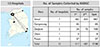
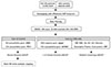
In order to collect a large number of the KD patients' clinical data and genomic deoxyribonucleic acid (DNA) samples for genetic studies of KD in Korea, the first KKDGC was formed in 2008 with 10 hospitals and 1 company (Seoul Clinical Laboratories, SCL). The consortium initially had set a goal to collect a total of 500 KD samples and achieved this goal in less than 2 years (May 2008 to February 2010) by collecting a total of 517 KD samples. The collected clinical data and genomic DNA samples were used for clinical data analysis and genetic studies of KD. However, it was realized that 500 KD case samples were not sufficient for large-scale genetic studies of KD. Thus, the second consortium was started in 2012 (April 2012 to September 2014). Through the first and second consortia, a total of 1198 KD case samples (genomic DNA and plasma samples) and clinical data were collected (Fig. 1). The collection of clinical data and genomic DNA samples is still ongoing by way of the second KKDGC.
Working Process for the Collection of Clinical Data and Genomic Deoxyribonucleic Acid Samples
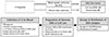
The KD patients were recruited from 13 tertiary academic hospitals in Korea that are currently participating in the KKDGC. All the KD patients were diagnosed by pediatricians, in accordance with the diagnostic criteria of the American Heart Association.2) At each participating hospital, approximately 5 mL of blood from a KD patient was sampled in an acid citrate dextrose or ethylenediamine tetraacetic acid tube after explanation and approval with the informed consents form. The collected patients' blood samples were used for genomic DNA preparation or establishment of Epstein-Barr Virus-transformed B cell lines at SCL (Fig. 2). Each patient's sample and clinical data were recorded by a new sample identification (ID) system that uses a two-digit disease symbol (KD), two-digit hospital ID, and a 3 digit-serial number (e.g., KD-AS-001). Pediatricians at each participating hospital collected all clinical data of the KD patients, including detailed clinical signs and treatment response. Finally, all the patients' clinical data and genomic DNA samples were deposited at Asan Medical Center at Seoul.
Standard Clinical Data Collection Form of Kawasaki Disease
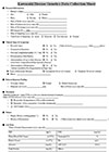
In order to characterize the clinical features of Korean children with KD prior to genetic studies, we constructed a standard clinical data collection form of KD with a total of 41 variables. The clinical data collection sheet is composed of: 1) patient's personal information, 2) clinical signs and symptoms, 3) echocardiogram findings, 4) treatment responses, 5) family history and recurrence, and 6) clinical lab data (Fig. 3). For genetic association studies of the KD subgroup, the clinical data were used to categorize the patient subgroups into a KD type (complete vs. incomplete KD), KD patients with or without CALs (normal vs. CAL), or IVIG responsiveness (responder vs. non-responder).
Clinical Data Analysis of Kawasaki Disease
Using the clinical data of 478 KD patients collected during the first consortium, there was an investigation of the clinical features and risk factors associated with CAL development and IVIG nonresponsiveness in Korean children with KD. We found that incomplete KD type, IVIG nonresponse, fever duration of 7 days or longer, and CC/AC genotypes of the rs7604693 single nucleotide polymorphism (SNP) in the PELI1 gene, were significantly associated with the development of CALs, with odds ratios (ORs) ranging from 2.06 to 3.04.4) This study also found that a serum albumin level of 3.6 g/dL or lower was significantly associated with nonresponsiveness to IVIG (OR=2.76; p=0.006).4)
Cell Proliferation Assay and Deoxyribonucleic Acid Microarray Studies in Kawasaki Disease Patient-Derived B Cell Lines after in vitro Immunoglobulin Treatment
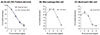
To examine the mechanism of IVIG-resistance in KD patients, we initially performed cell proliferation assays after in vitro immunoglobulin treatment in various cell lines, including KD patient-derived B cell lines (either IVIG responder or IVIG non-responder patient). Both the KD patient-derived B cell lines equally exhibited dose-dependent cell death by in vitro immunoglobulin treatment. However, no difference in cell death was observed in either B cell lines derived from the responder KD patient or non-responder KD patient (Fig. 4A). Other cell lines also underwent cell death by in vitro immunoglobulin treatment (Fig. 4B and C). This result suggests that the B cell may not play a role for IVIG responsiveness in KD patients. In addition, to understand the mechanisms of IVIG resistance in KD patients, we examined the gene expression profiles of B cell lines derived from IVIG responsive (n=2) or IVIG nonresponsive KD patients (n=3). We observed 34 upregulated (>2-fold) and 14 downregulated (<2 fold) genes after in vitro immunoglobulin treatment in B cells.5) Specifically, there was significant upregulation of gene expression in immune-related genes and downregulation in genes involved in cell-cycle after in vitro immunoglobulin treatment in B cell lines. However, there was no difference in the pattern of gene expression between IVIG responders and IVIG non-responders. This result suggests that the resistance of IVIG treatment in KD patients is not mediated by the change of quantitative gene expression in specific genes after IVIG treatment. Furthermore, in vitro immunoglobulin treatment in B cell lines did not increase the expression of the inhibitory FCGR2B gene. Therefore, this data suggest that the therapeutic effect of IVIG treatment in KD patients is not mediated by either inhibitory Fc receptor (FCGR2B) on B cells or B cell-mediated immune responses.
Genome-Wide Association Study of Kawasaki Disease
We performed a Genome-Wide Association Study (GWAS) using Affymetrix SNP array 6.0 in 186 KD patients and 600 healthy controls (Fig. 5). One SNP on chromosome 1p31 (rs527409) and a PELI1 locus on chromosome 2p13.3 (rs7604693) was associated with KD susceptibility and CAL formation, respectively.6) A subgroup analysis was also performed to identify IVIG response genes and CAL susceptibility using case samples only. It was found that a SNP (rs17136627) in the KCNN2 gene was significantly associated with CAL formation, particularly with large aneurysms (diameter >5 mm) (OR=12.6, p=1.96×10⁻⁸).7) This result indicates that the KCNN2 gene can have an important role in the development of coronary artery aneurysms in KD. Furthermore, we also found a significant association of a CRP promoter SNP (rs12068753) with a high CRP level in KD (beta=3.97, p=1.11×10⁻¹³) via the genetic association analysis of inflammatory biomarkers in KD patients.8) In addition, we collaborated with international KD genetics groups, including International KD Genetics Consortium, Taiwan KD Genetics Consortium, and Japan KD Genetics Consortium. In particular, we contributed to the studies validating the FCGR2A association with KD, performed by the international KD Genetics Consortium,9) and replication of B lymphoid tyrosine kinase association with KD that was carried out by Taiwan KD Genetics Consortium.10) In addition, the meta-analysis of Asian KD GWAS data is under way with the Japanese and Taiwanese groups.
Ongoing Projects and Future Plans
Currently, we are re-analyzing the clinical data of all collected KD patients (n=~1200) to evaluate the effect of age, gender, family history status, recurrence status, and KD types, on clinical features of KD. We also plan to study the interaction of clinical risk factors and genetic risk factors in KD. In order to identify new KD susceptibility and subphenotype loci, we are performing another GWAS using an Illumina Human Omni1 SNP chip with approximately 300 KD cases, concerning 16 cases with family history, 46 cases with recurrence, 119 cases with IVIG non-responsiveness, and 52 cases with CALs (diameter >5 mm). Multiple subsets of KD cases will be very useful to detect the loci associated with the subphenotypes of KD in GWAS data analysis. In the near future, we also plan to adopt the exome sequencing approach for KD genetic studies. As demonstrated above, consortium-based genetic studies are an effective way to identify the clinical risk factors and genetic risk factors of KD that may be affected by multiple genes and environmental factors. Furthermore, the KKDGC will facilitate and contribute to the understanding of pathophysiological mechanisms of KD and the development of novel diagnosis, as well as treatment and prevention strategies.
 XML Download