

 PDF
PDF ePub
ePub Citation
 Print
Print


Introduction
Deep vein thrombosis (DVT) is a rare complication following coronary angiography (CAG). A few clinical cases have been reported, especially in settings of venous compression with an enlarged hematoma, mechanical groin compression, prolonged procedure or hypercoagulable state. We present an unusual case of DVT after diagnostic CAG without any significant risk factors.
Case
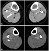
A 50-year-old woman presented with frequent retrosternal chest tightness. She also had a ten-year history of anxiety disorder. She was admitted and underwent CAG through the femoral artery approach using a 4 Fr sheath. We did not use prophylactic anticoagulation during diagnostic CAG. CAG showed no significant stenosis of the coronary artery. The femoral sheath was removed 30 minutes after the CAG, and hemostasis was achieved by manual compression for 15 minutes. After the patient had taken bed rest for 16 hours, she complained of pain and swelling in the right calf. There was no chest pain, cough, dyspnea, or shortness of breath, and the patient was stable, with a temperature 36.3℃, blood pressure of 120/80 mmHg, pulse of 80 beats/min, respiration of 18 breaths/min, and room air oxygen saturation 99%. All extremity pulses were unremarkable and the lower limbs appeared to be of equal temperature. Laboratory tests gave a white blood cell count 7300/mm3 with 53.2% neutrophils, hemoglobin 11.3 g/dL, platelet count 304×103/mm3, sodium 141 mEq/L, potassium 4.5 mEq/L, blood urea nitrogen 15 mg/dL, and creatinine 0.8 mg/dL. The plasma D-dimer level was 1140.0 ng/mL (normal range <250.0 ng/mL). Electrocardiography showed a normal sinus rhythm without focal abnormalities. Echocardiography revealed normal systolic function with good regional wall motion without right ventricle dilation with tricuspid regurgitation and elevation of pulmonary artery pressure. We performed a lower extremity CT angiography and found a thrombus extending from the right infrapopliteal vein to the anterior and posterior tibia vein, without any hematoma or pseudoaneurysm (Fig. 1). Based on the above results, a diagnosis of DVT regarding the right lower extremity was made.
The patient was further evaluated for the risk factors of DVT. She was a hairdresser, a non-smoker, and premenopausal. She had no history of hormone replacement therapy, other substance abuse, surgery, trauma or chronic systemic problems. Her anxiety disorder had been treated for ten years with fluoxetine 20 mg, diazepam 2 mg, alprazolam 0.5 mg, and tofisopam 25 mg. Her condition had been stable and there had been no change of medicine for at least six months. There was no family history of DVT. An extensive laboratory work-up was performed in search of any underlying disorders predisposed to DVT. Protein C activity 102% (70-130%), free protein S antigen 60.0% (50-150%), antithrombin III activity 86% (80-120%), antiphospholipid IgM (-), antiphospholipid IgG (-), international normalized ratio 0.85, activated prothrombin time 24 seconds (23-38 seconds), and homocysteine levels, were all in the normal range. Factor V Leiden and prothrombin 20210A mutations were also ruled out. The patient was treated with low molecular weight heparin and warfarin. Following these treatments, her symptoms improved. The patient was discharged uneventfully after ten days of hospitalization. She continued the anticoagulant therapy with oral warfarin and at a follow-up of three months the presenting leg pain had resolved and no further complications were evident.
Discussion
A recent retrospective review of all peripheral vascular complications after femoral artery catheterization gave the incidence of DVT as 0.05% (5 cases among 10450 cardiac diagnostic or therapeutic catheterizations).1) Although DVT is a rare complication after diagnostic transfemoral catheterization, its incidence may be higher, but most thrombotic and pulmonary embolic complications are not clinically evident, and their true incidence may be underestimated.2)
Risk factors demonstrated for venous thromboembolism (VTE) include old age, prolonged immobility, malignancy, major surgery, multiple trauma, prior VTE, chronic heart failure, and inherited or acquired thrombophilia.3) In addition, some medications such as oral contraceptives or hormone replacement therapy are also reported to be risk factors for VTE.3)4)
Prolonged bed rest, immobility of a catheterized extremity, groin compression, and large hematomas compressing the femoral vein, may all cause a predisposition to venous thromboembolic complications following femoral catheterization and may increase the incidence of such complications in combined left and right heart catheterization or therapeutic cardiac catheterization performed with large-sized sheaths.1)2)
The femoral approach is the most commonly used route for diagnostic CAG. Prophylactic anticoagulation may not be necessary in stable patients without other known risk factors who are undergoing elective diagnostic CAG expected to last less than 30 minutes. However, for a procedure thatis expected to last longer than 30 minutes, it may be advisable to administer an anticoagulant to prevent thrombus formation.5) Access site hemostasis is generally achieved by manual compression after sheath removal. Manual compression leads to immobilization of the patient for several hours.6) However, recent studies have suggested that the length of bed rest thought to be necessary after sheath removal appears to be decreasing, and early ambulation after removal of the sheath has been shown to be safe, in certain settings, and may improve patient comfort.7-9)
Previous reports have suggested that medication for psychiatric disorders may increase the risk of VTE.10) Many studies regarding the possible association between antipsychotic drugs and VTE have been published, and most of them have suggested that antipsychotics tend to increase the risk of VTE. In addition, a few clinical studies and isolated case reports have pointed to an association between the use of antidepressant drugs and VTE.11)12)
In the present case, DVT developed unusually after transfemoral diagnostic CAG of an apparently normal coronary artery in the absence of any risk factors for VTE. We suggest that compression of the groin and subsequent prolonged immobilization, as well as the use of antidepressant drugs, may be risk factors for DVT following CAG.
This case demonstrates that clinicians should consider the possibility of a VTE after diagnostic CAG even when patients lack significant risk factors. A protocol using smaller sheath sizes, close observation, and early ambulation after proper manual compression of the femoral artery may be a good approach to ensuring patient safety. Moreover, prophylactic anticoagulation during CAG may be helpful to prevent VTE in patients using antidepressants or anxiolytics.
 XML Download