

 PDF
PDF ePub
ePub Citation
 Print
Print


Introduction
Coronary cameral fistula (CCF) is the unusual anomalous communications between an epicardial coronary artery and a cardiac chamber. Coronary artery-left ventricular fistula is an extremely rare condition with a reported incidence of 1.2% of all coronary artery fistulas (CAF).1)2) There have been some case reports of myocardial ischemia, caused by CAF known as the "steal phenomenon".2)3) We present two cases of coronary artery-left ventricular fistulas, in which the hemodynamic significance was assessed by a fractional flow reserve (FFR).
Cases
Case 1
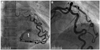
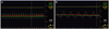
A 59-year-old man was referred to our department for further investigation of abnormal findings on an echocardiography. He had a history of hypertension and cerebral hemorrhage. He suffered distal interphalangeal joint deformity in his right third finger and underwent routine preoperative evaluation. His electrocardiogram (ECG) and chest X-ray were normal. Transthoracic echocardiography showed an abnormal Doppler color flow, through the left ventricle (LV) wall to the basal part of the ventricular cavity. Left ventricular systolic and diastolic functions were normal with normal chamber sizes. Selective coronary angiography demonstrated a normal right coronary artery. The left anterior descending artery (LAD) and the left circumflex artery (LCX) were large and tortuous without stenosis. They merged at the end of their course near the basal inferoposterior wall of the LV, and formed a huge fistulous track draining into the LV cavity (Fig. 1). Due to its large size, FFR was measured in both arteries. During hyperemia, obtained by an intravenous adenosine infusion (140 µg/kg/min), the FFR values were 0.93 in the LAD and 0.97 in the LCX (Fig. 2). The LV end-diastolic pressure was normal (11 mm Hg). He subsequently completed 17 minutes of a treadmill test (Bruce protocol) without chest pain and abnormal ECG changes. Due to the lack of symptom and evidence of hemodynamic compromise, we decided to leave the fistula alone and follow the patient closely. He had undergone an orthopedic surgery, uneventfully.
Case 2
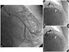
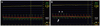
A 73-year-old woman was admitted to the hospital with dyspnea on exertion for one month. She underwent a total gastrectomy, due to advanced gastric cancer, 13 years ago. She had a history of diabetes mellitus and was on antianginal medication. Her resting ECG demonstrated a sinus rhythm with anterolateral and inferior T-wave inversions. The chest X-ray exhibited cardiomegaly. Transthoracic echocardiography showed a hypertrophied LV wall with an average thickness of 12 mm. The LV systolic function was normal with impaired relaxation patterns. Selective coronary angiography revealed normal right coronary artery and multiple coronary-left ventricular fistulas, arising from the left coronary artery system. A severe myocardial bridge, with total obstruction at the systolic phase, was observed in the mid segment of the LAD (Fig. 3). The FFR measurement was performed to evaluate the hemodynamic consequences, resulting from the myocardial bridge and the fistulas. The FFR value of the LAD was obtained by an intravenous adenosine infusion (140 µg/kg/min). During hyperemia, the FFR value was 0.92 in the LAD, beyond the bridging segment. The pressure pull back tracing showed that the FFR value increased from 0.92 to 0.94 in the segment that was proximal to the bridge (Fig. 4). Notably, the intracoronary systolic pressure overshooting was followed by the abrupt early diastolic pressure gradient in the distal segment beyond the bridge (Fig. 4B). Those findings disappeared immediately after the sensor proceeded through the bridging segment into the proximal segment of the LAD with a pull-back maneuver. The multiplicity of the fistulas and the lack of evidence of ischemia, we concurred to manage the patient medically.
Discussion
Coronary artery fistulas are observed in 0.1% of patients undergoing a diagnostic cardiac catheterization.4) Approximately, half of them arise from each coronary artery and more than 90% drain into the right-side of the heart.5) Coronary artery-left ventricular fistulas are rarely encountered. These fistulas may cause congestive heart failure, subacute bacterial endocarditis, myocardial ischemia, rupture of an aneurysmal coronary artery and thromboembolism.1)5)6) In symptomatic patients, closure of the fistula, by either surgical or transcatheter approach is advocated.7) However, fistula closures in asymptomatic adults remain controversial, even though elective closure in childhood has been recommended.6) Vavuranakis et al.4) reported that in a 6 years follow-up period, none of the patients developed any complications that are related to coronary artery fistula in 34 adults. Spontaneous occlusions, due to coronary atherosclerosis, were observed in two patients.
Whether the fistula is hemodynamically significant or not is the key point for management. Interestingly, one Doppler wire study demonstrated that the blood flow in the fistula decreased at adenosine-induced hyperemia, implicating an increased myocardial blood flow.8) We assessed such significance using a FFR in two different types of CCF, and successfully ruled out ischemia. In the presence of myocardial bridge, such as in Case 2, hyperemia induced with adenosine alone may be insufficient to detect a myocardial ischemia. Escaned et al.9) showed that intravenous dobutamine may induce more vigorous compression of the bridging segment, and reveal masked ischemia being used concomitantly with adenosine. However, in their study, none of those who had the mean FFR value of 0.90 or above, during hyperemia without dobutamine, had a decreased FFR value of less than 0.75, after a dobutamine challenge. In our case, her bridging segment was already compressed totally, without dobutamine, and the FFR value was over 0.90, which would not likely to fall below 0.75 with additional dobutamine.
The FFR measurement can be performed manageably and provide practical information on the spot, especially in the case of coexisting coronary artery diseases, including atherosclerosis and myocardial bridge. Unnecessary interventions to close the fistula and procedure-related risks could be avoided.
In the light of changes in the LV volume, the hemodynamic physiology of the coronary artery-left ventricular fistulas is similar to that of aortic regurgitation. Thus, strict follow-up monitoring is warranted.
In conclusion, the present cases demonstrated that the large coronary-left ventricular fistula was not associated with coronary steal phenomenon or myocardial ischemia in patients with insignificant epicardial stenosis.
 XML Download