Journal List > Korean Circ J > v.42(11) > 1016948

Jung: Pericardial Effusion and Pericardiocentesis: Role of Echocardiography

Abstract

Pericardial effusion can develop from any pericardial disease, including pericarditis and several systemic disorders, such as malignancies, pulmonary tuberculosis, chronic renal failure, thyroid diseases, and autoimmune diseases. The causes of large pericardial effusion requiring invasive pericardiocentesis may vary according to the time, country, and hospital. Transthoracic echocardiography is the most important tool for diagnosis, grading, the pericardiocentesis procedure, and follow up of pericardial effusion. Cardiac tamponade is a kind of cardiogenic shock and medical emergency. Clinicians should understand the tamponade physiology, especially because it can develop without large pericardial effusion. In addition, clinicians should correlate the echocardiographic findings of tamponade, such as right ventricular collapse, right atrial collapse, and respiratory variation of mitral and tricuspid flow, with clinical signs of clinical tamponade, such as hypotension or pulsus paradoxus. Percutaneous pericardiocentesis has been the most useful procedure in many cases of large pericardial effusion, cardiac tamponade, or pericardial effusion of unknown etiology. The procedure should be performed with the guidance of echocardiography.

Introduction

Normal pericardium is a double-walled sac that contains the heart and the roots of the great vessels. The pericardium is composed of two different layers; an outer fibrous parietal pericardium and an inner visceral pericardium.1) The inner visceral pericardium is a serous-type membrane and is located immediately outside of the myocardium.2)3) The pericardium prevents sudden dilatation of the heart, especially the right chamber, and displacement of the heart and great vessels, minimizes friction between the heart and surrounding structures, and prevents the spread of infection or cancer from the lung or pleura. The pericardium also contributes to diastolic coupling between the two ventricles.3)4) In between the parietal and visceral pericardium, there is a pericardial cavity filled with 10-50 cc of fluid, an ultrafiltrate of plasma that is produced by the visceral pericardium. Pericardial fluid acts as a lubricant between the heart and the pericardium. Excess fluid or blood accumulation in this cavity is called pericardial effusion.4)5)

Etiologies of Pericardial Effusion

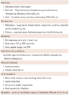
Pericardial effusion can develop in patients with virtually any condition that affects the pericardium, including acute pericarditis and a variety of systemic disorders.1)5) The clinical causes of pericardial effusion are very diverse and include malignancies of other organs, pulmonary tuberculosis, chronic renal failure, thyroid disease, autoimmune disease, and iatrogenic and idiopathic causes (Table 1).5-7)
The development of a pericardial effusion may have important implications for the prognosis (as in patients with intrathoracic malignancy) or diagnosis (as in myopericarditis or acute pericarditis), or both (as in dissection of the ascending aorta).6) When a pericardial effusion is initially or incidentally detected, a major concern for clinicians may be its etiology. In a majority of cases, the etiology of the effusion can be presumed from the underlying condition of the patient. Although the exact etiology of pericardial effusion can be identified by invasively-obtained pericardial fluid or tissue, an invasive procedure, like pericardiocentesis, is only indicated when the effusion is large or symptomatic, the effusion is accompanied by tamponade, or the cause of the effusion is questionable. There is some discrepancy in the etiologies of pericardial effusion, requiring pericardiocentesis between countries or centers. A review of many articles from throughout the world shows that the relative frequency of different etiologies is mainly affected by the decade, local epidemiology, hospital volume, and the diagnostic protocol.6)7) The etiology of pericardial effusion in Korea and other countries is summarized in Table 2.8-15) In Korea, lung cancer and pulmonary tuberculosis are predominant causes of large pericardial effusion.8-10) Sagrista-Sauleda et al.13) reported that when an etiology is not apparent, the clinical indices, including the size of the effusion, the presence of inflammatory signs, and cardiac tamponade, may be useful in suggesting a possible etiologic category. For example, severe effusions without inflammatory signs or tamponade were significantly associated with chronic idiopathic pericardial effusion. In contrast, cardiac tamponade without inflammatory signs was significantly associated with malignant effusions.

Transthoracic Echocardiography for Pericardial Effusion

M-mode and 2-dimensional Doppler echocardiography is the most effective technique, and is the gold standard for the diagnosis of pericardial effusion, because it is sensitive, specific, noninvasive, and easily made available at the bedside.16) Pericardial effusion can be detected as an "echo-free space" on 2-dimensional echocardiography (Fig. 1).1-5) Small collections of pericardial fluid, which can be physiologic (25 to 50 mL), may be visible during ventricular systole. When the amount of effusion is more than 50 mL, an echo-free space persists throughout the cardiac cycle. When a pericardial effusion is detected by echocardiography, the next step is to assess the size of the effusion, its location, hemodynamic importance, and associated diseases.4)17) A pericardial effusion is not always uniformly distributed around the heart. Initially, small effusions are evident over the posterobasal left ventricle; however, as the fluid volume increases, it gradually spreads anteriorly, laterally, and behind the left atrium. Ultimately, the separation becomes circumferential and appears as "swinging heart", which is a sign of a large pericardial effusion and possibly cardiac tamponade.4) Despite the fact that there is a large amount, the pericardial effusion does not surround the posterior portion of the heart. Therefore, the amplitude of the P wave is not changed, in spite of a large pericardial effusion or after pericardiocentesis (Fig. 2).18) Several grading systems have been developed, based on the size of the pericardial effusion. However, a generally accepted system is the effusion graded as minimal (scanty), small, moderate, or large. For circumferential pericardial effusions, any pericardial effusion with less than 5 mm of pericardial separation in diastole (corresponding to a fluid volume of 50 to 100 mL) is defined as minimal; 5 to 10 mm of separation as small (corresponding to a fluid volume of 100 to 250 mL); 10 to 20 mm of separation as moderate (corresponding to a fluid volume of 250 to 500 mL); and greater than 20 mm separation as large (corresponding to a fluid volume greater than 500 mL) (Fig. 3).4)7)8)13) This classification may be useful in daily clinical practice. However, even the diffused and circumferential effusion dimensions of the echo-free space may be different in the views examined; therefore, it is more correct and easier to measure and annotate the dimension of the effusion and to report where it has been evaluated (e.g., 12 mm in the left ventricular lateral wall in the apical four-chamber view; 10 mm along the right atrium in the subcostal view). This methodology not only facilitates the definition of effusion size, but also allows follow-up studies by detecting changes in the amount of pericardial fluid after therapy.17) Except for the epicardial fat, the abnormal masses attached on the epicardial surface or floating in the pericardial space must also be reported. It may be an infiltrative metastatic mass, inflammatory fibrin strands, pus, or a blood clot (Fig. 4). Other imaging modalities, such as computed tomography (CT) or magnetic resonance imaging (MRI), may be used to identify the characteristics of pericardial effusion and tamponade in the presence of a technically-limited echocardiographic study.6)7)19) However, the clinical utility of CT and MRI is questioned because of the high positive predictive value of echocardiography with potential safety concerns in acutely ill patients.
Currently, these modalities have adjunctive roles to echocardiography, especially in situations that show atypical hemodynamics, presence and severity of tamponade are doubtful, or when there are other unexplained conditions.19) For instance, in the case of pericardial effusion associated with intrathoracic malignancies, such as lung, breast, or esophageal cancer, chest CT might be useful for understanding the disease progression at a glance.

Cardiac Tamponade

Cardiac tamponade is a life-threatening, slow or rapid compression of the heart due to the pericardial accumulation of fluid, pus, blood, clots, or gas as a result of effusion, trauma, or rupture of the heart.20)21) The normal pericardium can stretch to accommodate increases in the pericardial volume. Once the pericardium has been stretched to its limit, by accumulating effusion, ongoing accumulation of pericardial fluid into a closed space increases the intrapericardial pressure. When the intra-pericardial pressure becomes high enough to impede cardiac filling, cardiac function is impaired. Therefore, the definition of cardiac tamponade is the state when the heart is compressed due to fluid accumulation and increased intrapericardial pressure, resulting in cardiogenic shock and circulatory collapse.20) In the physiology of tamponade development, a complex interaction of factors influences the ultimate physiologic consequences of pericardial effusion, in any given patient. These factors include the pericardial pressure/volume relationship, the rapidity of fluid accumulation, underlying cardiac pathology (particularly hypertrophy and shunts), and systemic volume status. It is well understood that small acute pericardial effusions (occurring most commonly post-intervention or traumatically) can lead to dramatic tamponade physiology when fluid accumulates rapidly; whereas, moderate-sized or even large pericardial effusions that accumulate slowly can be well tolerated, hemodynamically (Fig. 5).20)21) Therefore, large pericardial effusions or the swinging motion of the heart is not always present in cardiac tamponade. On the contrary, a localized compressing effusion may lead to dramatic hemodynamic consequences without producing classical echocardiographic indications of tamponade.4)22) The most common cause of cardiac tamponade reported is malignancy, which is involved in >50% of all tamponade cases. Especially lung cancer was involved in >70% of cardiac tamponade of malignant origin.21) Although cardiac tamponade is considered a clinical diagnosis, clinical findings like dyspnea, hypotension, tachycardia, elevated jugular venous pressure, and pulsus paradoxus, are known to have limited sensitivity and specificity.20)

Echocardiographic Findings of Cardiac Tamponade

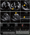
Echocardiography is the most useful diagnostic tool for evaluating patients with cardiac tamponade, and it should be performed without delay in patients if suspected.1)23) The most characteristic echocardiographic findings that suggest cardiac tamponade is diastolic collapse of the free walls of the right atrium, and/or the right ventricle.1)7)24) Any cardiac chamber can be collapsed, but it usually occurs on the right-sided chambers because both the right atrium and right ventricle are compliant and low pressure chambers.1) Right atrial compression for more than one third of the cardiac cycle (late diastole) is highly sensitive and specific for cardiac tamponade (Fig. 6A).1)7)22)25)26) Right ventricular compression in early diastole is less sensitive for the presence of cardiac tamponade than RA diastolic collapse, but is very specific for cardiac tamponade (Fig. 6B).1)7)27-30) RV collapse may not occur when the RV is hypertrophied or when its diastolic pressure is greatly elevated. Left atrial chamber collapse can be seen in about 25% of patients with hemodynamic compromise, and is very specific for cardiac tamponade. Left ventricular collapse is less common, since the wall of the left ventricle is more muscular, but can be seen under special conditions, such as localized postsurgical tamponade or severe pulmonary hypertension. Reciprocal changes in the left and right ventricular volumes occur with respiration in cardiac tamponade. Doppler echocardiography has been used to estimate respiratory variation in mitral and tricuspid diastolic inflow velocity. In accordance with pulsus paradoxus, a more exaggerated respiratory variation in inflow velocity, representing an inspiratory decrease in peak mitral inflow velocity ≥30%, and an inspiratory increase of peak tricuspid inflow velocity ≥50%, would suggest an increased ventricular interdependence and presence of cardiac tamponade (Fig. 6C).1)7)31)32) Dilatation of the inferior vena cava (IVC) and <50% reduction in the diameter of the dilated IVC during inspiration, so called IVC plethora, reflecting a marked elevation in the central venous pressure is frequently seen in patients with cardiac tamponade.1)7)33) IVC plethora was associated with pulsus paradoxus and was present in 92% of those with pericardial effusion, who required pericardial drainage. Collapse of the right-sided chambers is a sensitive indicator of tamponade, but abnormalities in cardiac filling are a more specific finding of tamponade.34) Although cardiac tamponade may be diagnosed, using the above findings, clinicians should be aware that echocardiography has some limitations for diagnosis and follow-up of cardiac tamponade. For example, severe hypovolemia or large pleural effusions can cause diastolic chamber collapse. Additionally, disease states that increase right-sided cardiac pressures, such as pulmonary hypertension or pulmonary thromboembolism, may prevent diastolic chamber collapse.35) Cardiac tamponade is not an "all-or-none" phenomenon, but rather a spectrum of findings.6) Therefore, for the final diagnosis of cardiac tamponade, clinicians should correlate the echocardiographic signs of tamponade (RV collapse, RA collapse, respiratory alteration of mitral and tricuspid flow, and IVC plethora) with the symptoms and signs of clinical tamponade (dyspnea, tachycardia, jugular venous distension, pulsus paradoxus, hypotension, and shock).6)

Echocardiography Guided Pericardiocentesis

Percutaneous needle pericardiocentesis has been the most useful therapeutic procedure for the early management of large, symptomatic pericardial effusion or cardiac tamponade, and it continues to be used as a diagnostic procedure in some patients with asymptomatic pericardial effusion, such as chronic idiopathic effusion.7)17)36)37) Before, during, and after the percutaneous pericardiocentesis, transthoracic echocardiography guidance is essential.36)38) If the pericardiocentesis is performed at bedside without echocardiographic guidance, the risk of threatening complications like bleeding or shock have been reported to be as high as 20%. In contrast, echocardiographic guidance increases the success rate of pericardiocentesis by reducing these complications.7)11) In Korea, the success rate of echocardiography-guided pericardiocentesis was about 99%.8)9)
When deciding to perform pericardiocentesis, many clinical and echocardiographic factors must be comprehensively considered. These factors include the amount and location of the effusion, hemodynamics on echocardiography, sufficient margins of the echo free space so as to prevent laceration, clinical indication and urgency, underlying etiologies, and bleeding tendency. Recently, Halpern et al.39) suggested a "pericardial effusion scoring index" for deciding whether to perform pericardiocentesis. The scoring index consists of 3 components obtained at initial presentation; effusion size on echo, echocardiographic assessment of hemodynamics, and etiology of effusion. Authors reported that percutaneous pericardiocentesis could be performed when the score was 4 or above.
Echocardiography-guided pericardiocentesis can be performed in the following sequences.
  • 1) Check clinical indications and medical history, such as taking anti-platelet or anti-coagulation drugs, and get a consent form.

  • 2) Positioning and echocardiographic imaging

    • a) Position the patient in the semi-fowler position in bed.

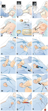
    • b) Perform 2D echocardiographic imaging at the apical view (Fig. 7A), subxyphoid view (Fig. 7B), and left para-sternal view (Fig. 7C) to gain insight into the effusion.40)41)

  • 3) Determination of puncture site

    • a) Choose sites with the biggest echo-free space for safe needle entry, and mark it using a pen or nail tip on the site (Fig. 7D).

    • b) Image and determine the 3-dimensional direction of needle entry.

  • 4) Preparation of puncture site

    • a) Prepare all requisite tools for the centesis on the table (Fig. 7E) and perform the sterile skin preparation (Fig. 7F).

    • b) Cover the patient with a surgical drape and the patient's eyes with an eye patch to reduce tension and anxiety (Fig. 7G).

    • c) Perform sufficient local anesthesia at the puncture site (Fig. 7H).

  • 5) Pericardiocentesis

    • a) Perform a preliminary exploration with the local anesthetic needle (21 gauge) to confirm the direction of the needle approach and to feel the nature of the effusion.

    • b) Put the puncture needle (18 gauge) to the tip of the puncture syringe.

    • c) Gently insert and advance the puncture needle from the skin of the puncture site toward the heart in the breath holding state. The puncture needle must maintain the direction that was determined during the preliminary exploratory test (Fig. 7I).

    • d) Feel the "pop" moment at the puncture and observe negatively drained fluid in the syringe (Fig. 7J).

    • e) Stop the advancing puncture needle and hold the needle with your fingers to prevent further advancement. Insert a smooth tip guidewire through a back-hole in the syringe, sufficiently into the pericardial space (Fig. 7K).

    • f) Remove the puncture syringe with the needle (Fig. 7L), keep ing the guidewire in the pericardial space (Fig. 7M).

    • g) After dilatation of the pathway (Fig. 7N), insert a double lu men indwelling catheter over the guidewire into the pericardial space (Fig. 7O).

    • h) Remove the guidewire with the remaining indwelling catheter in the pericardial space.

    • i) Confirm the success of the procedure by manual drainage with a syringe (Fig. 7Q) or a saline bubble test with echocardiography. The patient's reduced or relieved symptoms soon after the drainage, even by a small amount, is additional evidence of success.

    • j) Suture the catheter on the skin and continue to drain the effusion into a bottle (Fig. 7R).

There are 3 approaches to needle entry during pericardiocentesis; left parasternal, subxyphoid, and left apical.37)41) Among these, the left parasternal approach seems to be preferred in the era of echocardiography-guided pericardiocentesis, because the left parasternal view provides a more direct route to the largest echo free space and better safety than other approaches. Using the left parasternal approach, the puncture needle is inserted close to the sternum, usually in the left 5th or 6th intercostal space. This approach is associated with possibly a higher risk of pneumothorax than with the subxyphoid approach.41)42) The puncture needle during the subxyphoid approach is inserted at an angle between the xiphisternum and left costal margin, towards the left shoulder at a 15 to 30 degree angle to the skin.20) This approach has been performed when echocardiography is not available, in the cardiac catheterization room with ECG monitoring, or in emergent situations. This route is extrapleural and avoids the coronary, pericardial, and internal mammary arteries. Injuries to the pleura, liver, or stomach, irritation to the diaphragm and phrenic nerve resulting in bradycardia and shock, and higher procedure-related mortality have been reported with this approach than with others.7)41)
In the left apical approach, the puncture needle is inserted into the intercostal space outside of the left nipple, where the left ventricular apical beating is touched, and is toward the right shoulder. During this approach, the apex can easily be pierced, and evoke ventricular fibrillation; thus, it is not indicated in emergent situations and must be performed under the guidance of echocardiography.41) The choice of direction of approach must be decided by an experienced clinician, after considering if there is a safe enough margin of echo free space, the patient's position, and clinical situation. A major complication of percutaneous pericardiocentesis includes laceration of the heart and coronary arteries, hemothorax and pneumothorax, ventricular tachyarrhythmia, and vasovagal response.7) During pericardiocentesis, clinicians must be attentive to the important anatomic structures in the way of the puncture needle approach, including the internal thoracic arteries (located 0.5 to 2 cm lateral to the sternal edge), intercostal vessels (inferior border of the rib) and nerves, phrenic nerve in the diaphragm, pleura, lung parenchyma, and free wall of the right or left ventricle.41) Aortic dissection is a major contraindication of pericardiocentesis. Relative contraindications include uncorrected coagulopathy, anticoagulant therapy, and thrombocytopenia <50000/mm3, and small, posterior, and loculated effusions.7) Surgical drainage, rather than percutaneous pericardiocentesis is preferred in the following situations: traumatic hemopericardium, purulent pericarditis, recurrent malignant effusion, loculated effusion in the posterior side of the heart, a need for pericardial biopsy with drainage for diagnosis, and coagulopathy or thrombocytopenia.43-45)

Conclusions

Pericardial effusion can develop in patients with virtually any condition that affects the pericardium, including acute pericarditis, malignancies, pulmonary tuberculosis, chronic renal failure, thyroid disease, autoimmune disease, or iatrogenic and idiopathic causes. The causes of large pericardial effusions, requiring invasive pericardiocentesis or pericardiotomy, vary according to the times, status of the countries, and the location, size, and facilities of the hospital. Especially in Korea, pulmonary tuberculosis and lung cancer are predominant causes. Transthoracic echocardiography is the most important tool for the diagnosis, grading, pericardiocentesis procedure, and follow up for pericardial effusion. Cardiac tamponade is a kind of cardiogenic shock and a medical emergency. Clinicians should understand the tamponade physiology, especially cardiac tamponade that can develop without large pericardial effusion or swinging heart. In addition, clinicians should correlate the echocardiographic findings of tamponade, such as right ventricular collapse, right atrial collapse, respiratory variation in the mitral and tricuspid flow, and IVC plethora, with the signs of clinical tamponade, such as hypotension or pulsus paradoxus. Pericardiocentesis is lifesaving for cardiac tamponade, and indicated in cases where there is a large volume of pericardial effusion. The procedure should be performed with the guidance of echocardiography.

Figures and Tables

Fig. 1
Representative images of echo-free space on 2-dimensional echocardiography.
kcj-42-725-g001
Fig. 2
Schematic showing that pericardial effusion mainly accumulates around the 2 ventricles, rather than the 2 atria; therefore, the p waves are not changed much after pericardiocentesis, compared with the Q wave (Jung HO et al. Am J Cardiol 2010;106:441 with permission).18) RA: right atrium, LA: left atrium, RV: right ventricle, LV: left ventricle.
kcj-42-725-g002
Fig. 3
Echocardiographic grading of pericardial effusion as scanty (A), mild (B), moderate (C), and large (D).
kcj-42-725-g003
Fig. 4
Echocardiography showing abnormal masses on the epicardial surface. Final diagnoses are (A) post-operative pericarditis and (B) metastatic infiltration of lung cancer to pericardium.
kcj-42-725-g004
Fig. 5
Pericardial pressure-volume relationships in patients who have rapidly (left curve) and gradually (right curve) developed a pericardial effusion. In rapid accumulating effusion (left curve), even a small effusion volume can exceed the limit of parietal pericardial stretch and finally causes a steep rise in pressure. In contrast, slow accumulating effusion (right curve) requires a long time and a large volume to exceed the limit of pericardial stretch because of the activating compensatory mechanisms (Spodick DH. N Engl J Med 2003;349:685 with permission).20)
kcj-42-725-g005
Fig. 6
Representative echocardiographic images of cardiac tamponade. A: diastolic collapse of the free wall of the right atrium from late diastole to early systole is observed on the apical 4 chamber view (middle) and magnified view (right), compared with the image of early diastole (left). B: collapse of the right ventricle in early diastole is observed on the parasternal long axis view (middle) and M-mode (right), compared with the image of systole (left). C: marked respiratory variation is observed on the transmitral (left) and transtricuspid (right) Doppler flow. RA: right atrium, RV: right ventricle, LV: left ventricle, MV: mitral valve, TV: tricuspid valve.
kcj-42-725-g006
Fig. 7
Illustrations of the procedure of echocardiography guided percutaneous pericardiocentesis.
kcj-42-725-g007
Table 1
Major etiologies of pericardial effusion
kcj-42-725-i001

CMV: cytomegalovirus, HIV: human immunodeficiency virus, ICD: implantable cardiac defibrillator, CRT: cardiac resynchronization therapy, CPR: cardiopulmonary resuscitation

Table 2
Summary of the frequency of etiologies with large volumes of pericardial effusion requiring pericardiocentesis reported in Korea and other countries
kcj-42-725-i002

Collagen VD: Collagen vascular diseases, HIV: human immunodeficiency virus, MI: myocardial infarction, Tbc: tuberculosis, thyroid D: thyroid diseases

Acknowledgments

The author gratefully acknowledges the assistance of Jung Won Lee, RDCS, Jung Yeon Chin, MD, and Jung Eun Lee, MD.

Notes

The author has no financial conflicts of interest.

References

1. Little WC, Freeman GL. Pericardial disease. Circulation. 2006. 113:1622–1632.
2. Braunwalt E. Fausi AS, Braunwalt E, Kasper DL, editors. Pericardial disease. Harrison's internal medicine. 2008. 17th ed. New York: McGraw Hill Medical;1488–1492.
3. Oh JK, Seward JB, Tajik AJ. Oh JK, Seward JB, Tajik AJ, editors. Pericardial diseases. The echo manual. 2007. 3rd ed. Philadelphia: Lippincott Williams and Wilkins (Wolters Kluwers);289–292.
4. Munt BI, Moss RR, Grewal J. Otto CM, editor. Pericardial disease. The practice of clinical echocardiography. 2011. 4th ed. Philadelphia: Saunders/Elservier;565–578.
5. Manning WJ. Goldman L, Ausiello D, editors. Pericardial disease. Cecil Medicine. 2008. 23rd ed. Philadelphia: Saunders/Elservier;548–552.
6. Sagristà-Sauleda J, Mercé AS, Soler-Soler J. Diagnosis and management of pericardial effusion. World J Cardiol. 2011. 3:135–143.
7. Maisch B, Seferović PM, Ristić AD, et al. Guidelines on the diagnosis and management of pericardial diseases executive summary; The Task force on the diagnosis and management of pericardial diseases of the European society of cardiology. Eur Heart J. 2004. 25:587–610.
8. Kil UH, Jung HO, Koh YS, et al. Prognosis of large, symptomatic pericardial effusion treated by echo-guided percutaneous pericardiocentesis. Clin Cardiol. 2008. 31:531–537.
9. Cho BC, Kang SM, Kim DH, et al. Clinical and echocardiographic characteristics of pericardial effusion in patients who underwent echocardiographically guided pericardiocentesis: Yonsei Cardiovascular Center experience, 1993-2003. Yonsei Med J. 2004. 45:462–468.
10. Kim DY, Park JH, Shin JD, et al. Long-term follow-up results and clinical manifestations of patients with a moderate to large amount of pericardial effusion. Korean J Med. 2008. 74:154–161.
11. Tsang TS, Enriquez-Sarano M, Freeman WK, et al. Consecutive 1127 therapeutic echocardiographically guided pericardiocenteses: clinical profile, practice patterns, and outcomes spanning 21 years. Mayo Clin Proc. 2002. 77:429–436.
12. Kabukcu M, Demircioglu F, Yanik E, Basarici I, Ersel F. Pericardial tamponade and large pericardial effusions: causal factors and efficacy of percutaneous catheter drainage in 50 patients. Tex Heart Inst J. 2004. 31:398–403.
13. Sagristà-Sauleda J, Mercé J, Permanyer-Miralda G, Soler-Soler J. Clinical clues to the causes of large pericardial effusions. Am J Med. 2000. 109:95–101.
14. Levy PY, Corey R, Berger P, et al. Etiologic diagnosis of 204 pericardial effusions. Medicine (Baltimore). 2003. 82:385–391.
15. Reuter H, Burgess LJ, Doubell AF. Epidemiology of pericardial effusions at a large academic hospital in South Africa. Epidemiol Infect. 2005. 133:393–399.
16. Horowitz MS, Schultz CS, Stinson EB, Harrison DC, Popp RL. Sensitivity and specificity of echocardiographic diagnosis of pericardial effusion. Circulation. 1974. 50:239–247.
17. Pepi M, Muratori M. Echocardiography in the diagnosis and management of pericardial disease. J Cardiovasc Med (Hagerstown). 2006. 7:533–544.
18. Jung HO, Seung KB, Madias JE. Electrocardiographic changes resulting from pericardial effusion drainage. Am J Cardiol. 2010. 106:437–441.
19. Verhaert D, Gabriel RS, Johnston D, Lytle BW, Desai MY, Klein AL. The role of multimodality imaging in the management of pericardial disease. Circ Cardiovasc Imaging. 2010. 3:333–343.
20. Spodick DH. Acute cardiac tamponade. N Engl J Med. 2003. 349:684–690.
21. Ariyarajah V, Spodick DH. Cardiac tamponade revisited: a postmortem look at a cautionary case. Tex Heart Inst J. 2007. 34:347–351.
22. Kronzon I, Cohen ML, Winer HE. Cardiac tamponade by loculated pericardial hematoma: limitations of M-mode echocardiography. J Am Coll Cardiol. 1983. 1:913–915.
23. Tsang TS, Barnes ME, Hayes SN, et al. Clinical and echocardiographic characteristics of significant pericardial effusions following cardiothoracic surgery and outcomes of echo-guided pericardiocentesis for management: Mayo Clinic experience, 1979-1998. Chest. 1999. 116:322–331.
24. Singh S, Wann LS, Schuchard GH, et al. Right ventricular and right atrial collapse in patients with cardiac tamponade--a combined echocardiographic and hemodynamic study. Circulation. 1984. 70:966–971.
25. Gillam LD, Guyer DE, Gibson TC, King ME, Marshall JE, Weyman AE. Hydrodynamic compression of the right atrium: a new echocardiographic sign of cardiac tamponade. Circulation. 1983. 68:294–301.
26. Kochar GS, Jacobs LE, Kotler MN. Right atrial compression in postoperative cardiac patients: detection by transesophageal echocardiography. J Am Coll Cardiol. 1990. 16:511–516.
27. Gaffney FA, Keller AM, Peshock RM, Lin JC, Firth BG. Pathophysiologic mechanisms of cardiac tamponade and pulsus alternans shown by echocardiography. Am J Cardiol. 1984. 53:1662–1666.
28. Armstrong WF, Schilt BF, Helper DJ, Dillon JC, Feigenbaum H. Diastolic collapse of the right ventricle with cardiac tamponade: an echocardiographic study. Circulation. 1982. 65:1491–1496.
29. Engel PJ, Hon H, Fowler NO, Plummer S. Echocardiographic study of right ventricular wall motion in cardiac tamponade. Am J Cardiol. 1982. 50:1018–1021.
30. Reydel B, Spodick DH. Frequency and significance of chamber collapses during cardiac tamponade. Am Heart J. 1990. 119:1160–1163.
31. Appleton CP, Hatle LK, Popp RL. Cardiac tamponade and pericardial effusion: respiratory variation in transvalvular flow velocities studied by Doppler echocardiography. J Am Coll Cardiol. 1988. 11:1020–1030.
32. Burstow DJ, Oh JK, Bailey KR, Seward JB, Tajik AJ. Cardiac tamponade: characteristic Doppler observations. Mayo Clin Proc. 1989. 64:312–324.
33. Himelman RB, Kircher B, Rockey DC, Schiller NB. Inferior vena cava plethora with blunted respiratory response: a sensitive echocardiographic sign of cardiac tamponade. J Am Coll Cardiol. 1988. 12:1470–1477.
34. Mercé J, Sagristà-Sauleda J, Permanyer-Miralda G, Evangelista A, Soler-Soler J. Correlation between clinical and Doppler echocardiographic findings in patients with moderate and large pericardial effusion: implications for the diagnosis of cardiac tamponade. Am Heart J. 1999. 138(4 Pt 1):759–764.
35. Nagdev A, Stone MB. Point-of-care ultrasound evaluation of pericardial effusions: does this patient have cardiac tamponade? Resuscitation. 2011. 82:671–673.
36. Tsang TS, Freeman WK, Barnes ME, Reeder GS, Packer DL, Seward JB. Rescue echocardiographically guided pericardiocentesis for cardiac perforation complicating catheter-based procedures. The Mayo Clinic experience. J Am Coll Cardiol. 1998. 32:1345–1350.
37. Seferović PM, Ristić AD, Imazio M, et al. Management strategies in pericardial emergencies. Herz. 2006. 31:891–900.
38. Lindenberger M, Kjellberg M, Karlsson E, Wranne B. Pericardiocentesis guided by 2-D echocardiography: the method of choice for treatment of pericardial effusion. J Intern Med. 2003. 253:411–417.
39. Halpern DG, Argulian E, Briasoulis A, Chaudhry F, Aziz EF, Herzog E. A novel pericardial effusion scoring index to guide decision for drainage. Crit Pathw Cardiol. 2012. 11:85–88.
40. Goodman A, Perera P, Mailhot T, Mandavia D. The role of bedside ultrasound in the diagnosis of pericardial effusion and cardiac tamponade. J Emerg Trauma Shock. 2012. 5:72–75.
41. Loukas M, Walters A, Boon JM, Welch TP, Meiring JH, Abrahams PH. Pericardiocentesis: a clinical anatomy review. Clin Anat. 2012. 25:872–881.
42. Tsang TS, Freeman WK, Sinak LJ, Seward JB. Echocardiographically guided pericardiocentesis: evolution and state-of-the-art technique. Mayo Clin Proc. 1998. 73:647–652.
43. Spodick DH. Braunwald E, Zipes DP, Libby P, editors. Pericardial diseases. Heart disease. 2001. 6th ed. Philadelphia: W.B. Saunders;1823–1876.
44. Allen KB, Faber LP, Warren WH, Shaar CJ. Pericardial effusion: subxiphoid pericardiostomy versus percutaneous catheter drainage. Ann Thorac Surg. 1999. 67:437–440.
45. McDonald JM, Meyers BF, Guthrie TJ, Battafarano RJ, Cooper JD, Patterson GA. Comparison of open subxiphoid pericardial drainage with percutaneous catheter drainage for symptomatic pericardial effusion. Ann Thorac Surg. 2003. 76:811–815. discussion 816.
TOOLS
Similar articles