

 PDF
PDF ePub
ePub Citation
 Print
Print


Introduction
Heart rate recovery (HRR) is defined as the decline in heart rate immediately following exercise. The rapid deceleration of the heart rate immediately following exercise is dependent on a complex interplay between various intrinsic, neural, and humoral factors.1-3) The decrease in heart rate immediately after exercise is a function of the reactivation of the parasympathetic nervous system.2) There are cross-sectional associations of HRR with glucose, insulin, high-density lipoprotein-cholesterol (HDL-C), triglyceride (TG), blood pressure (BP), low-density lipoprotein-cholesterol (LDL-C).4-8) Decreased HRR is a predictor of a poor cardiovascular prognosis, independent of and in addition to other risk factors.9-11)
Brain natruretic peptide (BNP) is a hormone that is released from the ventricles in response to an increase in left ventricular pressure and volume. Plasma BNP levels play a crucial role in the diagnosis and prognosis of heart failure (HF) patients.12)13) Increased BNP levels have been observed in patients with hypertension (HTN), atrial fibrillation, valvular heart disease, ST-segment elevation myocardial infarction and myocardial ischemia without extensive necrosis.14)15)
Higher BNP levels are consistently observed in patients with HF and coronary artery disease (CAD) with blunted HRR.15)16) Also, the correlation between BNP and cardiac autonomic function has been studied in type 2 diabetic patients.17) However, there is limited data on the relationship between HRR and BNP levels in patients without HF. Also, there have been few studies of BNP levels before, after, and during exercise. The purpose of this study was to examine the association between HRR and plasma BNP levels before, after, and during exercise in patients with a normal systolic function and to determine important associated factors.
Subjects and Methods
Subjects
This study was conducted at the Kangbuk Samsung Hospital and included 105 patients who had complained of chest pain but had no symptoms at admission, and had a probable diagnosis of coronary heart disease. Coronary angiography (CAG) and an exercise test were conducted on all patients. Of these patients, 40 were in the CAD group. We excluded patients with an echocardiographic ejection fraction (EF) of ≤50%, respiratory difficulty, pulmonary abnormalities, myocardial infarction or valvular disease, a history of cardiac surgery or percutaneous transluminal coronary angioplasty (PTCA) or kidney disease. Patients were hospitalized and treated with aspirin and clopidogrel according to the attending physician's judgment with regard to CAD. Other prescriptions, such as angiotensin converting enzyme inhibitor and beta-blockers, were continued and arranged to avoid duplicating existing HTN or diabetes drugs. No additional medications were taken. Informed consent was obtained from each patient in accordance with the Helsinki Declaration.
Stress testing
A stress test was conducted on patients after over-night fasting. All patients underwent symptom-limited treadmill exercise testing according to the Bruce protocol and patients were continuously monitored by 12-lead electrocardiography. BP and heart rate were measured at rest, at 3-minute intervals during exercise, during peak effort and at 1-minute intervals during recovery. Testing was interrupted for the following reasons: exhaustion, a sustained drop in systolic blood pressure (SBP), clinical manifestations of intense typical chest pain, a ST segment depression of ≥2 mm, a ST segment elevation of ≥2 mm in a lead without the presence of the Q wave or the development of arrhythmias. Patients with a horizontal or down-sloping ST segment depression of ≥1 mm for at least 60 milliseconds to 80 milliseconds after the end of the QRS complex in two adjacent leads were considered positive for CAD.18)
Heart rate recovery
After achieving the peak workload, all patients spent at least 2 minutes in a cool-down period during treadmill testing at a speed of 2.4 km per hour and a grade of 2.5 percent. This period was considered the recovery period. The value for the recovery of heart rate was defined as the difference in the heart rate from the peak rate during exercise to the rate at 2 minutes after cessation of exercise. A cutoff HRR value of 24 beats per 2 minutes was considered abnormal, reflecting a desired HRR value of 12 beats per minute. However, one minute is not sufficient for patient arrangement, inflation and deflation of the cuff to measure BP and HRR after the treadmill test. Also, a 2 minutes HRR may decrease more than a 1 minute HRR.20)
Coronary angiography and extent of severity
Two cardiologists reviewed the angiograms independently of each other in a blinded manner. Stenosis exceeding 50% of the major epicardial coronary artery diameter was considered significant.
Brain natruretic peptide measurements
All subjects were instructed to fast overnight before BNP level measurements. Plasma BNP levels were measured before exercise and after 5 minutes at sitting rest after completion of exercise. The BNP change is the absolute value of the difference between pre- and post-exercise BNP levels. A polyethylene tube containing ethylene diamine tetra-acetic acid was filled with 5 mL of blood. The plasma BNP concentrations were determined using the Triage BNP test kit (Biosite, San Diego, CA, USA), a fluorescent immunoassay method with a detection limit of 5 pg/mL.
Echocardiography
M-mode echocardiography was performed under 2-dimensional echocardiography guidance using an Agilent Sonos 5500 (Hewlett Packard, Palo Alto, CA, USA) equipped with a 2.5-MHz transducer. Two-dimensional images taken from longitudinal or transverse parasternal views were used to ensure that all measurements were obtained at the same level. The following measurements were performed according to the recommendations of the American Society of Echocardiography: intraventricular septal thickness at end-diastole (IVSTd), left ventricular internal dimension at end-diastole (LVIDd) and posterior wall thickness at end-diastole (PWTd). The left ventricular ejection fraction (LVEF) was measured according to the modified Simpson's rule. The mitral flow velocities (E: early filling velocity, A: late filling velocity) were measured using a pulsed-wave Doppler, with the sample volume placed between leaflet tips. The Devereux and Reichek formula was used to calculate the left ventricular mass index (LVMI): LVMI={1.04×[(LVIDd+3PWTd+LVIDd)3-LVIDd3]-13.6}/body surface area.
Statistical analysis
Statistical analysis was conducted using Statistical Package for the Social Sciences (SPSS) version 12.0 software (SPSS corporation, Chicago, IL, USA) with advice from the statistics department in our hospital. Continuous variables are presented as mean values with standard deviation. The comparisons between the two groups of patients with abnormal HRR after exercise were performed using a t-test. A p≤0.05 (less than or equal to 0.05) was considered statistically significant. Multivariate regression analysis was used to identify possible independent variables associated with impaired HRR after exercise in patients with normal systolic function.
Results
Clinical characteristics
The study group consisted of 105 subjects (64 men and 41 women). The base-line characteristics of the patients according to their HRR are shown in Table 1 and 2. The 105 enrolled patients met all of the inclusion criteria. A cutoff value of ≤24 beats for the first 2 minutes of the HRR was found to maximize the log-rank test statistic. An abnormal value for the HRR was found in 44 patients (42%). There was no significant difference in age, HTN, diabetes mellitus (DM) prevalence, body weight, total cholesterol, LDL-C, TG level, or BNP change between the normal HRR group and the abnormal HRR group (p>0.05). The abnormal HRR group had significantly higher pre- and post-exercise BNP levels than the normal HRR group (p=0.017). Seven patients (4 in normal HRR groups, and 3 in abnormal HRR groups) were taking beta-blockers but there was no significant difference in the two groups. In addition, the abnormal HRR group had a significant higher prevalence of CAD (p=0.011), which is probably due to the fact that women, but not men, had a significantly higher CAD prevalence in the abnormal HRR group (p=0.01 and p=0.24, respectively).
Correlation between brain natruretic peptide and heart rate recovery
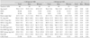
Correlation analysis showed that the HRR had a significant negative association with pre-exercise BNP levels (p<0.01) (Table 3, Model 1). After correcting for the peak heart rate, CAD, age, HTN, and DM sequentially, the pre-exercise BNP level was significantly correlated with HRR (Table 3, Model 2). When the peak heart rate, CAD, age, HTN, and DM were corrected together, there was a significant negative correlation between the pre-exercise BNP level and HRR (p<0.05) (Table 3, Model 3).
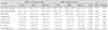
Table 4 and 5 show correlation analysis for post-exercise BNP and BNP change, respectively. HRR had a significant negative association with the post-exercise BNP level (p<0.01) (Table 4, Model 1), but the association was not as strong as that with the pre-exercise BNP level. Models 2 and 3 show adjusted associations for each parameter. The BNP change was not significantly correlated with HRR in any of the three models (Table 5, Models 1, 2, 3). Based on our correlation analysis, we determined that the pre-exercise BNP level is the most reliable measurement of the three BNP measurements used in this study.
Discussion
BNP is a hormone that is secreted from the cardiac ventricles in response to an increase in left ventricular pressure and volume. Moreover, elevated plasma BNP levels are highly sensitive and specific for the diagnosis and prognosis of HF, and for the prognosis of acute myocardial infarction.12)13)15) Patients with HF have blunted HRR concurrent with the presence of higher BNP levels.16) But, the BNP level is typically measured before exercise; there is little data on post-exercise BNP levels and BNP change during exercise, and few studies have considered gender differences in BNP level and HRR. We analyzed patients with normal systolic function, and found that a rapid decrease in HRR had a significant negative association with pre- and post-exercise BNP levels, but not with BNP change.
HRR is defined as the maximum heart rate during exercise, minus the heart rate after exercise, which is a measure of the actual decrease in heart rate relative to the peak heart rate during exercise. Previous studies on HRR vary in non-HF patients; the optimal prognostic cutoff values for HRR (defining normal vs. abnormal HRR) are based on specific statistical considerations or are arbitrarily selected. The most commonly used cutoff value is 12 beats per minute.14) We found that patients with abnormal HRR values (≤24 beats first 2 minutes of HRR) had lower HDL-C, lower peak heart rate, and higher pre- and post-exercise BNP levels than patients with normal HRR values. In addition, the patients with abnormal HRR had a significantly higher prevalence of CAD. The higher pre- and post-exercise BNP levels in the abnormal HRR group can be explained by 1) parasympathetic dysfunction, 2) diastolic dysfunction, and 3) CAD.
First, heart rate variability (HRV) is one of the best methods to detect a quantitative relationship between the cardiovascular and autonomic nervous system (ANS). The ANS-mediated response, in particular parasympathetic reactivation, is a major determinant of HRR. In healthy individuals, the parasympathetic system is dominant at rest; exercise is associated with parasympathetic withdrawal and, as the intensity of exercise is increased, with sympathetic activity.19) Savin et al.19) have observed significantly reduced HRR values in individuals whose parasympathetic systems were blocked with atropine after exercise, suggesting that parasympathetic reactivation, not sympathetic withdrawal, is responsible for the reduced heart rate in the recovery phase. Abnormal HRR is considered a manifestation of altered autonomic responsiveness and abnormal vagal tone, possibly leading to an increased heart rate at rest.20)
Second, an increase in BNP reportedly reflects an early stage of cardiac dysfunction.21) Increased levels of plasma BNP are related to cardiac reflex parasympathetic dysfunction. The early stage of cardiac function and parasympathetic dysfunction may be associated with increased plasma BNP levels.17) In our study, there was a significant correlation between delayed HRR and two BNP measurements, pre- and post-exercise. Multivariate analysis revealed that impaired HRR after exercise is significantly related with age and diastolic dysfunction.22) But in this study, the abnormal HRR group showed a significantly lower E/A ratio than the normal HRR group. The discrepancy between this study and other studies may be explained by differences in ventricular function. All subjects in this study have normal systolic function. In systolic failure, the heart overfills to compensate for the reduced pump function, which leads to chamber dilation and remodeling. However, in many older patients with HTN and ventricular hypertrophy that develop HF, dilatation does not occur.23) This results in the EF remaining in the normal range with a limited cardiac output reserve.24) The beneficial effects of BNP may be particularly important in hypertensive patients with decompensated HF, who demonstrated markedly reduced maximum exercise capacity compared with the reference values from healthy subjects.25)26) The E/A ratio included in this study can reflect only partially the diastolic dysfunction. This study compared the BNP levels of patients with a normal systolic function at rest and the HRR while they were exercising, and we found a difference in the E/A ratio according to HRR. Therefore, diastolic dysfunction could have substantially affected the results of this study. Although there is a significant correlation between BNP and HRR, even after correcting for confounding factors, the possibility of a difference in the HRR due to diastolic dysfunction cannot be ruled out. There are a number of potential areas in which the investigation of HRR and diastolic dysfunction could be expanded in future studies.
Third, patients with CAD had a significantly slower decrease in HRR.27) There was a strong correlation between the chronotropic variables (peak HR, percent peak HR and HRR after exercise) in the normal and CAD groups.18) In our study, patients with CAD also had a reduced HRR. The abnormal HRR group had a significantly higher prevalence of CAD than the normal HRR group (52% vs. 28%, respectively, p=0.011), as shown in Table 1. Also, in Table 3 and 4, pre- and post-exercise BNP levels had a negative correlation with HRR. This correlation was still significant after adjusting for CAD, but the correlation values decreased more than for other parameters such as age, HTN, and DM. This result is consistent with previous findings related to the relationship between BNP and CAD.15)25)28)
A comparative study on N-terminal pro-BNP concentrations indicated that the patients with unstable angina had higher BNP blood levels than stable patients.18) Changes in serum natruretic peptide levels in patients without HF are closely correlated with the presence of exercise-induced myocardial ischemia.28) Revascularization can partially restore HRV during the 6 months following PTCA, suggesting that ischemia is one cause of the HRV.29) In this study, we performed CAG in patients with normal systolic function. CAD affected BNP and HRR, but there was a significant correlation between pre- and post-exercise BNP levels, and with HRR, even after correcting for CAD. This result suggests that the BNP level is affected more by HRR than by CAD, but further study is still needed.
This study has several limitations. First, our study was a cross-sectional survey and could not determine cause-and-effect relationships. Second, our study subjects may not be fully representative of the general adult population because they were enrolled based on the presenting complaint of chest pain. Third, our study had a relatively small sample size available for analysis. Furthermore, the study group consisted of carefully selected patients, recruited based on specific inclusion and exclusion criteria. Our findings need to be confirmed in a larger, community-based, non-selective HF cohort. Further, this study did not take into account diastolic dysfunction, which substantially affects the HRR and BNP. The E/A ratio cannot measure diastolic function fully. Lastly, this study did not investigate the smoking history of the subjects. Sidney et al.30) have reported that smoking is associated with a faster HRR, possibly because smoking-related beta-receptor down-regulation results in a blunted HR response to exercise.
In conclusion, HRR was independently associated with pre- and post-exercise BNP levels, but not BNP changes during exercise, even in patients with normal systolic function. The strongest association was between HRR and pre-exercise BNP levels. BNP change was less important than pre- and post-exercise BNP levels; therefore, pre-exercise BNP levels are the most powerful parameter in this study. BNP is a marker for HF, HTN, and myocardial infarction, as well as potentially for autonomic function. Prospective studies and research are still necessary for conformation.


 XML Download