

 PDF
PDF ePub
ePub Citation
 Print
Print


Introduction
Diastolic dysfunction refers to abnormalities of the process encompassing active myocardial relaxation and passive ventricular filling, and can often be asymptomatic.1) Therefore, diastolic dysfunction is more prevalent than diastolic heart failure, for which the symptoms are indistinguishable from those of systolic heart failure. Patients who have diastolic dysfunction have an intolerance to certain types of hemodynamic stress such as hypertension, which can result in heart failure. Whereas many studies have compared the pathophysiology of diastolic heart failure to normal hearts, there have been very few pathophysiologic studies comparing diastolic dysfunction without overt symptoms of heart failure to normal hearts.
We hypothesized that the characteristics of diastolic dysfunction without overt symptoms of heart failure would be located, on the clinical continuum, between a normal heart and diastolic heart failure. In turn, the characteristics could manifest as differences in myocardial deformation and rotation in speckle tracking echocardiography, which has recently been used to quantify myocardial deformation in the longitudinal, radial, and circumferential directions, and to measure the left ventricular (LV) twist. Therefore, we examined the characteristics of myocardial deformation and rotation in patients presenting with diastolic dysfunction without any overt symptoms of heart failure and compared them to characteristics of patients with normal hearts.
Subjects and Methods
Subjects
Participants in an annual health examination program were enrolled in the present study. Subjects with a history of overt symptoms of heart failure or a left ventricular ejection fraction (LVEF) of less than 50% were deemed unsuitable for the present study according to previously validated diagnostic criteria.2) Subjects who had valvular heart disease, atrial fibrillation, or bundle branch block were also excluded. As a result, 102 subjects (47 males and 55 females) were included in our study. They were examined for myocardial deformation and rotation using the two-dimensional speckle tracking imaging (2D-STI) technique after completion of a standard echocardiographic study.
Standard echocardiographic study including pulsed-wave and tissue doppler imaging
All subjects were scanned with two-dimensional gray-scale imaging using a GE Vivid 7 ultrasound system to acquire the standard parasternal and apical images; these were used for measurement of the cardiac dimensions and ejection fraction (EF). The LV ejection fraction was obtained using a modified biplane Simpson's method from apical two and four chamber views. The LV dimension was measured in M-mode. The frame rates were 70-110 frames per second. Using pulsed-wave Doppler, the mitral inflow profiles such as the E- and A-waves, E-deceleration time (DT), isovolemic relaxation time (IVRT), and E/A velocity ratio were measured according to previously validated and recommended methods.3)
At the mitral annulus on the septal side, tissue Doppler imaging (TDI) was performed with a 1-2 mm sample volume in the apical 4-chamber view to obtain values for the systolic (S') and early diastolic annular motion (E'). For estimating the evaluation of LV filling pressure, the ratio of the transmitral early peak velocity over the early diastolic mitral annulus velocity (E/E' ratio) was preferred in the present study. Subjects were allocated into three groups according to their E/E' ratio. Subjects were placed in the normal E/E' ratio group when the E/E' ratio was less than 9, in the elevated E/E' ratio group when it was greater than 15, and in the intermediate group when it was between 9 and 15.
Analysis of the myocardial deformation and rotation using two-dimensional speckle tracking image
Analysis was conducted offline using an EchoPac workstation without any clinical information on the subjects. Measurement of myocardial deformation was performed using 2D-STI. In each of the apical 4-, 2-, and long-axis views, global longitudinal strain curves were obtained and all myocardial segments were considered to be regions of interest. Then, the average value of the peak systolic longitudinal strain from the three apical views was calculated as the global LV longitudinal strain (LSG). From each of the three short-axis views, the global circumferential strain curves were obtained. The average of these global circumferential strains was then calculated as the global LV circumferential strain (CSG). Radial strain was measured in all 16 segments in the three short-axis views, and averaged for the global LV radial strain (RSG).
Additionally, basal and apical cardiac rotations from the parasternal short-axis view were measured using 2D-STI according to the method of a previous study.4) Clockwise rotation was presented as a negative value and counterclockwise rotation as a positive value when viewed from the apex. The LV torsion (LVtor) was then calculated as the difference between the apical and basal rotation, and the peak LVtor was considered to be the maximal difference.
Statistical analysis
Continuous data are presented as the mean value±standard deviation (SD) unless otherwise stated. Comparisons of continuous data among the groups were performed using ANOVA with a post hoc analysis. The chisquare test or fisher's exact test was used for comparing categorical data. Relationships between the continuous variables were analyzed using regression analysis, and selection of the most powerful factor was performed using multivariable analysis {statistical package for social science (SPSS) version 12}. A p≤0.05 was used to define a significant result.
Results
Clinical characteristics
The mean age of the three groups increased according to the grade of the E/E' ratio from the normal E/E' ratio group to the elevated E/E' ratio group (Table 1) (p=0.001), and there were more males in the normal group than in the two other groups (p=0.033). However, the body mass index (BMI), heart rate and EF exhibited no differences among groups. The prevalence of hypertension and diabetes among the groups showed no difference.
The mean values of the LV mass and left atrial dimension (LAD) significantly increased according to the grade of the E/E' ratio from the normal group to the elevated E/E' ratio group (p=0.046 and p=0.001, respectively).
Parameters of the transmitral flow velocity and annular velocity
The mean values of the E- and A-wave velocities gradually increased according to the grade of the E/E' ratio as in the above sequence (Table 1) (p=0.000); the DT and IVRT exhibited no differences among groups. The mean value of the E/A ratio in the normal group, which was more than 1.0, was significantly greater than that in the two other groups (p=0.026), and the E/A ratio gradually decreased with age (r=0.577 and p=0.000).
The mean value of S' in the normal group was greater than that in the two other groups (p=0.041), but S' was not related to age. The mean value of E' among the three groups differed significantly (p=0.000), and E' had a tendency to gradually decrease with age (r=0.69 and p=0.000). The E/E' ratio gradually increased with age (r=0.36 and p=0.000).
Parameters of myocardial deformation and rotation according to the degree of E/E' ratio elevation
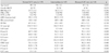
Mean values of the longitudinal strains, which were obtained from the apical 4-chamber view (LS4), 2-chamber view (LS2), long-axis view (LSL), and LSG, showed no differences among the three groups (Table 2). However, the mean values of the circumferential strains, which were obtained from the base level (CSbase), papillary muscle level (CSmid), apical level (CSapex) of the parasternal short-axis view, and CSG exhibited significant differences among the three groups (p=0.047, p=0.026, p=0.030, and p=0.019, respectively). The mean values were smallest in the normal group, and greatest in the elevated E/E' ratio group, except for the CSapex case. Furthermore, the CSbase, CSmid, and CSG varied in direct proportion to the increment in the E/E' ratio (r=0.259 and p=0.011; r=0.247 and p=0.015; r=0.242 and p=0.017, respectively).
The mean values of the radial strain obtained from the apical level (RSapex) were significantly different among the three groups (p=0.002), and showed a positive relationship with the E/E' ratio (r=0.291 and p=0.004). However, the mean values of the radial strain obtained from the basal level (RSbase) and papillary muscle level (RSmid), as well as the RSG, exhibited no statistical differences among the three groups. The mean values of the basal and apical rotations and LVtor exhibited no differences among the three groups.
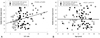
Adjustment with age
From the above results, the circumferential strains (CSbase, CSmid, and CSG) and RSapex were found to be related to the E/E' ratio. Since advancing age induces various physiologic changes in the LV myocardium, it is understandable that the above results would vary with age. After performing an adjustment for age (Table 3), the RSapex still had a positive relationship (Fig. 1) (r=0.304 and p=0.003); whereas, the CSbase, CSmid, and CSG did not maintain a relationship with the E/E' ratio. Instead, the circumferential strains were significantly related to age (CSbase: r=0.301 and p=0.003; CSmid: r=0.309 and p=0.002; CSG: r=0.380 and p=0.000). The basal rotation and LVtor also exhibited a relationship with age (r=0.344 and p=0.000; r=0.318 and p=0.001, respectively), but were not related to the E/E' ratio.
Discussion
Diastolic dysfunction is increasingly recognized in daily clinical practice as an important aspect of the pathophysiology of heart failure because heart failure can be anticipated in spite of the absence of decreased left ventricular EF. Diastolic heart failure is often presumed from the presence of symptomatic heart failure with a preserved EF that is over 50%, or alternatively, can be inferred from a raised serum brain natriuretic peptide with a preserved EF.5) The prevalence of diastolic heart failure relative to that of systolic heart failure increases with advancing age and myocardial hypertrophy, and is more common in females.6) In the present study, advancing age, left atrial enlargement, and increased LV mass, as well as a female preponderance, were frequently observed in the elevated E/E' ratio group but not in the normal group.
The hemodynamic characteristics of diastolic dysfunction are manifested as an elevated tangent (dp/dv) at any point on the LV pressure-volume curve during the diastolic phase, which is reflected by an increase in the pulmonary capillary wedge pressure.7-9) However, cardiac catheterization is limited by its invasiveness, even though it is ideal for assessing diastolic dysfunction. It is widely accepted that hemodynamic characteristics can be evaluated non-invasively by analyzing myocardial tissue motion and transmitral flow dynamics using Doppler.10-12) In the present study, the E/E' ratio was used to non-invasively assess impairment of diastolic function; the E/E' ratio value used in our study was validated in previous studies, and two cut-off values were applied-less than 9 for the normal group and more than 15 for the elevated E/E' ratio group.13-15) These values had a very low and high probability, respectively, for the presence of diastolic dysfunction.
In the study using TDI, Yu et al.16) reported that there was a gradual progression of a systolic abnormality as S' decreased in diastolic heart failure, and to a much lesser extent, in isolated diastolic dysfunction. In the above study, 52% of the patients with diastolic heart failure and 14% of those with isolated diastolic dysfunction had a reduced longitudinal systolic velocity. In the present study, S' had a tendency to be reduced according to the degree of E/E' ratio elevation; the longitudinal strain did not present any differences when the long-axis systolic function was assessed using 2D-STI. Vinereanu et al.17) reported long-axis systolic dysfunction in isolated diastolic dysfunction, in which the mean value of the E/E' ratio in the control was 5.8±1.4, which was much lower than the mean value of the E/E' ratio in the present study (7.5±1.0). Therefore, this heterogeneity of control values might explain why the longitudinal strain using 2D-STI did not reveal any differences with respect to the degree of E/E' ratio elevation, but S' did show a difference in the present study.
In a study using 2D-STI, Wang et al.18) reported that the longitudinal strain and radial strain were depressed in patients with diastolic heart failure when compared to control, but the circumferential strain and LVtor were preserved. When the present study was performed in patients who had diastolic dysfunction and a preserved LVEF but who did not have overt symptoms of heart failure, the radial strain at the apical level was found to be augmented in the elevated E/E' ratio group compared to the normal group. Other values for myocardial deformation and rotation did not show any differences according to the degree of E/E' ratio elevation, and the circumferential strain had a tendency to increase with advancing age as well as with LVtor. Takeuchi et al.19) observed that LVtor was significantly enhanced with advancing age in healthy volunteers because the subendocardial fibers weakened on counteraction against subepicardial fibers, which resulted in augmentation of LV myocardium rotational movement. The major difference between the present study and the study of Wang et al.18) was whether there were symptoms of heart failure. Therefore, we speculate that systolic function of the LV myocardium is relatively well preserved in isolated diastolic dysfunction rather than in diastolic heart failure, even though the EF seems similar in both.
Unexpectedly, we found that the radial strain at the apical level had a tendency to increase according to the E/E' ratio in the investigated subjects, but other values of radial strain did not show any differences. There are at least two explanations for this. 1) As a consequence of LV remodeling, the apex may be transformed somehow into a spherical shape, which results from an increased left ventricular end diastolic pressure. In turn, this change in apical shape can exaggerate the radial strain at the apical level because the myocardium is transected more tangentially, rather than vertically, by the echo beam. Even without considering LV remodeling, the closer to the cardiac apex the echo beam approaches in the parasternal short-axis view, the more tangentially the myocardium is transected. 2) In another explanation, which uses the myocardial single band model,20) the cardiac apex is a folded point as the descendant segment of the LV myocardial band becomes the ascendant segment, which is more likely to show an exaggerated bulging and shrinking motion than other parts of the LV myocardium. Matsuzaki et al.21) reported the non-uniformity of the thickening of both the inner and outer halves of the ventricular wall. Akagawa et al.22) observed that the inner half of the apical myocardium showed the most remarkable changes in wall thickness when dobutamine infusion was done or afterload increased. Thus, further study is necessary to determine whether augmentation of the radial strain at the apical level is a unique feature in isolated diastolic dysfunction.
Study limitations
Cardiac catheterization was not performed in the present study to estimate the severity of diastolic dysfunction because it was judged to be too invasive for subjects who did not have any symptoms of heart failure. The exact location of the basal and apical plan varied from person to person, which might result in measurement error.
In conclusion, except for the radial strain at the apical level, there were no differences in the values of LV myocardial deformation and rotation according to the degree of E/E' ratio elevation when there was no history of diastolic heart failure. However, the systolic mitral annular velocity decreased as a function of the severity of diastolic dysfunction. Additionally, in clinical situation such as isolated diastolic dysfunction, advancing age has a relatively greater influence on characteristics of LV myocardial deformation and rotation rather than on the E/E' ratio.


 XML Download