

 PDF
PDF ePub
ePub Citation
 Print
Print


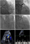
A 65-year old male was referred to the emergency room for an acute ST segment elevation myocardial infarction (ASTEMI) that occurred 5 hours previously. The patient complained of persistent chest pain after thrombolysis at the local hospital. An electrocardiogram (ECG) revealed ST segment elevation on the precordial leads (V2-V5) and the inferior leads (II, III, aVF). There was an increase in both the creatine kinase-MB fraction (256 µg/L) and cardiac troponin-I level (12.2 ng/mL). Based on these results an emergency coronary angiography was performed. The angiography revealed the presence of a total occlusion of the middle left anterior descending coronary artery (LAD) and a fistula originating from the proximal LAD to the main pulmonary artery (Fig. A and B). The patient had a small right coronary artery, a large LAD and a large left circumflex coronary artery. Therefore, a primary percutaneous coronary intervention (PCI) was performed in the middle LAD with a paclitaxel eluting stent (TAXUS, Boston Scientifics, Natick, MA, USA). No abnormalities were found in the distal LAD after the PCI (Fig. C). The follow up coronary angiography showed a fistula from the septal branches of the distal LAD to the right ventricle (RV) and mild aneurysmal changes at the stent site in the middle LAD after 9 months (Fig. D).1)2) The transthoracic echocardiography revealed a focal diastolic flow within the RV chamber (Fig. E and F). The patient had no discomfort. The presence of a fistula from coronary artery to RV after an ASTEMI and aneurysmal changes in a paclitaxel eluting stent are very rare. This may have developed as a result of myocardial necrosis after the ASTEMI and the left dominant coronary system may have contributed to the fistula from the distal LAD to the RV.3)4)
Figures and Tables
Fig. 1
The coronary angiography revealed a total occlusion of the middle left anterior descending coronary artery (LAD) and a fistulae originating from the proximal LAD to the main pulmonary artery (A and B). No abnormalities were observed in the distal LAD after the PCI (C). The follow up coronary angiography showed a fistulae from the septal branches of the distal LAD to the right ventricle (RV) and mild aneurysmal changes at the stent in the middle LAD after 9 months (D). The transthoracic echocardiography revealed a focal diastolic flow within the RV chamber (E and F). PCI: percutaneous coronary intervention.
References
1. Yu R, Sharma B, Franciosa JA. Acquired coronary artery fistula to the left ventricle after acute myocardial infarction. Am J Cardiol. 1986. 58:557–558.
2. Cheong ER, Park HS, Yang DH, et al. Clinical characteristics of the patients with myocardial rupture after acute myocardial infarction. Korean Circ J. 2002. 32:467–472.
3. Schanzenbächer P, Bauersachs J. Acquired right coronary artery fistula draining to the right ventricle: angiographic documentation of first appearance following reperfusion after acute myocardial infarction, with subsequent spontaneous closure. Heart. 2003. 98:e22.
4. Ryan C, Gertz EW. Fistual frome coronary arteries to left ventricle after myocardial infarction. Br Heart J. 1977. 39:1147–1149.
 XML Download