

 PDF
PDF ePub
ePub Citation
 Print
Print


Introduction
Restenosis has become the major therapeutic challenge for interventional cardiology since the introduction of percutaneous coronary intervention.1-5) Drug-eluting stents (DES) have dramatically reduced the risk of restenosis, and they have made a significant impact on the practice of interventional cardiology. Initial studies have shown that these devices reduce the target vessel revascularization rates to ~5% in simple lesions, and this rate approaches the target vessel revascularization rate of bypass surgery.1-5) However, restenosis remains a major clinical issue in real-world patients due to the complexity of many lesions. In addition, DES may increase the risk of late stent thrombosis due to delayed endothelialization, which results in the need for prolonged dual antiplatelet therapy.6) Further advances in stent technology may be required before a 'true' cure for coronary artery disease is established. The present review discusses the clinical, lesional and procedural predictors of angiographic restenosis after DES implantation, and this may help guide the practical use of DES.
Balloon Angioplasty
Restenosis is a major limitation of balloon angioplasty. A number of clinical and anatomic factors had been reported to be predictive of restenosis after percutaneous coronary balloon angioplasty.7-10) Diabetic patients have a much higher risk of restenosis than do non-diabetic patients, with the reported restenosis rates ranging from 50-70%. Lesion- and procedure-related factors, including ostial disease, diffuse disease, a heavy plaque burden and a small post-intervention lumen diameter, have also been reported to be predictive factors. However, the predictive power of these latter factors is very limited.
Atherosclerotic plaques can be compressed or stretched; this results in severe laceration during revascularization, and restenosis can result from such injury. Constrictive arterial remodeling rather than neointimal hyperplasia is a major mechanism of restenosis after balloon angioplasty.11)
Bare-metal Stents
Coronary stenting prevents arterial remodeling by placing mechanical scaffolding in the vessel. While the use of bare-metal stents has led to improved acute and long-term outcomes, this success has been limited by the development of restenosis.12)13) Several reports have evaluated the impact of baseline and procedural characteristics on the risk of subsequent restenosis after bare metal stent implantation, and a number of high-risk parameters such as diabetes, lesion length and vessel size have been consistently identified in most studies (Table 1).14-16) Intravascular ultrasound variables, including the in-stent area, the extent of the preexisting plaque, stenting of total occlusions and a history of diabetes mellitus, as well as implantation of a long stent, have all been shown to predict in-stent restenosis.17) In addition, the thickness of the stent strut has also been found to play an important role in the development of restenosis, with thin-strut stents reported to cause less neointima proliferation.18) Of these variables, the relationship between the final lumen size and restenosis has been well validated, leading to the principle of "bigger is better".19) However, the final lumen size predicted the occurrence of restenosis in only 30% of patients, and the disadvantage of a larger lumen is exaggerated neointimal hyperplasia. In contrast to restenosis after balloon angioplasty, in-stent restenosis is exclusively caused by intimal hyperplasia because stents prevent the remodeling process.20) Thus, the factors that affect neointima formation primarily influence in-stent restenosis, an observation which has led to the development of DES.
Drug-eluting Stents
DES has revolutionized the treatment of coronary atherosclerosis by dramatically reducing restenosis.1-5) After their initial success in stable patients with simple, de novo lesions, the use of DES has been extended to high-risk patients and complex lesions. The clinical impact of DES for treating small vessel disease, diffuse disease, multivessel disease or for patients with diabetes awaits further evidence from multicenter trials. While Food and Drug Administration(FDA) approval of DES applied to only a narrow patient population, so-called "off-label" use now accounts for at least 60% of DES implantion.6) Such use has lacked thorough analysis, and it may be associated with a higher risk of stent thrombosis, death or myocardial infarction compared to on-label use. In addition, the risk of restenosis increases with the wide, unrestricted use of DES. In a real-world population, the restenosis rate is not negligible, and identification of the patients at high risk for restenosis is still required to better guide therapy. Restenosis is a complex phenomenon that may be associated with a variety of stent-, drug-, patient- and lesion-related factors.
Stent-related factors
The stent delivery system is composed of polymer and the drug, and these are integral components of DES. Drugs should be evenly delivered if a stent expands, and a stent platform with regular strut spacing appears to be optimal for uniform drug delivery. Two drugs, sirolimus and taxol, have been extensively investigated as components of DES, and they have FDA approval for clinical use.2) Paclitaxel inhibits the assembly of tubulin into stable microtubules, which is essential for cellular division and cell migration, and both cellular division and cell migration are involved in the restenosis process. Likewise, sirolimus inhibits smooth muscle cell proliferation, matrix production and inflammation. Other drugs currently being tested include the rapamycin analogs everolimus and ABT-578, and these appear to be very effective in inhibiting restenosis. In addition to the drug type and polymer composition, the optimal dosing and release kinetics for drugs may also affect restenosis and vascular healing.
Two types of DES are currently widely used in clinical practice: the paclitaxel-eluting Taxus stent and the sirolimus-eluting Cypher stent. While both these DESs are durable and effectively prevent restenosis, there is ongoing debate as to the potential superiority of one device over the other.21) Several studies have compared the efficacy of the two DES when they are used as the primary therapy for coronary artery disease, and some conflicting observations have resulted. The REALITY trial22) is the largest prospective, randomized, multicenter comparison study of the two DES. The trial enrolled 1,353 patients with de novo native coronary lesions, and the primary endpoint was the in-lesion binary stenosis rate at 8 months. The trial found that the Cypher stent was associated with less late loss than the Taxus stent(0.09±0.43 vs. 0.31±0.44 mm, respectively, p<0.001). There was no difference between the stents in terms of the restenosis rate(9.6% vs. 11.1, respectively, p=0.31) and clinical outcomes(10.7% vs. 11.4, respectively, p=0.73). The SIRTAX trial23) was a randomized, 1,012-patient, single blind comparison of Cypher and Taxus stents. In contrast to the REALITY trial, the SIRTAX study found that the Taxus stent was associated with a higher major adverse cardiac event rate and a higher target lesion revascularization rate. Likewise, the ISAR-DIABETES randomized trial24) that compared Cypher and Taxus stents in 250 diabetic patients with de novo lesions found the restenosis rate associated with the Cypher stent was lower than that associated with the Taxus stent(6.9% vs. 16.5%, respectively, p=0.03). That study also showed that the two stents did not differ in terms of the target lesion revascularization rates(6.4% vs. 12.2%, respectively, p=0.13). We recently reported the results of the Long-DES II trial,25) which compared the use of Cypher and Taxus stents in 500 patients with long(≥25 mm) native coronary lesions. The study found that the Cypher stent was associated with a lower in-segment binary restenosis rate than the Taxus stent(3.3% versus 14.6%, respectively, relative risk: 0.23, p<0.001). The in-stent late loss of the lumen diameter was 0.09±0.37 mm in the Cypher group and 0.45±0.55 mm in the Taxus group(p<0.001). There was no significant difference between the stents in terms of the incidence of death or myocardial infarction after 9 months of follow-up. These findings suggest that for long native coronary artery disease, the Cypher stent is superior in terms of restenosis and target vessel failure.
Kandzari et al.26) compared the recently introduced zotarolimus-eluting stent with the sirolimus-eluting stent in terms of the relative clinical efficacy, the angiographic outcomes and safety in 436 patients with de novo native coronary lesions. The study found that the zotarolimus-seluting stent was associated with greater angiographic in-segment late lumen loss(0.34±0.44 mm vs. 0.13±0.32 mm, respectively, p<0.001) and in-segment binary angiographic restenosis(11.7% vs. 4.3%, respectively, p=0.04) compared with the sirolimus-eluting stent. The two stents were found to be similar in terms of clinically driven target lesion revascularization(6.3% zotarolimus vs. 3.5% sirolimus, p=0.34) and target vessel failure (12.0% zotarolimus vs. 11.5% sirolimus, p=1.0).
Overall, the current available data indicates that using the Cypher stent results in less late lumen loss than using the Taxus or Endeavor stents.21) These differences may reflect the metal platform design, the polymer and/or the pharmacological agents. The extent of late loss correlates with target lesion revascularization, and its influence on restenosis is related to the baseline risk of restenosis. Therefore, in terms of restenosis, while the Cypher stent may provide better outcomes than the Taxus or Endeavor stents for complex lesions that have a high risk for restenosis, such a difference may not exist in low risk lesion groups.
Patient-related factors
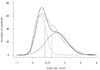
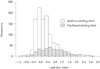
The frequency-distribution curves of the angiographic indices of restenosis after bare-metal stent placement have a bimodal pattern, indicating the existence of two distinct populations with different propensities for restenosis(Fig. 1). Kastrati et al.27) also reported that the risk of a lesion developing restenosis after stent implantation was 2.5 times higher if a companion lesion had restenosis, and this was independent of the analyzed patient risk factors.28) These findings suggest that unidentified patients factors may exert a critical influence on the development of restenosis after baremetal stent implantation. However, the pattern of angiographic late loss differs between thse lesions treated with DES and and the lesions treated with bare-metal stents. The distribution of late loss for the Cypher stent appeared largely skewed to the right with a Gaussian distribution.29) In addition, we found that late loss was higher in the Taxus stent group than in the Cypher stent group, demonstrating that late lumen loss tended to favor the Cypher stent over the Taxus stent(Fig. 2).30) However, it remains uncertain whether the likelihood of restenosis for a lesion is greater when a companion lesion has developed restenosis after DES implantation.
There are conflicting results from the previous reports regarding the effect of diabetes on restenosis after DES implantation.31-33) While some authors have concluded that diabetes is an independent predictor of restenosis, others have reported that diabetes per se may not be an independent risk factor for repeat revascularization. Local variables such as small vessels and diffuse disease may be more important for predicting restenosis than simply diabetes. However, only a relatively small number of diabetic patients, and even fewer insulin-dependent diabetics, have been studied, so further studies are required to ascertain whether diabetes is a predictor of restenosis in the DES era. Overall, the reports in the literature suggest that clinical variables are not strong predictors of which patients will or will not develop restenosis after DES implantation.
Procedure- and lesion-related factors
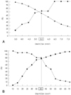
The post-intervention final lumen area has been documented to be the most powerful predictor of restenosis after both balloon angioplasty and bare-metal stent implantation. Several studies have shown that in patients receiving DES implants, the post-intervention final lumen area is the most important determinant of restenosis, suggesting that a greater stent area contributes to a decreased rate of restenosis, even in those patients with implanted DES.31) Intravascular ultrasound studies have revealed that the independent predictors of angiographic restenosis after Cypher stent implantation are the post-procedural final minimum stent area and the stented length of the artery. We have shown that the angiographic restenosis rate is highest in lesions with a stent area <5.5 mm2 and a stent length >40 mm(Fig. 3).33) Non-uniform strut distribution also contributes to intimal hyperplasia after Cypher stent implantation in de novo lesions, and this suggests a gap between the stent struts may be associated with a decrease in local drug delivery, which may then contribute to the development of restenosis.34) In addition, stent fracture is rarely related to very focal intrastent restenosis despite complete abolition of intimal hyperplasia in the remainder of the stented segment.35) Overall, the published reports have suggested that residual stenosis is a significant component of the restenosis problem, and this indicates that achieving a larger lumen area with adequate stent expansion remains an important strategy for reducing restenosis, even in the DES era.
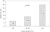
While the lesion length and stent length correlate with restenosis, lesion length is an independent predictor of restenosis.36) A small increase in the ratio of the in-stent length to the lesion length has a profound effect in reducing that margin effect. These findings indicate that stent length has less influence on restenosis when using DES compared with using bare metal stents,36)37) and this supports the current strategy of complete lesion coverage. However, a full metal jacket approach(stented length >60 mm) in small vessels has been linked with a high risk of DES failure(Fig. 4).38)
Conclusions
DES have been widely used as a revascularization strategy for a broad variety of clinical and anatomic situations, and restenosis remains a major clinical issue. Small vessel disease, diffuse disease and long stents have been shown to be predictive variables for restenosis after DES implantation. However, there is a poor correlation between these variables and the risk of subsequent restenosis. Further studies are required in order to identify the factors that can be used to reliably predict whether an individual patient will experience restenosis a fter DES implantation.

 XML Download