

 PDF
PDF ePub
ePub Citation
 Print
Print


Total knee arthroplasty (TKA) has been performed mainly for treatment of elderly patients who have severe degenerative arthritis with varus deformity. The varus knee is the most common deformity in patients presenting for TKA. A knee joint with severe varus deformity is followed by progressive contracture of the medial soft tissues and attenuation of the lateral soft tissues.1)
The goal of TKA is to obtain a balanced and well-functioning knee. To achieve this result, release of contracted tissues and removal of peripheral osteophytes are necessary to correct the deformity. For successful TKA, postoperative mediolateral (ML) soft tissue balancing is essential.2) A widely-used technique for the correction of varus deformity relies on the progressive release of the medial soft tissue until its length equals that of the lateral ligamentous structures.3)
Although in the majority of varus knees, medial release can be accomplished with a standard approach, a small percentage of primary knee arthroplasties will require additional sequential release of the medial soft tissues. In severe varus deformity, the release of superficial medial collateral ligament (MCL), semimembranosus tendon, posteromedial (PM) capsule, and pes tendons is required, leaving the medial side barren.45) This extensive direct medial soft tissue release increases the likelihood of overcorrection and may necessitate more extensive releases and the possible need for a constrained implant.67)
To overcome these shortcomings, the method of bone resection of the medial proximal tibia (MPT) with minimal medial soft tissue release can be considered as an alternative to avoid iatrogenic medial soft tissue injury.8) This bone resection technique may reduce the incidence of excessive soft tissue release required to balance the knee for coronal stability, minimizing the risk of medial overrelease. The authors hypothesized that bone resection of the MPT for soft tissue balancing would be an effective and safe method to obtain a well-balanced knee in TKA.
The purpose of this study was to investigate the effect of this present technique for ML balancing and the quantification of method of bone resection of the MPT as an alternative technique for achieving soft tissue balancing in TKA.
METHODS
Between September 2011 and March 2013, 88 primary TKAs were performed consecutively by a single surgeon (JHA) in patients with degenerative osteoarthritis of the knee. Inclusion criteria were patients who needed bone resection after initial minimal medial soft tissue release during TKA for degenerative arthritis with varus deformity with a minimum 2-year follow-up. Exclusion criteria were loss to follow-up for minimum 2 years, knee osteoarthritis with valgus deformity and no need for an additional bone resection technique during TKA. Ten cases were excluded due to valgus deformity (n = 5) and no need for an additional bone resection technique for balancing (n = 5). Finally, 78 TKAs performed in 60 patients (9 men and 51 women) were enrolled in the present study. The average patient age at TKA was 70.8 years (range, 59.1 to 83.5 years). The femorotibial angle was measured in the long leg anteroposterior view X-ray in standing position. Preoperatively, the average mechanical axis was 11.86° varus (range, 7.23° to 22.61° varus). In preoperative range of motion, the average flexion contracture was 4.92° (range, 0° to 12.61°) and the average flexion was 113.38° (range, 89.27° to 142.69°). In postoperative range of motion at the final follow-up, the average flexion contracture and average flexion were 1.82° (range, 0° to 5.63°) and 135.26° (range, 117.37° to 162.43°), respectively. All patients were retrospectively evaluated through measurement of the ML gap balance and thickness of the resected bone by using the present technique during TKA. The mechanical axis of the femorotibial angle was measured using the whole lower extremity radiograph in standing position preoperatively and at the final follow-up. The mean follow-up period was 32.6 months (range, 24 to 42 months).
Surgical Technique
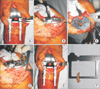
Posterior cruciate-substituting components were used in all cases (Genesis ll Oxinium; Smith & Nephew Inc., Memphis, TN, USA). The Genesis ll Oxinium is designed to have a thinner posterior medial condyle than the posterior lateral condyle of the femoral component to compensate for the trapezoidal flexion space. This maintains the joint line perpendicular to the long axis of the tibia. Under tourniquet control, a standard midline skin incision and a mid-vastus approach was used in all cases. The peripheral osteophytes of the distal femur and proximal tibia were removed and then the medial release of deep MCL and PM capsule was performed without superficial MCL release. The distal femoral resection was performed at 6° of valgus, and the external rotation of the femoral cuts was determined with a plane parallel to the femoral posterior condyle by using the measured resection technique. Periosteal stripping for deep MCL release was not beyond 2 cm from the joint line distally in all cases. The position of the proximal tibial cut was determined using an intramedullary instrument, with a 4° posterior slope and a 2 mm resection referenced off the medial tibial plateau. Both cruciate ligaments were removed for a posterior cruciate-substituting system. The femoral component was then trialed and ML balancing was assessed. After femoral component trial insertion with the knee extended and flexed, we measured the ML gap using a Gapper bi-compartmental ML gap measurement device (B. Braun, Melsungen, Germany) to achieve ML soft tissue balancing (Fig. 1A). The medial and lateral gaps were distracted by Scott Spreader (Innomed Inc, Savannah, GA, USA) with the same tension. If there was reduced medial gap balance, we first positioned the trial tibial component as laterally as possible (Fig. 1B). In the second step, we performed bone resection of the MPT vertically along the tibial long axis with a step-by-step resection technique using 2 mm thickness (Fig. 1C). After bone resection (Fig. 1D), the same procedure was performed after measuring the ML gap using the Gapper device and spreader. No additional medial soft tissue release, including the superficial MCL and the pes tendon group, was required, and a balanced, stable TKA with less than 2 mm ML gap imbalance was achieved in all cases (Fig. 1E). After achieving a balanced knee, we estimated the total thickness of the resected bone by calipers (Fig. 1F).
Correlation between the Thickness of MPT Bone Resection and ML Gap Change
Intraoperatively, the ML gap balance was measured using the Gapper device after initial deep MCL release and 2 mm step-wise MPT resections until the ML gap balance was within 2 mm in knee extension and flexion. The patella was not everted in all measurements, but just subluxated laterally. We measured and calculated the total thickness of MPT resection measured by calipers and the ML gap change during knee extension and flexion. For analysis of bivariate correlations between thickness of bone resection and ML gap change during knee extension and flexion, we used Pearson's test with significance set at p < 0.05. Statistical analysis of the data was performed by IBM SPSS ver. 21.0 (IBM Co., Armonk, NY, USA).
Quantification of the Effect of MPT Resection on ML Gap Change
To quantify the effect of MPT resection thickness, we tried to calculate the correlation between the two variables, total thickness of bone resection and ML gap change, during knee extension and flexion. We used simple linear regression without a constant to predict the bone resection thickness required to widen the gap for equal balance within 2 mm.
Radiologic and Clinical Evaluation
The mechanical axis of the femorotibial angle was measured on the whole lower extremity radiograph in standing position preoperatively and at the final follow-up. The change in mechanical axis was measured and the correlation between the thickness of resected MPT and the change in the mechanical femorotibial axis was evaluated statistically. Clinical assessment was performed using the Knee Society clinical rating system preoperatively and at the final follow-up.
RESULTS
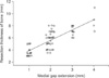
We analyzed the correlation between the thickness of bone resection and ML gap change during knee extension and flexion (Table 1). The average thickness of bone resection was 8.25 ± 1.92 mm. During knee flexion, the average preresection medial gap was 11.49 ± 1.93 mm and the average preresection lateral gap was 12.91 ± 1.80 mm. During knee extension, the average preresection medial gap was 8.94 ± 1.69 mm and the average preresection lateral gap was 12.79 ± 1.70 mm. In the flexion gap, 10 of the 78 cases had a ML gap imbalance beyond 3 mm. In the extension gap, all 78 cases had ML gap imbalance beyond 3 mm. After the vertical resection of MPT bone to balance the ML within 2 mm, the average postresection medial gap was 12.35 ± 1.92 mm and the average postresection lateral gap was 13.00 ± 1.69 mm during knee flexion. During knee extension, the average postresection medial gap was 11.88 ± 1.67 mm and the average postresection lateral gap was 12.85 ± 1.64 mm. The average flexion-medial gap change was 0.85 ± 1.00 mm, the average flexion-lateral gap change was 0.08 ± 0.61 mm, and the average extension-medial gap change was 2.94 ± 0.87 mm and the average extension-lateral gap change was 0.05 ± 0.58 mm. The correlation between the thickness of the resected bone and the change in medial and lateral gaps in flexion and extension was analyzed. The thickness of MPT resection correlated significantly with the changes in medial gap during knee extension (r = 0.695, p = 0.000). In the other gap changes, there was no significant correlation with the thickness of resected bone. Even in the medial gap change in flexion, there was no statistical significance (r = 0.214, p = 0.059). To quantify the effect of MPT resection on the femorotibial gap change, a correlation equation was formulated using simple linear regression without a constant in extension. Medial gap change displayed a statistical correlation. The correlation equation between the two variables was Y = 2.80X (R2 = 0.64) (Fig. 2). When the medial tibia was resected at an average thickness of 8.25 ± 1.92 mm, the medial gap during extension was increased by 2.94 ± 0.87 mm. In simple linear regression without a constant, MPT resection at a thickness of 2.80 mm was predicted to be necessary to increase the medial gap by 1.00 mm during knee extension.
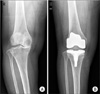
The mean mechanical axis after bone resection changed from 11.86° ± 3.47° varus to 1.31° ± 2.46° varus, and the mean variation was 10.55° ± 2.74° (Fig. 3). No significant correlation was identified between the thickness of resected MPT and the change in mechanical femorotibial axis (r = 0.111, p = 0.335).
The mean Knee Society knee score improved significantly from 56.3 points (range, 25 to 70 points) preoperatively to 89.4 points (range, 80 to 100 points) at the final follow-up (p = 0.000). In the enrolled TKAs, there were no postoperative complications including instability and periprosthetic infection at the final follow-up.
DISCUSSION
It is widely accepted that appropriate ligament balance is a cornerstone for good function and survival after TKA. However, it is problematic that there is no consensus on the gold standard for attaining a ML balanced knee during the arthroplasty procedure. Several principles of medial release for achieving ligament balance during TKA have been developed, but they do not clarify the definite and generalized surgical method.9101112) The present study had two purposes. The first purpose was assessment of the possibility of MPT bone resection technique as a reasonable alternative method for ML balancing in knee arthroplasty. The second purpose was quantification of the method of bone resection by calculating the coefficient ratio between the thickness of the resected bone and the change in the femorotibial gap.
In TKA, the conventional method for achieving coronal plane stability is the release of medial soft tissue including distal attachment of the superficial MCL. The sequence of the medial soft tissue release method was reported by several authors with some variations.10131415) But, the adequate amount of soft tissue release has not been extensively studied.16) The proper amount of medial release for achieving a balanced knee is dependent on a surgeon's subjective experience and decision, and not on an objective guideline.1718192021) A definite disadvantage of the medial soft tissue release technique is the risk of medial over-release that may result in coronal plane instability after knee arthroplasty.22) The coronal plane instability after medial over-release may be the critical cause of early failure of knee arthroplasty due to component loosening and wear.2324) This disastrous complication stems from the absence of an objective guideline for the procedure of ML soft tissue balancing.2526)
We estimated the statistical correlation between the width of the resected bone and the change in ML gap and knee alignment. We expected that the medial gap would increase according to the amount of resected bone thickness; the correlation between the two factors was proven statistically. The change in mechanical knee axis did not have a significant correlation with the thickness of resected MPT. The mechanical axis is a static image which does not reflect the reducibility of varus deformity and this may be the reason why the mean mechanical axis did not show a significant correlation with the thickness.27)
The limitation in the first topic of this study was the possibility of variations in the first step of soft tissue balancing. The amount of initial minimal capsule release may affect the change in perioperative ML balancing. To prevent the unexpected effect of initial medial soft tissue release, we limited the extent of initial medial release within 2 cm. In all cases, the minimal periosteal release of medial capsule including the PM capsule was performed as the first step and minimal release was limited within 2 cm from the knee joint line before the method of bone resection of MPT.
In the second topic, we attempted to derive a coefficient ratio to explain the quantified relationship between the thickness of resected bone and the measured gap change. In medial gap change during extension, the coefficient ratio was 2.80, which indicated a 1.00 mm medial gap increase in knee extension and the need for bone resection of the MPT at a thickness of 2.80 mm. On the basis of this quantification, the MPT bone resection technique could be applied in the clinical practice for achieving ML balancing during knee arthroplasty. Using the quantified coefficient 2.80, the complications such as medial overrelease and coronal plane instability could be avoided. We did not consider the thickness of the saw blade, which may be a limitation in the calculation of the quantified relationship in our present study.
As an alternative method for the conventional medial soft tissue release technique, the multiple needle puncturing pie-crust method has been reported.928) But, none of the studies have assessed how many repetitions are needed for achieving adequate balancing for the quantification of the pie-crust technique. The strength of our present study is the obtainence of a coefficient ratio by quantification between medial bone resection technique and ML gap change. Engh and Ammeen29) reported excellent results with medial epicondylar osteotomy as another alternative method, but this technique was more difficult and complex in which bone union at the osteotomy site was confirmed postoperatively.
The overall limitation of our study was the limited effect on medial gap change in knee flexion. After initial release of PM capsule and femoral cut, the average difference in ML gap in flexion was 1.42 mm (range, 0 to 4 mm), and only 10 of the 78 cases had a ML gap imbalance beyond 3 mm. The reason why there were few cases with ML gap imbalance beyond 3 mm in flexion than in extension may be the typical design of the femoral implant of Genesis ll. Heesterbeek and Wymenga12) reported the effects of releasing medial structures including the superficial MCL, semimembranosus tendon and PM capsule were the greatest for the extension gap; this result is in contrast with the previously published studies that reported greater effects in flexion than in extension with distraction of the gaps. In the study by Heesterbeek and Wymenga,12) when the first released structure was the superficial MCL, the release of this structure led to 4° median change in alignment in extension and 1.5° median change in femoral rotation in flexion. In our study, the average change in medial gap in extension and flexion was 2.94 mm and 0.85 mm, respectively. In these two studies, the ratios of medial gap changes in extension and flexion were 2.66 (4°/1.5°) and 3.45 (2.94/0.85 mm). During TKA, the position of tight medial soft tissue in knee flexion may be shifted backward with limited contact with the MPT after medial release of the deep MCL and PM capsule, compared with that in knee extension. This can be the possible explanation for the limited effect on medial gap change in knee flexion in our present study. In a previous study that assessed the effect of removal of femoral osteophytes on releasing the MCL, Pottenger et al.30) showed that varus-valgus angle increased by 3.6° for each centimeter of ipsilateral femoral osteophyte width removed with sufficient force to obtain a firm end point. The position of the patella during measurement of the femorotibial gap was also a limitation since patellar positioning may affect knee joint gap measurement. To deal with this limitation, we performed gap measurement with patellar lateral translation without patellar eversion to minimize the effect of patellar position on gap measurement.
Ahn and Back31) reported a comparative study between medial soft tissue release and bone resection of the proximal medial tibia with a technique similar to our technique with better ML balance during TKA. Dixon et al.8) reported the correction of severe varus deformity in TKA by tibial component downsizing and resection of uncapped proximal medial bone in 12 knees of 10 patients, which was similar to the surgical method used in our study. But, they applied the technique only in severe varus knee with deformity exceeding 20°, and in all the cases they performed downsizing and lateralizing of the tibial component with subsequent removal of the proximal medial tibia flush with the downsized component. In our study, we applied our surgical method to the knees with varying degrees of varus deformity the average mechanical axis before TKA was 11.86° varus (range, 7.23° to 22.61° varus) in 78 knees, and usually did not perform downsizing of the tibial component, except in two cases with downsizing of the tibial component for increased bone resection width. We lateralized the tibial component and resected the uncapped proximal medial bone to achieve balance without downsizing the tibial component in most cases. We attempted to develop a simple and practical surgical technique that could be applied to knee arthroplasty in most varus knees. There was a possibility of femoraltibia component size mismatch with downsizing of the tibial component in a previous study,8) while in our present study, there was no component size mismatch, even in the two cases with downsizing of the tibial component. Particular attention should be paid to the safety of size mismatching during TKA with a procedure similar to that used in our study.
The significant benefits of vertical bone resection are achieving ML soft tissue balance through vertical bone resection of the MPT without releasing the superficial MCL, and predicting the ML gap balance with vertical bone resection of the MPT.
In conclusion, The method of bone resection of the MPT can be considered effective with predictable results for achieving soft tissue balancing in terms of quantification during TKA.

 XML Download