

 PDF
PDF ePub
ePub Citation
 Print
Print


The association between spondylolysis and spondylolisthesis is well known. Of the 5 types of spondylolysis, isthmic spondylolysis is the most common to present symptomatically, having a reported incidence of 3% to 6% in the general population. The majority of defects (85% to 95%) occur at L5, with much of the remainder occurring at L4. On the other hand, spondylolytic defects involving multiple vertebral levels are extremely rare. Park et al.1) have reported an asymmetric four level lumbar spondylolysis. Here, we present a case of isthmic spondylolysis with consecutive lower lumbar levels of T12 to L5, and our approach of treating him as multilevel spondylolysis.
CASE REPORT
A 24-year-old male has presented to our outpatient clinic with persistent back pain since several weeks. He had no history of back injury or other medical illness, and the pain was aggravated by physical activities. On initial evaluation, the visual analogue scale (VAS) score was 7 points. Physical examinations revealed decreased range of lumbar spinal motion, resulting in pain. The straight leg test was negative on both sides. No abnormality in sensory distribution was observed. Motor strength was 5/5, reflexes were equal and symmetric, and all other examined parameters were within normal limits.
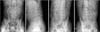
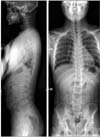
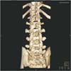
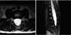
Anteroposterior (AP), neutral lateral, and both the oblique views (Fig. 1) revealed Meyerding grade 1 spondylolytic spondylolisthesis at L4 on L5, with mild dome-shaped superior endplate and consecutive multilevel spondylolysis at T12-L5. Standing AP and lateral views of the whole spine (Fig. 2) revealed normal balance of sagittal and coronal alignment. A computed tomography scan revealed bilateral spondylolysis at T12-L4, left unilateral spondylolysis at L5, and spina bifida at L5 to sacral region (Fig. 3). Magnetic resonance imaging revealed mild dural ectasia at the lower lumbar region (Fig. 4).
Without any accompanying neurological symptoms, conservative management was employed. The patient was rested a few weeks with a corset brace, and physiotherapy was started. After 4 weeks after the conservative treatment, the patient showed an improvement in the back pain, the VAS score decreased from 7 to 2, and the brace was discontinued, and he was able to return to normal activity.
DISCUSSION
Based on reported incidences at the time, Ravichandran2) calculated that only 1.48% of patients with back pain were diagnosed with upper lumbar spondylolysis. He concluded that although trauma is an important factor in producing multiple pars defects in a single patient, genetics may also play a role. A higher rate of multilevel spondylolysis (as well as single-level spondylolysis) has been found in native Alaskans.3) It is widely accepted that spondylolysis has a genetic predisposition since there is a difference in the incidence among races, and familial aggregation. However, since radiographic studies of newborns have shown the incidence to be 0%, it proves that this is clearly not a congenital defect. In our case, the patient had no family medical history and no spinal trauma.
Spondylolysis is known to occur less frequently in the upper lumbar spine, but this is commonly seen in cricket fast bowlers as well as in gymnasts.4) It is known that the thickness and strength of pars interarticularis increases in the lower lumbar spine depending on the size, and that the extension and flexion forces produce a maximum shear stress at the L5 level. Therefore, the rarity of the spondylolysis of the upper lumbar spine has been considered to be due to the lower mechanical stress in this area. Sairyo et al.5) reports multilevel spondylolysis may occur due to genetic and not biomechanical reasons since the biomechanical stress concentration at the L4 pars may not be the main cause of the newly developed L4 spondylolysis following L5 spondylolysis. It is interesting that our case shows more progressive spondylolysis in upper lumbar spine than lower, i.e. bilateral spondylolysis of L4 and unilateral spondylolysis of L5.
Pelvic incidence is significantly higher in isthmic spondylolisthesis patients as compared with controls, but was not clearly correlated with the grade of slipping. Increased lumbar lordosis associated with spondylolisthesis is secondary to the high pelvic incidence and is an important factor causing high shear stresses at the pars interarticularis. Differential stresses on the anterior versus the middle and posterior parts of its growth plate could modify the bone growth rate distribution, according to the Hueter-Volkman principle; this may lead to a doming of the superior endplate.6) Our patient had normal sagittal and coronal alignments with Meyerding grade 1 spondylolisthesis, and mild dome shaped superior end plate of L5.
Spina bifida is the most common neural tube defect, resulting from the failure of the fusion of the vertebral arches. Blackburne and Velikas4) found that the risk of a progressive vertebral slip was greater in patients with a midline lumbosacral defect. The presence of dysplastic or deficient posterior elements in the spina bifida defect may put an increased load on the pars. Studies have shown that the incidence of spondylolysis was significantly higher in patients with spina bifida occluta (SBO) than in those without SBO.7) However, Sakai et al.7) reported that SBO does not alter lumbar biomechanics with respect to stress and range of motion. He concluded the high coincidence of spondylolysis in spines with SBO may not be due to the mechanical factors. During his 10 year of study, Mesfin et al.8) also reported that there was no development or progression of spondylolisthesis or spondylolysis, and no increase in dural ectasia size in patients with dural ectasia and Marfan syndrome. Our patient had a dural sac ratio (dural sac diameter/vertebral body diameter) of 0.58.
The optimum treatment for multilevel spondylolysis with spondylolisthesis remains controversial. Historically, symptomatic spondylolysis or low-grade spondylolisthesis have been treated non-operatively by decreasing physical activities, strengthening of the back and abdominal muscles, and sometimes the requirement of a brace. Activity modification includes cessation of sports activities and treatment with non-steroidal anti-inflammatory agents, combined with an exercise regimen aimed principally at the reduction of lumbar lordosis as well as at the treatment of hip flexion and hamstring contracture.9) Park et al.10) reported a multiple spondylolysis case was treated surgically by posterior and posterolateral fusion at L2-3-4 with intersegmental fixation using pedicle screws and an auto iliac bone graft.
To conclude, we report here an unusual case about multilevel spondylolysis with spondylolisthesis and spina bifida. At the latest follow-up, his lower back pain had resolved and he returned to normal activity. We, therefore, think that conservative management should be considered a viable way to treat multilevel spondyloly with low-grade spondylolisthesis. Further studies, involving a larger sample of patients, are required to analyze this approach in treating multilevel spondylolysis.
 XML Download