

 PDF
PDF ePub
ePub Citation
 Print
Print


The mechanism of injury for mallet fractures is usually hyperflexion or axial loading of the distal interphalangeal (DIP) joint. Displaced mallet fractures are known to cause extension lag and swan neck deformities.1) Surgical and non-surgical methods have been used to treat mallet fractures. As non-surgical treatments may cause secondary degenerative arthritis, loss of movement, and poor cosmetic outcomes, accurate reduction of the articular surface and stable fixation by surgery have been recommended.2,3) Indications for surgery are a mallet fracture involving more than one-third of the articular surface or palmar subluxation of the DIP joint.4)
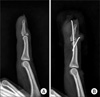
Surgical methods include DIP joint pinning,5) tension band wiring,2,3,6) extension block pinning,7-10) compression pin fixation, and screw fixation.11,12) Among these, extension block pinning, introduced by Ishiguro et al.,8) is an effective and convenient technique that is commonly used. However, extension block pinning performed more than five weeks after injury cannot completely reduce fractures.8) The range of motion of the DIP joint is also likely to be limited because a wire penetrating the joint does not allow for joint motion for several weeks and it can also injure the articular cartilage.8) Moreover, this technique is an indirect reduction method which involves inserting a wire into the dorsal aspect of the DIP joint and extending the distal phalanx to reduce a bony fragment. This does not allow for a complete anatomic reduction in some cases, which causes minimal postoperative pain (Fig. 1). For this reason, we consider that a modified technique has become necessary for cases in which extension block pinning does not achieve a complete reduction of mallet fractures and the intrafocal pinning technique using the first K-wire as a hinge to compress the fracture fragment can produce a more accurate reduction of the mallet fracture.
We applied the intrafocal pinning to compress the bony fragments for mallet fractures and obtained anatomic reduction in cases where extension block pinning alone did not anatomically reduce fracture fragments. We report the details of this operative technique and its efficacy.
METHODS
Among 52 patients who underwent operations for mallet fractures between March 2004 and August 2009, 14 digits of 14 patients for whom extension block pinning alone could not achieve anatomic reduction of fracture fragments were treated using the intrafocal pinning technique and extension block pinning concurrently. There were eight men and six women with an average age of 34 years (range, 15 to 53 years). The right hand was involved in five cases and the left hand in nine cases. Five injuries occurred in the dominant hand. The fingers affected were five small fingers, four middle and ring fingers, and one index finger. The injury mechanisms included slip downs in six cases, being struck by a ball in three cases, bumping up against a wall in four cases, and a crush injury in one case. There were no compound fractures or open fractures. The average articular surface involvement was 52% (range, 40 to 70%). Subluxation of the distal phalanx was observed in ten of the 14 fractures (71%). The fractures were classified by the Wehbe and Schneider1) method. There were four type IB, nine type IIB, and one type IIC fractures. The mean time from injury to surgery was 23 days (range, 3 to 62 days) broken down into the following groups: within 10 days in four cases, between 11 and 30 days in six cases, and over 31 days in four cases. The average follow-up was 16 months (range, 6 to 38 months). The congruity of the articular surface and the degree of anatomic reduction were confirmed on plain radiographs taken postoperatively and at the last follow-up. Functional outcomes were determined by using Crawford's classification.13) Indications for surgery included a displaced large fragment involving more than one-third of the articular surface or fractures associated with palmar subluxation of the distal phalanx. Inaccurate reduction was defined as the fracture gap or the joint step off was more than 0.5 mm.
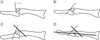
All procedures were performed under digital block anesthesia. Under C-arm image intensifier control, extension block pinning was attempted as described by Ishiguro et al.8) If anatomic reduction of the fracture fragment and congruity of the articular surface were not observed on plain radiographs, the inserted K-wire was removed as it prevented the intrafocal pinning. Next, the intrafocal pinning technique and extension block pinning were performed concurrently. First, a 0.7 mm K-wire was inserted dorsally, aiming at the fracture site with the DIP joint in the neutral position. When the K-wire reached the fracture site, it was pushed slightly forward (Fig. 2A). Using the distal cortical bone of the fracture site as a hinge, the outside of the K-wire was tilted proximally to 40-45°, compressing the fracture fragment towards the palmar side. Next, the K-wire was passed through the palmar cortical bone (Fig. 2B). The DIP joint was then flexed to 30° and a 0.9 mm extension block K-wire was inserted into the head of the middle phalanx at 45° (Fig. 2C). The distal phalanx was extended to obtain reduction of the fracture fragment. After reduction was verified by image intensifier control, a 0.9 mm K-wire that crossed the DIP joint was inserted to fix the joint (Fig. 2D).
After surgery, an aluminum splint was applied to fix the DIP joint while active motion of the proximal interphalangeal joint and metacarpophalangeal joint was encouraged. Pin disinfection was performed twice a week. Fracture union was defined as bridging trabeculae and a nontender DIP joint on plain radiographs. When bone union was achieved, the K-wires were removed and passive and active exercises of the DIP joint were encouraged.
RESULTS
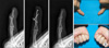
There was no cortical breakage of the distal phalanx, pull out of additional intrafocal K-wire, or displacement of the fracture fragment during follow-up. According to Crawford's classification, nine patients had excellent results, five had good results, and none had fair or poor results. The DIP joint demonstrated a mean range of 78° (range, 70 to 85°) in flexion and 1.8° (range, 0 to 5°) of extension loss (Fig. 3). Bone union was achieved in all cases by a mean postoperative day of 38.4 (range, 29 to 43 days). In one case, infection occurred at the pin site at postoperative week five, which was resolved by pin removal and dressing. Other complications, such as dermal necrosis, fractures of bone fragments, and nail-plate deformities, were not observed.
DISCUSSION
Treatment methods for mallet fractures are divided into closed and open fixation techniques. Open fixation is technically challenging due to the small sizes of the fracture fragments and the fact that the articular surface of the DIP joint is not easily observed. Additionally, complications, including early avascular necrosis, nail growth deformities, soft tissue scar formation, infection, implant failure, and subsequent joint stiffness, may occur.2,3,10) Percutaneous pinning was developed in an attempt to reduce the risk of complications from open fixation and still obtain anatomic reduction of the fracture fragments. The extension block pinning technique is an effective and convenient technique that is widely accepted and it gives good results.2,7,8,10)
Although extension block pinning is an easy and reliable method, when the dorsal fragment is large, markedly displaced, or rotated, anatomic reduction of the fracture is difficult to obtain and the fragment can sometimes be displaced during follow-up exams. Hofmeister et al.9) retrospectively reviewed 24 fractures treated with an extension block pin and reported there were 2 instances of slight displacement of the reduction. Various modified methods to improve the accuracy of reduction and stability of fixation have been suggested. One modified technique involved the first K-wire inserted proximal to the fractured fragment in a 45° proximodistal direction and then tilted 90° distally to reduce the fracture fragment.14) Another is the anatomic reduction of the fracture fragment using two extension blocking K-wires.15) Hofmeister et al.9) have reported an anatomic reduction by manipulating the dorsal translation of the DIP joint during extension block pinning. Ishiguro et al.8) have recommended inserting the tip of an injection needle into the dorsal aspect of the fracture to freshen the fracture surface when fractures are greater than two weeks old.
We have used the extension block pinning as a primary method of the reduction for mallet fractures since 2004. Of 52 mallet fractures, 14 were not anatomically reduced with extension block pinning. We analyzed the 52 mallet fractures to determine which factors have prevented the anatomic reduction in extension block pinning for the mallet fractures, and found that anatomic reduction was more challenging when the time from injury to operation was longer, the subluxation of the DIP joint was present, the gap of the fractured site was larger, and the fracture was dislocated to the distal site of fracture fragments (p < 0.05). Therefore, for reasons of inaccurate reduction during extension block pinning, we considered the organizing fracture hematoma, insufficient manipulation, or rotation of the fracture fragment that cannot be corrected by extension block pinning. To achieve a more accurate anatomic reduction in 14 cases, we additionally attempted intrafocal pinning aimed at compressing the fractured site and observed satisfactory reduction in all the 14 cases, including 3 with fractures older than 6 weeks. All the cases demonstrated good or excellent results at the last follow-up. We suggest that the additional intrafocal pins inserted at the fracture site compressed the distal side of the fragment, which prevented its rotation by the terminal tendon, thereby achieving a better reduction and maintenance. Theoretically, an intrafocal pin may produce the proximal migration of dorsal fragment because of the thickness of the pin if the pin is inserted to the fracture gap. However, it probably does not matter because the inserted pins are thin.
Using two extension block pins, Lee et al.15) reported transient nail ridging after vigorous manipulation or repetitive reduction maneuvers in patients with marked displacement of fracture fragments or older fractures of more than three weeks after injury. In our series, nail-plate deformities were not observed, likely because anatomic reduction was obtained without vigorous manipulation and the K-wire used in the intrafocal pinning technique is only 0.7 mm thick and is inserted into the proximal side of the nail bed.
We recommend the additional intrafocal pinning technique as an alternative and useful method to obtain anatomic reduction of mallet fractures in cases where extension block pinning alone is insufficient to restore the anatomic configuration of the articular surface.
 XML Download