

 PDF
PDF ePub
ePub Citation
 Print
Print


INTRODUCTION
Breast cancer is a heterogeneous and complex genetic disease that is characterized by altered expression of several coding and noncoding genes. Despite recent advances in early detection and therapies, breast cancer is still the leading cause of cancer related death among women worldwide. For that reason, unraveling the molecular mechanisms underlying the initiation and progression of breast tumors may lead to promising translations towards better diagnoses, prognoses, and targeted therapies [1].
The accumulation of a variety of genetic and epigenetic alterations in somatic cells can drive tumorigenesis in different tissues. Among the genetic changes, gene amplification and deletions are most frequently observed in several kinds of cancers. Specifically, the amplification of oncogenes and deletion of tumor suppressor genes have been investigated in genome-wide analyses, which have revealed some of the mech-anisms of carcinogenesis. Similar to other cancers, breast cancer is initiated by a progressive accumulation of genetic and epigenetic aberrations [23].
Loss of heterozygosity (LOH) on the short arm of chromosome 17 (17p) is a frequent event in breast cancer, where deletions in 17p are observed in 49% of breast cancers [2]. Particularly, in several LOH studies, a high frequency of deletions in two distinct regions of 17p (17p13.1 and 17p13.3) have been reported. The p53 tumor suppressor gene is located on chromosome band 17p13.1, and it is thought that the 17p13.3 region encodes other tumor suppressor genes with potential regulatory roles in tumorigenesis [245].
Three microRNAs (miRNAs), namely miR-22 and the miR-212/132 family, have been identified in the chromosomal region 17p13.3, and are involved in normal breast tissue development. These miRNAs have vital roles in normal developmental processes such as epithelial-stromal interactions and expansion of mammary progenitor cell populations [678]. miRNAs are a new class of endogenous, small (19–25 nucleotide length), noncoding RNAs, which posttranscriptionally regulate gene expression. miRNAs are evolutionarily conserved, and are involved in several developmental and cellular processes including the cell cycle, cell proliferation, and tumorigenesis [9]. The discovery of miRNAs has also led to new strategies for disease diagnosis and therapy [10].
miRNAs exert their functions primarily by targeting the 3' untranslated region of their target messenger RNAs (mRNAs), by which they induce translational silencing, either by cleaving the mRNA or blocking translation. Therefore, miRNAs may act as either tumor suppressor genes or oncogenic miRNAs (oncomiRs) in various types of cancers [1112]. Several studies have revealed that there is aberrant expression of miRNAs in breast cancer initiation and progression. Moreover, several miRNAs have been considered as potential tumor markers for breast cancer [1314].
In the present study, we evaluated the expression of miR-22, miR-132, and miR-212 in tumor and nontumor breast tissues. Moreover, we investigated whether expression alterations in these miRNAs were correlated with the malignant state of the tumors.
METHODS
Human clinical samples
Formalin-fixed paraffin-embedded (FFPE) tissues were obtained from the pathology center of Shariati Hospital in Tehran, Iran. The tissues were collected from female patients with invasive ductal carcinoma as determined through evaluation of histopathological parameters according to the World Health Organization (WHO) criteria for histologic grade and the TNM system for stage classification. A total of 36 tumor samples and 36 matched nontumor samples from the same patients were obtained for this study. This study was approved (#91715) by the Ethics Committee of Tarbiat Modares University, Tehran, Iran.
RNA extraction, complimentary DNA synthesis, and real-time polymerase chain reaction
RNA extraction from FFPE samples can be troublesome because of different chemical modifications and degradation over time. Specifically, in FFPE samples, the RNA is heavily degraded during the fixation process, fragment lengths vary from single bases to several hundred bases, and thus the yield of RNA extracted from FFPE tissues is much lower than that from fresh tissues. Paraffin from FFPE tissue sections was removed by the xylene-ethanol method, and then total RNA was isolated using TRIzol Reagent (Invitrogen, Paisley, UK) according to the manufacturer's instructions. The RNA yield and A260/280 ratio were determined by a NanoDrop spectrophotometer (Thermo Fisher Scientific, Waltham, USA). Subsequently, the samples were treated with RNase-free DNase to remove possible traces of genomic DNA. Complimentary DNA (cDNA) synthesis was then performed on 2 µg total RNA using the MiR-Amp Kit (ParsGenome, Tehran, Iran), in which the polyA tailing method with syn primers was used to synthesize specific cDNA from miRNAs. Briefly, the samples were incubated for 10 minutes at 37℃ to form the polyA tails. Next, the samples were incubated for 60 minutes at 43℃. Lastly, heat-inactivation of reverse transcriptase was carried out by incubation for 1 minute at 85℃.
Real-time polymerase chain reaction (RT-PCR) was performed using 1 µL cDNA product, miR-specific primers, and EvaGreen dye (Biotium, Hayward, USA). The 5s rRNA gene was used as an internal control. RT-PCR was carried out in an ABI-7500 real-time quantitative PCR instrument (Applied Biosystems, Foster City, USA) with the following conditions: 95℃ for 15 minutes, followed by 40 cycles of 95℃ for 15 seconds, 63℃ for 25 seconds, and 72℃ for 35 seconds. All RT-PCR amplifications were performed in duplicate, and a negative control was included for each reaction for quality control.
Product verification
The authenticity of the amplified RT-PCR products was evaluated by the following two methods: agarose gel electrophoresis and DNA sequencing. Due to the small length of miRNAs, cloning is necessary for accurate sequencing, and thus TA cloning was carried out using the InsTAclone PCR Cloning Kit (Fermentas, Hanover, USA) prior to sequencing.
Statistical analyses
The reaction efficiencies for each primer pair were determined by LinRegPCR software (Academic Medical Centre, Amsterdam, The Netherlands). RT-PCR data analyses were performed with REST 2009 software (Qiagen, Hilden, Germany) and GraphPad Prism 6 software (GraphPad Software Inc., SanDiego, USA). The miRNA expression levels in each sample were normalized to those of 5s rRNA. Then, normalized miRNA expression levels in the tumor samples were normalized to those of the matched nontumor samples, by using the 2-ΔΔCt method. The results were analyzed by performing Student t-tests, with p<0.05 considered statistically significant. All experiments were carried out at least two times. Samples with a two threshold cycle difference across duplicates were discarded from gene expression analyses.
RESULTS
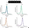
We used specific primers and a quantitative RT-PCR approach to amplify miRNAs located in the 17p13.3 chromosomal region, which is frequently deleted in breast cancer. After the addition of polyA tails to the given miRNAs, universal and miR-specific primers amplified ~70 base pair products. The uniqueness and authenticity of the amplified products were confirmed by agarose gel electrophoresis, the presence of single, sharp melting curves, and direct DNA sequencing (Figure 1).
miR-132 is significantly downregulated in breast tumors
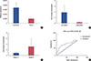
We determined the relative expression of miR-132 in 36 paired tumor/nontumor FFPE breast tissue samples. The relative expression levels of miR-132, adjusted to those of the 5s rRNA internal control, in tumor samples were normalized to those of the nontumor controls obtained from the same patient. Our data revealed that there was a significant downregulation of miR-132 expression levels in tumor samples (0.328-fold decrease in expression) compared to those of their matched nontumor controls (p<0.001) (Figure 2A). As shown in Figure 2B, a noticeable decrease in miR-132 expression was also observed in high histologic grade tumors compared to that of tumor samples with low histologic grade. However, this observed difference was not statistically significant, which may have been due to the small sample size (20 samples of high-grade tumors and 16 samples of low-grade tumors). Similarly, the levels of miR-132 expression in stage III tumors were higher than those in stages I and II tumors (Figure 2C). Nevertheless, this observed difference was also not statistically significant. We also employed a receiver operating characteristic (ROC) curve analysis to evaluate the sensitivity and specificity of miR-132 expression to discriminate between tumor and nontumor breast tissue samples. The total area under the curve (AUC) for miR-132 expression was 73% (p<0.001), suggesting that it may be suitable as a potential tumor marker for breast cancer (Figure 2D).
miR-212 is significantly downregulated in breast tumors
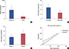
We determined the relative expression of miR-212 in 31 paired tumor/nontumor FFPE breast tissue samples. The relative expression levels of miR-212, adjusted to those of 5s rRNA, in tumor samples were normalized to those of nontumor specimens obtained from the same patient. A significant downregulation of miR-212 expression levels was observed in tumor samples (0.281-fold decrease in expression) compared to those of their matched nontumor controls (p=0.043) (Figure 3A). Similar to miR-132, the apparent expression alterations of miR-212 were not statistically associated with tumor grades (Figure 3B) or stages (Figure 3C). Compared to miR-132, the ROC curve analysis revealed a lower sensitivity and specificity for miR-212 expression (AUC=63%, p=0.061) to discriminate between tumor and nontumor breast tissue samples (Figure 3D).
miR-22 is significantly upregulated in breast tumors
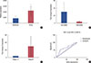
Our data revealed that there was a significant upregulation of miR-22 expression levels in tumor samples (2.183-fold increase in expression) compared to those of their matched nontumor controls (p=0.040) (Figure 4A). As shown in Figure 4B, a noticeable decrease in the expression of miR-22 was observed in high-grade tumors compared to that in low-grade tumors. However, this observed difference was not statistically significant, which may have been due to the small sample size (20 samples of high-grade tumors and 16 samples of low-grade tumors). In contrast, miR-22 expression levels in stage III tumors were higher than those in stages I and II tumors (Figure 4C). Nevertheless, this observed difference was also not statistically significant. ROC curve analysis revealed an AUC of 64% (p=0.047) for miR-22 expression in discriminating between tumor and nontumor breast tissue samples, suggesting that it may be moderately suitable as a potential marker for breast cancer (Figure 4D).
DISCUSSION
Deletions or LOH are frequent events in cancers. For instance, frequent deletions in 17p13.3 have been reported for breast cancer [245]. It is postulated that at least one or two tumor suppressor genes reside within this region [4]. As three breast development-related miR-NAs, namely miR-132, miR-212, and miR-22, are located within this region, we hypothesized that theses miRNAs may have tumor suppressor roles in breast tumorigenesis. Thus, we expected to observe downregulation of these miRNAs in breast tumor tissues.
Recently, both downregulated and upregulated expression of miR-132, miR-212, and miR-22 have been reported for different malignancies. These prior investigations have suggested that the aforementioned miRNAs can act as either oncogenes or tumor suppressor genes by targeting different mRNAs in multiple cellular pathways [15161718192021]. The versatile expression patterns of these miRNAs in various cancers point to their diverse cellular functions and the difficulty in determining their regulation and potential targets.
In the current study, we found that the miRNAs of interest were expressed in all tumor and nontumor samples. As expected, a significant downregulation of miR-132 and miR-212 was observed in breast tumor samples, suggesting that they have potential tumor-suppressor roles in breast tumorigenesis. These results are in contrast with those in a report by Park et al. [19], in which increased expression of both these miRNAs was observed in pancreatic cancer. However, our data are consistent with those of Zhang et al. [22], whose findings demonstrated the downregulation of miR-132 in pancreatic cancer.
One logical explanation for the conflicting reports on miRNA expression in similar cancer types may be that they were the result of using samples with different stages or grades of malignancy. As we have shown here, the expression levels of these miRNAs varied greatly in different grade and stage subgroups. Interestingly, the pattern of expression in the different grade and stage subgroups was very similar for miR-132 and miR-212, which both differed from that of miR-22. This observation seems to be associated with the chromosomal locations of these miRNAs. Specifically, while miR-132 (17: 1953202–1953302) and miR-212 (17:1953565–1953674) are closely packed together, miR-22 is located ~336 kb away from them (17:1617197–1617281). Therefore, the distance between them could lead to their involvement in separate genetic and epigenetic events during the initiation and progression of breast tumorigenesis.
As mentioned above, we found a different pattern of expression for miR-22 in breast tumors. In contrast to what we hypothesized and to what we observed for miR-132 and miR-212, we found a significant upregulation of miR-22 expression in breast tumors compared to that in their marginal nontu-mor counterparts. Nevertheless, miR-22 expression levels in different grades and stages of malignancy were similar to those of miR-132 and miR-212. Interestingly, a conflicting role for miR-22 as an oncomiR [23] and as a tumor suppressor [242526] has been reported. In addition to the possible effects of sample grade and stage on the outcome of results, another possibility should be taken into account that may explain these conflicting reports. As we have already reported for miR-21, the stromal content of samples can affect the out-come of expression results [27]. This may be explained as miRNA expression is not confined to tumor cells, and thus some altered expression may be attributed to stromal cells within the tumor microenvironment, including cancer-associated fibroblasts [28]. Therefore, the amount of stromal content in tumor samples may affect the outcome of results [28].
Our data revealed a consistent and noticeable downregulation of miR-132, miR-212, and miR-22 in high-grade samples, as well as a noticeable upregulation in stage III breast tumors. The latter finding is in agreement with a recent report by Hanieh [29], in which antimetastatic properties of the miR-212/132 cluster through SOX4 suppression in human tumor cells were demonstrated. In the case of miR-132, our data are consistent with those in a report by Zhang et al. [30], which indicated a critical role for miR-132 in breast cancer by suppressing cell proliferation, invasion, migration, and metastasis via negative regulation of HN1.
Taken together, our data revealed significant expression alterations of miR-132, miR-212, and miR-22 in breast tumors, and that miR-132 may be utilized to differentiate between tumor and nontumor states of breast tissue samples.
 XML Download