

 PDF
PDF ePub
ePub Citation
 Print
Print


INTRODUCTION
Breast cancer has become the second most frequent cause of female deaths and the major causes of treatment failure and/or death are tumor invasion and metastasis in Europe.
Recent concepts for cancer development suggest that a minority population of cancer stem-like cells (CSCs) may determine the biologic behavior of tumors [1-3], including response to therapy [4]. Recently it was demonstrated a consistent presence of CSCs in residual breast cancers after both neoadjuvant chemotherapy, and endocrine therapy [5].
The initial reports about breast cancer stem cells described the use of CD44+CD24-/low cell-surface antigen signature to select CSCs [6,7]. However, recent data show the detection of a CD44+CD24-/low subpopulation in only 31% of 240 human breast cancer samples analyzed, with a strong association with the basal-like phenotype [8]. Moreover, it was recently shown that CD44+CD24-/low phenotype detection is not sufficient, alone, to characterize breast CSCs [9].
Cancer stem cell research has recently included Prominin-1, CD133, a pentaspan transmembrane glycoprotein with a molecular weight of 120 kDa, that was initially considered to be a marker of hematopoietic stem cells [10-12]. Recently it has been reported the detection of CD133, PAX2, ESA, and GPR30 expression in invasive ductal breast carcinomas [13] and it was shown, mainly in triple-negative invasive ductal breast carcinoma patients, that the expression of CD133 protein could be correlated with tumor size, metastasis of the axillary lymph nodes and the clinical stage [14].
In all cases it was documented that the percentage of stem cells selected by CSCs markers immunostaining varies generally from 2% to 40% and is strongly connected to grade and aggressiveness [15].
In this paper we have identified a low grade tubulolobular variant of breast cancer showing an aberrant expression of prominin-1 marker. The hyperexpression of CD133 was evaluated by flow cytometry analysis, confirmed by immunohistochemistry and for gene expression by quantitative real-time polymerase chain reaction (PCR).
METHODS
Patients and specimens
Twelve patients, of which 5 underwent mastectomy for treatment of invasive ductal breast and 7 underwent quadrantectomy during March/April 2011 at the National Cancer Institute "Giovanni Pascale" of Neaples were enrolled in this study. We also selected 8 tubulolobular variants from our archive of paraffin samples. All cases were reviewed according to World Health Organization classification criteria, using standard tissue sections and appropriate immunohistochemical slides.
Medical records were reviewed for clinical information, including histologic parameters were determined from the H&E-stained slides. Clinicopathologic parameters evaluated for each tumor included patient age at initial diagnosis; tumor size; histologic subtype; histologic grade; nuclear grade; nodal status; number of positive lymph nodes; tumor stage; tumor recurrence or distant metastasis; type of surgery.
Moreover, all specimens were characterized for all routine diagnostic immunophenotipic parameters.
Flow cytometric analysis
The fresh tissue obtained from surgery was disaggregated mechanically and immediately tested by flow cytometry. The cell suspension obtained was counted and washed in PBS 1x. At least 200,000 cells were incubated with 1 µg/mL of fluorescent-labelled monoclonal antibodies and respective isotype controls at 4℃ in the dark room. After washing steps, the labelled cells were analyzed by flow cytometry using Facs ARIA II (Becton & Dickinson, Mountain View, USA). The antibodies used were: CD44 FITC conjugated, CD326 PE conjugated, all provided by BD Pharmingen, Buccinasco, Italy, and CD133/2PE mouse anti-Human CD133 (Miltenyi Biotec Inc., Auburn, USA). All data were analyzed using a CellQuest software (BD Biosciences, San Jose, USA).
Immunohistochemical analysis
For each patient, immunohistochemical staining was done on 7 slides from formalin-fixed, paraffin embedded tissues, to evaluate the expression of CD133, estrogen receptor (ER), progesterone receptor (PR), c-erbB-2, Ki67, E-cadherin, and CD44 markers. Paraffin slides were then deparaffinized in xylene and rehydrated through graded alchols. Antigen retrieval was performed with slides heated in 0.01 M citrate buffer (pH 6.0 for CD133, PR, c-erbB-2, Ki67, E-cad, CD44) or Tris-EDTA (pH 9 for ER) in a bath for 20 minutes at 97℃. After antigen retrieval, the slides allow to cool. The slides were rinsed with TBS and the endogenous peroxidase was inactivated with 3% hydrogen peroxide. After protein block (BSA 5% in PBS 1x), the slides were incubated with primary antibody to human CD133 (Myltenyi Biotec CD133/1 [AC 133] pure human 1:150) for 1 hour, and to human ERα (DAKO Monoclonal Mouse Anti-Human ERα Clone ID5 1:35), PR (DAKO Monoclonal Mouse Anti-Human PR Clone 636 1:50), c-erbB-2 (DAKO Polyclonal Rabbit Anti-Human Oncoprotein 1:300), Ki67 (DAKO Monoclonal Mouse Anti-Human Ki67 Ag Clone MIB-1 1:75), CD44 (Novocastra Lyophilized Mouse Monoclonal Antibody CD44 Variant 3, 1:35) for 30 minutes and to human E-cad (DAKO Monoclonal Mouse Anti-Human E-cadherin Clone NCH-38 1:75) for 20 minutes. The sections were rinsed in TBS and incubated for 20 minutes with Novocastra Biotinylated Secondary Antibody (RE7103), a biotin-coniugated secondary antibody formulation that recognized mouse and rabbit immunoglobulins. Then the sections were rinsed in TBS and incubated for 20 minutes with Novocastra Streptavidin-HRP (RE7104) and then peroxidase reactivity was visualized using a 3,3'-diaminobenzidine (DAB). Finally, the sections were counterstained with hematoxylin and mounted. Results are interpreted using a light microscope.
Evaluation of immunohistochemistry
Antigen expression was evaluated independently by two pathologists (M.DB. and G.B.) using light microscopy. Observers were unaware of the clinical outcome. For each sample, at least five fields (inside the tumor and in the area exhibiting tumor invasion, 400×400) and >500 cells were analyzed. Using a semiquantitative scoring system microscopically and referring to each antigen scoring method in other studies, two observers evaluated the intensity, extent and subcellular distribution of CD133, CD44, E-cad, ER, PR, c-erbB-2, and Ki67. In scoring CD133, CD44, and E-cad proteins expression, both the extent and intensity of immunopositivity in the cell membrane and cytoplasm were considered. The cutoff used to distinguish 'positive' from 'negative' cases was ≥1% ER/PR positive tumor cells. Immunohistochemical analyses of c-erbB-2 expression describe the intensity and staining pattern of tumor cells. Only membrane staining intensity and pattern were evaluated using the 0 to 3+ score as illustrated in the HercepTest kit scoring guidelines. The FDA-recognized test, the Herceptest™ (DAKO), describes four categories: no staining, or weak staining in fewer than 10% of the tumor cells (0); weak staining in part of the membrane in more than 10% of the tumor cells (1+); complete staining of the membrane with weak or moderate intensity in more than 10% of the neoplastic cells (2+); and strong staining in more than 10% (3+). Scores of 0 or 1+ were considered negative for HER2/neu-expression, 2+ was uncertain, and 3+ was positive. Cases 2+ undergo fluorescence in situ hybridization analysis.
RNA extraction and analysis
Total RNA was isolated from frozen biopsies, from our Institutional Bio-Bank, using RNeasy Mini Kit (Qiagen GmbH, Hilden, Germany) following the manufacturer's instructions. Samples were treated with RNase-free DNase (Qiagen GmbH) to prevent amplification of genomic DNA. A total of 1 µg RNA was subjected to cDNA synthesis for 1 hour at 37℃ using the Ready To Go You-Primer First-Strand Beads kit (cod. 27-9264-01; Amersham Biosciences Europe GmbH, Freiburg, Germany) in a reaction mixture containing 0.5 µg random hexamers (GeneAmp RNA PCR Random Hexamers Set N808-0127; Applied Biosystems, Foster City, USA).
Real-time PCR
Quantitative RT-PCR was performed in a LightCycler system (Roche Molecular Biochemicals, Mannheim, Germany) using TaqMan® analysis. In this system, all reactions were run in glass capillaries with The LightCycler TaqMan Master Mix (cod. 04735536001; Roche Molecular Biochemicals), 10 µL, in a volume of 20 µL containing 2 µL of cDNA and 1 µL of specific TaqMan Gene Expression Assays for human CD133 (RealTime Designer Assay cod. 05583055001; Roche Molecular Biochemicals), according to the manufacturer's directions. All reactions were performed in triplicate. The thermal cycling conditions included a step of 20 seconds at 95℃ followed by a 40 cycles of 95℃ for 1 second and 60℃ for 20 seconds. The comparative Ct method was employed to determine the human CD133 gene variation, using as reference gene TaqMan Endogenous Controls Human ACTB (β-actin) Endogenous Control (RealTime Designer Assay cod. 05532957001; Roche Molecular Biochemicals). We identified a calibrator cell line that represents the unitary amount of the target of interest, consequently the samples express n-fold mRNA relative to the calibrator. Final amounts of target were determined as follows: target amount=2-Ct, where Ct=[Ct (CD133)-Ct (ACTB)]sample-[Ct (CD133)-Ct (ACTB)]calibrator.
Statistical analysis
The association between CD133 expression with other clinicopathological parameters was conducted using the κ2 and T Student's test.
The Pearson κ2 test was used to determine whether a relationship exists between the variables included in the study. The level of significance was defined as p<0.05. All the statistical analyses were carried out using the SPSS version 8.0 software (SPSS Inc., Chicago, USA).
RESULTS
Clinicopathological characteristics of breast cancer patients and tumors
In our casuistry of fresh tissues are included 12 samples of breast cancers, 10 invasive ductal breast carcinomas (83.3%), 1 lobular breast carcinoma (8.3%), and 1 tubulolobular breast carcinoma (8.3%).
After, we selected 8 tubulolobular variants cases from our archive of paraffin samples.
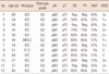
The age of the breast cancer patients ranged 27-85 years, with an average of 52 years. Tumors larger than 2 cm were present in 30% (4/20) of patients and lymphatic metastasis was found in 40% (8/20) of patients at operation. Thirty-five percent (7/20) of tumors were graded as grade 3, 25% (5/20) as grade 2, 40% (8/20) as grade 1. Of the 20 patients, 12 showed high expression (>66%), 4 low expression (<66%) and 4 no expression of estrogen receptor. The expression of progestin receptors was high (>66%) in 8 cases, low (<66%) in 6 cases, nothing in 6 cases. The score of c-erbB-2 was 3+ in 5 cases, 2+ in 2 cases, 1+ in 6 cases and 0 score in 7 samples. The expression of proliferation factor Ki67 was high (>20%) in 9 cases, and low (<20%) in 11 cases.
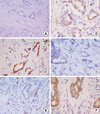
Expression of CD133 and CD44 in paraffin-embedded breast samples of fresh casuistry
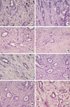
Expression of CD133 and CD44 was detected by immunohistochemistry in 12 paraffin-embedded breast cancer specimens. Also in this case the percentages of CD133+ cells were very heterogeneous in all cases. In samples 5, 6, 9, 10, 11 the expression was very low or absent, while it was moderate (5/15%) in samples 1, 2, 3, 4, 12 (Figure 2A and 2B). In case 7 the expression was >20% (Figure 2A and 2B) and only in tubulolobular variant breast cancer the expression of CD133 was about 35% (Figure 3).
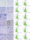
Cell sorting of breast samples with regard to CSCs markers expression
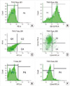
FACS was performed using all fresh breast cancers. We analyzed the expression of CD133, CD44, CD326. The results showed that the mean percentage of CD133 positive cells was 13.75% (range, 1.90-69.00%) while the mean percentage of CD44 positive cells was 17.90% (range, 2.30-52.30%). Moreover all cells analyzed were positive for CD326 (EpCAM), the antigen expressed on the basolateral surface of epithelial cells (data not shown). The flow cytometry analysis with regard to CD133 and CD44 expression is shown in Figure 2 and for tubulolobular sample in Figure 4. The percentages of CD133+ cells were very heterogeneous between all examinated cases. In samples 4, 10, and 11 the percentage was very low for CD133+ cells (3/7%), while in samples 1, 2, 3, 6, 7, 9, 12 was moderate (11/25%) (Figure 2A and 2B).
In tubulolobular breast variant the percentages of CD133+ cells was very high, about 70% (Figure 4).
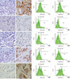
CD133 mRNA quantification in fresh breast samples
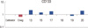
CD133 gene expression was evaluated on all selected breast tissues by real-time PCR quantification. In samples 3, 4, 5, 6, 10, 11 CD133 mRNA expression appeared very low or absent, while in 1, 2, 7, 9, 12 samples the expression was moderate (Figure 4).
Only in Tubulolobular breast specimen (sample 8) it was observed a significant increase in CD133 mRNA expression (Figure 5).
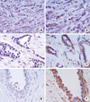
Expression of CD133 in archive breast tubulolobular variant samples
Expression of CD133 was detected by immunohistochemistry and real-time PCR quantification in 8 paraffin-embedded tubulolobular breast cancer specimens. Immunohistochemical detection shows a percentages of CD133+ cells moderate in 13 sample, very low in two cases 17 and 20, or absent in 14, 15, 16, 18, 19 samples (Figure 6).
CD133 gene expression was evaluated on selected breast tubulolobular tissues by real-time PCR quantification.
In samples 13, 16, 17, 20 CD133 mRNA expression appeared very low while in 18 and 19 samples was absent (Figure 7).
CD133 expression relates with Ki67 in breast cancers
From statistical elaboration of CD133 protein expression analysis with the other clinicopatological parameters in breast cancers selected for this study, it was shown that CD133 expression was significantly associated to Ki67 expression (p=0.010) (Table 3).
Moreover, we detected an association between CD133 expression and ductal breast cancer histotype (p=0.049) and a trend of statistical correlation between CD133 expression and tumor grade (p=0.057) (Table 3).
DISCUSSION
In this paper we have evaluated the rise of CD133 antigen as a suitable marker for CSCs detection in breast carcinomas. The analysis was carried out by different methods, flow cytometry, immunohistochemistry and investigate CD133 gene expression by real-time PCR quantification.
Our data were confirmed by all three methods, and the results are generally overlapping. Only in two cases the percentage of CD133-expressing cells detected by real-time PCR and flow cytometry, is found to be higher than that by immunohistochemistry.
There are possible explanations for the discordances between methods. First, fresh tumor tissue was used in the real-time PCR and flow cytometry measurements and paraffin-embedded tissue was used in immunohistochemistry. The staining pattern in the paraffin-embedded tissues could be altered by changes in tissue fixation conditions and antigen retrieval methods. Moreover, differences in immunostaining methodology, antibody concentrations, specificity of the antibodies used to localize the protein, and antibody cross-reactions must be considered [16].
All tumoral samples have a percentage of CD133 positive cells about 2-20%, except one tubulolobular breast variant that shows hyperexpression of the CSCs markers.
Tubulolobular carcinoma is a rare subtype of mammary carcinoma, representing less than 3% of all breast cancers, exhibiting features of both tubular and lobular differentiation. The prognosis of these tumors appears to be excellent, especially in those cases that are unilateral and less than 2 cm in size [17], and for this variant no information about the existence of stem cells detection was reported.
The hypothesis that mammary carcinogenesis results from the deregulation of normal stem cell self-renewal pathways suggests that components of these pathways might provide attractive targets for therapeutic development [18,19]. This idea is supported by recent description that the residual breast tumor cell populations surviving after conventional treatment may be enriched for subpopulations of cells with self-renewal features [5].
In the last years it was shown that the elective cancer stem cell marker for breast tumors is Prominin-1 which expression is not only strongly associated to stem cell phenotype but also to prognosis in invasive ductal breast cancer [14,15].
The possibility of targeting breast CSCs by CD133 inhibition provides a novel therapeutic approach for treatment of breast tumors [20].
In this study we show the strong overlap between gene and protein expression of CD133 marker in breast carcinomas. This might suggest the possibility of perturbing CD133 expression at transcription level, as decribed in malignant melanoma, where the shRNA-mediated downregulation results in alteration in cell growth and mobility, and reduces the capacity to metastatize in vivo [21], and at proteic level as previously shown in hepatocellular and gastric cancer xenografts [22].
Since there are no indications concerning the presence of stem cells compartments in tumors with good prognosis such as tubulolobular variant of breast cancer, our data suggest the importance of detecting stem cell niches, by CD133, in all phenotypic variants of breast carcinomas, to define prognostic value of these cells, as well as to establish novel therapeutic strategies targeted cancer stem cell self-renewal.

 XML Download