

 PDF
PDF ePub
ePub Citation
 Print
Print


Abstract
Statement of problem
Since the introduction of the concept of osseointegration in dental implants, high longterm success rates have been achieved. Though the use of dental implants have increased dramatically, there are few studies on domestic implants with clinical and objective longterm data.
Purpose
The aim of this retrospective study was to provide longterm data on the Implantium® implant, which features a sandblasted and acid-etched surface and internal connection with microthreads.
Material and methods
106 Implantium® implants placed in 38 patients at Yonsei University Hospital were examined to determine the effect of various factors on implant success and marginal bone loss, through clinical and radiographic results during a 6 to 30 month period.
Results
1. Out of a total of 106 implants placed in 38 patients, one fixture was lost, resulting in a 99.1% cumulative survival rate. 2. Among the 96 implants which were observed throughout the study period, the survival rates were 97.0% in the maxilla and 100% in the mandible. The survival rate in the posterior regions was 98.9% and 100% in the anterior regions. 3. The mean bone loss during the first year after prosthesis placement was 0.17 mm, while the mean annual bone loss after the first year was 0.04 mm, which was statistically less than during the first year (P < .05). 4. There was no significant difference in marginal bone loss according to age during the first year (P> .05), but after the first year, the mean annual bone loss in patients above 50 years was significantly greater (P < .05) compared with patients under 50 years. 5. No significant difference in marginal bone loss was found according to the following factors: gender, jaw, location in the arch, type of implant (submerged or non-submerged), presence of bone grafts, type of prostheses, and type of opposing dentition (P < .05).
Conclusion
Based on these results, the sole factor influencing marginal bone loss was age, while factors such as gender, jaw, location in the arch, type of implant, presence of bone grafts, type of prostheses and type of opposing dentition had no significant effect on bone loss. In the present study, the success rate of the Implantium® implant with a SLA surface and internal connection with microthreads was satisfactory up to a maximum 30 month period, and the marginal bone loss was in accord with the success criteria of dental implants.
REFERENCES
1.Adell R., Lekholm U., Rockler B., Bra ° nemark PI. A 15-year study of osseointegrated implants in the treatment of the edentulous jaw. Int J Oral Surg. 1981. 10:387–416.

2.Buser D., Mericske-Stern R., Bernard JP., Behneke A., Behneke N., Hirt HP., Belser UC., Lang NP. Longterm evaluation of non-submerged ITI implants. Part 1: 8-year life table analysis of a prospective multicenter study with 2359 implants. Clin Oral Implants Res. 1997. 8:161–72.
3.Albrektsson T., Dahl E., Enbom L., Engevall S., Engquist B., Eriksson AR., Feldmann G., Freiberg N., Glantz PO., Kjellman O. Osseointegrated oral implants. A Swedish multicenter study of 8139 consecutively inserted Nobelpharma implants. J Periodontol. 1988. 59:287–96.
4.Becker W., Becker BE., Alsuwyed A., Al-Mubarak S. Longterm evaluation of 282 implants in maxillary and mandibular molar positions: a prospective study. J Periodontol. 1999. 70:896–901.
5.Bahat O. Bra ° nemark system implants in the posterior maxilla: clinical study of 660 implants followed for 5 to 12 years. Int J Oral Maxillofac Implants. 2000. 15:646–53.
6.Wennerberg A., Albrektsson T., Andersson B., Krol JJ. A histomorphometric and removal torque study of screw-shaped titanium implants with three different surface topographies. Clin Oral Implants Res. 1995. 6:24–30.
7.Cochran DL. A comparison of endosseous dental implant surfaces. J Periodontol. 1999. 70:1523–39.
8.Norton MR. Marginal bone levels at single tooth implants with a conical fixture design. The influence of surface macro- and microstructure. Clin Oral Implants Res. 1998. 9:91–9.
9.Abrahamsson I., Berglundh T. Tissue characteristics at mi-crothreaded implants: an experimental study in dogs. Clin Implant Dent Relat Res. 2006. 8:107–13.
10.Lekholm U., Zarb GA. Patient selection and preparation, in Bra ° nemark P-I, Zarb GA, Albrektsson T (eds). Tissue-Integrated Prostheses. Osseointegration in Clinical Dentistry. Chicago, Quintesseence Publ Co;1985. p. 199–209.
11.Cochran DL., Buser D., ten Bruggenkate CM., Weingart D., Taylor TM., Bernard JP., Peters F., Simpson JP. The use of reduced healing times on ITI implants with a sandblasted and acid-etched (SLA) surface: early results from clinical trials on ITI SLA implants. Clin Oral Implants Res. 2002. 13:144–53.
12.Seo JY., Shim JS., Lee KW. Clinical and radiographical evaluation of implant-supported fixed partial prostheses. J Korean Acad Prosthodont. 2006. 44:394–404.
13.An HS., Moon HS., Shim JS., Cho KS., Lee KW. Clinical and radiographic evaluation of Neoplant implant with a sandblasted and acid-etched surface and external connection. J Korean Acad Prosthodont. 2008. 46:125–36.
14.Romeo E., Lops D., Margutti E., Ghisolfi M., Chiapasco M., Vogel G. Longterm survival and success of oral implants in the treatment of full and partial arches: a 7-year prospective study with the ITI dental implant system. Int J Oral Maxillofac Implants. 2004. 19:247–59.
15.Blanes RJ., Bernard JP., Blanes ZM., Belser UC. A 10-year prospective study of ITI dental implants placed in the posterior region. I: Clinical and radiographic results. Clin Oral Implants Res. 2007. 18:699–706.
16.Lindquist LW., Rockler B., Carlsson GE. Bone resorption around fixtures in edentulous patients treated with mandibular fixed tissue-integrated prostheses. J Prosthet Dent. 1988. 59:59–63.
17.Albrektsson T. Isidol. Consensus report of session IV. Lang N.P., Karring Y., editorsProceedings of the 1st European Workshop on Periodontology. 1994. p. 365–9. London: Quintessence Publishing Co., Ltd.
18.Hansson S. The implant neck: smooth or provided with retention elements. A biomechanical approach. Clin Oral Implants Res. 1999. 10:394–405.
19.Wennstrom JL., Ekestubbe A., Grondahl K., Karlsson S., Lindhe J. Implant-supported single-tooth restorations: a 5-year prospective study. J Clin Periodontol. 2005. 32:567–74.
20.Lee DW., Choi YS., Park KH., Kim CS., Moon IS. Effect of microthread on the maintenance of marginal bone level: a 3-year prospective study. Clin Oral Implants Res. 2007. 18:465–70.
21.Palmer RM., Smith BJ., Palmer PJ., Floyd PD. A prospective study of Astra single tooth implants. Clin Oral Implants Res. 1997. 8:173–9.
22.Norton MR. Marginal bone levels at single tooth implants with a conical fixture design. The influence of surface macro- and microstructure. Clin Oral Implants Res. 1998. 9:91–9.
23.Moy PK., Medina D., Shetty V., Aghaloo TL. Dental implant failure rates and associated risk factors. Int J Oral Maxillofac Implants. 2005. 20:569–77.
24.Chung DM., Oh TJ., Lee J., Misch CE., Wang HL. Factors affecting late implant bone loss: a retrospective analysis. Int J Oral Maxillofac Implants. 2007. 22:117–26.
25.Naert I., Koutsikakis G., Duyck J., Quirynen M., Jacobs R., van Steenberghe D. Biologic outcome of implant-supported restorations in the treatment of partial edentulism. part I: a longitudinal clinical evaluation. Clin Oral Implants Res. 2002. 13:381–9.
26.Wyatt CC., Zarb GA. Bone level changes proximal to oral implants supporting fixed partial prostheses. Clin Oral Implants Res. 2002. 13:162–8.
27.Naert I., Koutsikakis G., Quirynen M., Duyck J., van Steenberghe D., Jacobs R. Biologic outcome of implant-supported restorations in the treatment of partial edentulism. Part 2: a longitudinal radiographic study. Clin Oral Implants Res. 2002. 13:390–5.
28.Bryant SR., Zarb GA. Crestal bone loss proximal to oral implants in older and younger adults. J Prosthet Dent. 2003. 89:589–97.
29.Lang NP., Mombelli A., Tonetti MS., Bragger U., Hammerle CH. Clinical trials on therapies for peri-implant infections. Ann Periodontol. 1997. 2:343–56.
30.Wyatt CC., Zarb GA. Treatment outcomes of patients with implant-supported fixed partial prostheses. Int J Oral Maxillofac Implants. 1998. 13:204–11.
31.Ericsson I., Randow K., Glantz PO., Lindhe J., Nilner K. Clinical and radiographical features of submerged and non-submerged titanium implants. Clin Oral Implants Res. 1994. 5:185–9.
32.Cecchinato D., Bengazi F., Blasi G., Botticelli D., Cardarelli I., Gualini F. Bone level alterations at implants placed in the posterior segments of the dentition: outcome of submerged/non-submerged healing. A 5-year multicenter, randomized, controlled clinical trial. Clin Oral Implants Res. 2008. 19:429–31.
33.Becker W., Becker BE., Ricci A., Bahat O., Rosenberg E., Rose LF., Handelsman M., Israelson H. A prospective mul-ticenter clinical trial comparing one- and two-stage titanium screw-shaped fixtures with one-stage plasma-sprayed solid-screw fixtures. Clin Implant Dent Relat Res. 2000. 2:159–65.
34.Mayfield L., Skoglund A., Nobreus N., Attstrom R. Clinical and radiographic evaluation, following delivery of fixed reconstructions, at GBR treated titanium fixtures. Clin Oral Implants Res. 1998. 9:292–302.
35.Zitzmann NU., Scharer P., Marinello CP. Longterm results of implants treated with guided bone regeneration: a 5-year prospective study. Int J Oral Maxillofac Implants. 2001. 16:355–66.
36.Palmqvist S., Sondell K., Swartz B., Svenson B. Marginal bone levels around maxillary implants supporting overdentures or fixed prostheses: a comparative study using detailed narrow-beam radiographs. Int J Oral Maxillofac Implants. 1996. 11:223–7.
37.Enkling N., Nicolay C., Utz KH., Johren P., Wahl G., Mericske-Stern R. Tactile sensibility of single-tooth implants and natural teeth. Clin Oral Implants Res. 2007. 18:231–6.
38.Pjetursson BE., Sailer I., Zwahlen M., Hammerle CH. A systematic review of the survival and complication rates of all-ceramicand metal-ceramic reconstructions after an observation period of at least 3 years. Part I: Single crowns. Clin Oral Implants Res. 2007. 18:73–85.
39.Galasso L. Proposed method for the standardized measurement of marginal bone height on periapical radiographs with the Branemark System. Clin Implant Dent Relat Res. 2000. 2:147–51.
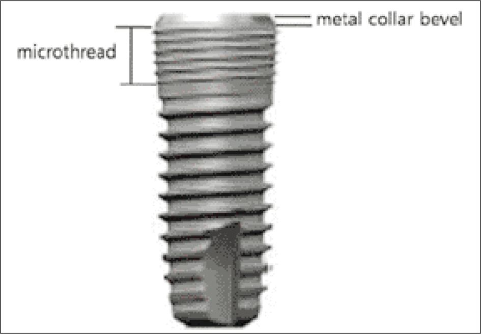
Fig. 2.
References used to measure actual marginal bone loss. (a): junction between implant machined collar bevel and rough surface (b): implant to marginal bone contact level (c): inter-thread distance of six threads
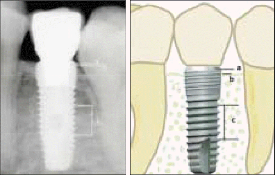
Table I.
Number of implants placed according to bone quality
| Type I | Type II | Type III | Type IV | Unknown | Total | |
|---|---|---|---|---|---|---|
| Maxilla | - | 3 | 20 | 11 | 3 | 37 |
| Mandible | - | 34 | 26 | 3 | 6 | 69 |
| Total | - | 37 | 46 | 14 | 8 | 106 |
Table II.
Number of implants placed according to bone quantity
| A | B | C | D | E | Unknown | Total | |
|---|---|---|---|---|---|---|---|
| Maxilla | - | 13 | 21 | - | - | 3 | 37 |
| Mandible | - | 21 | 39 | 3 | - | 6 | 69 |
| Total | - | 34 | 60 | 3 | - | 8 | 106 |
Table III.
Life table analysis showing cumulative survival rates
Table IV.
Number of implants placed according to implant diameter
| Implant diameter (mm) | ||||||
|---|---|---|---|---|---|---|
| 3.4 | 3.8 | 4.3 | 4.8 | 4.8W | Total | |
| Maxilla | - | 8 (1) | 10 | 11 | 3 | 32 (1) |
| Mandible | 7 | 9 | 17 | 26 | 6 | 65 |
| Total | 7 | 17 (1) | 27 | 37 | 9 | 97 (1) |
Table V.
Number of implants placed according to implant length
| Implant length (mm) | |||||
|---|---|---|---|---|---|
| 8 | 10 | 12 | 14 | Total | |
| Maxilla | 1 | 16 | 15 (1) | - | 32 (1) |
| Mandible | 12 | 27 | 26 | - | 65 |
| Total | 13 | 43 | 41 (1) | - | 97 (1) |
Table VI.
Marg period ginal bone loss arou und implants accord ding to observation
Table VII.
Comparison of marginal bone loss around implants according to observation period
Table VIII.
Comparison of marginal bone loss between older and younger adults
Table IX.
Comparison of marginal bone loss between male and female
Table X.
Number of implants placed according to location in arch
| Location | ||||||||
|---|---|---|---|---|---|---|---|---|
| Central incisor | Lateral incisor | Canine | 1st premolar | 2nd premolar | 1st molar | 2nd molar | Total | |
| Maxilla | - | - | - | 3 | 7 (1) | 16 | 7 | 33 (1) |
| Mandible | 3 | 3 | 3 | 6 | 8 | 23 | 18 | 64 |
| Total | 3 | 3 | 3 | 9 | 15 (1) | 39 | 25 | 97 (1) |
Table XI.
Survival rates according to respective region in arch and jaw
| Anterior | Posterior | Survival rate (%) | |
|---|---|---|---|
| Maxilla | 0 | 33 (1) | 97 |
| Mandible | 9 | 55 | 100 |
| Survival rate (%) | 100 | 98.9 |
Table XII.
Comparison of marginal bone loss between maxilla and mandible
Table XIII.
Comparison of marginal bone loss according to region in arch
Table XIV.
Comparison of marginal bone loss according to submerged and non-submerged implants
Table XV.
Comparison of marginal bone loss according to presence of bone grafts
Table XVI.
Comparison of marginal bone loss according to type of prostheses
Table XVII.
Comparison of marginal bone loss according to state of opposing dentition
| Opposing dentition | Number of implants observed | Bone resorption (mm) (Mean ±SD) | one-way ANOVA test P-value | |
|---|---|---|---|---|
| Sur-B | Natural tooth | 70 | 0.32 ±046 | 0.879 |
| Implant | 24 | 0.35 ±0.38 | ||
| Removable | 2 | 0.20 ±0.04 | ||
| Prosthesis | ||||
| B-1 Year | Natural tooth | 63 | 0.20 ±0.28 | 0.340 |
| Implant | 23 | 0.11 ±0.20 | ||
| Removable | 2 | 0.28 ±0.15 | ||
| Prosthesis | ||||
| After 1 Year | Natural tooth | 33 | 0.05 ±0.09 | 0.381∗ |
| Implant | 10 | 0.02 ±0.03 | ||
| Removable | ||||
| Prosthesis |
 XML Download