

 PDF
PDF ePub
ePub Citation
 Print
Print


INTRODUCTION
Due to the increasing demands for esthetic restorations and biocompatibility concerns, all ceramic restorations have been widely used in the last few decades.1 Recently, the use of computer-aided design/computer-aided manufacturing (CAD/CAM) systems for producing all ceramic restorations has been growing rapidly. The aim of this technology is to produce restorations with higher mechanical properties in a shorter period of time compared to conventional methods as well as generating new materials and systems for fabrication of dental restorations.23 Use of yttria-stabilized tetragonal zirconia polycrystalline (Y-TZP) for fabrication of all ceramic frameworks by means of CAD/CAM is common due to its unique characteristics including excellent biocompatibility, low plaque accumulation and unsurpassed mechanical properties.4
In addition to esthetic, strength, and biocompatibility, marginal accuracy is one of the fundamental requirements for clinical assessment and success of dental restorations.5 Inaccurate marginal fit causes a space between restoration and prepared tooth, which accelerates the dissolution of luting agent.6 Subsequently, oral bacteria and food debris accumulate in this space, leading to secondary caries, pulpal lesions, postoperative sensitivity, periodontal disease and marginal discoloration.78910 According to McLean and von Fraunhofer, the maximum acceptable marginal opening is 120 µm.11 The mean marginal discrepancy for all ceramic restorations reported in former studies was between 3.7 µm to 174 µm; and the majority of the reported values were less than or equal to 120 µm.5 In CAD/CAM restorations, it is claimed that due to the reduction in human errors and material imperfections, minimal acceptable marginal gap was less than 100 µm.12
Marginal fit of the crown is assessed by measuring the gap between the abutment and inner surface of the restoration. The assessment of the marginal gap in the path of placement and removal of the restoration is defined as vertical marginal discrepancy.1314 Several methods have been employed for measuring the marginal fit of restorations including direct microscopic view, cross-sectional view, replica technique, laser videography, and x-ray microtomography.515161718 As the direct view is a nondestructive technique, it is a proper method for evaluation of the marginal stability during the fabrication procedures of the restorations.15
Zirconia frameworks are usually veneered using the conventional layering technique. In recent years, some new veneering techniques have been introduced, aiming to reproduce stronger veneers to reduce debonding and chipping of zirconia veneers.19202122 Press-over technique and CAD-on technology are two new veneering methods which have shown higher mechanical properties compared to conventional layering technique.19202122 In press-over technique after application of a special liner to the zirconia framework, the veneer is waxed upon it. Alternatively, the wax or resin replica of the veneer could be produced by CAD/CAM technology, connected to the framework and invested using fluorapatiteglass-ceramic ingots.
In CAD-on technique, veneer is designed using CAD software and milled from Lithium-disilicate ingots (IPS e. max CAD). Then veneer connects to the framework by using a low fusion glass ceramic. A sinter bond firing acts as crystallization of Lithium-disilicate and fusion process simultaneously.
Given the importance of the fitting accuracy of restoration, there has been much debate on the consequence of veneering porcelain on the marginal fit of all-ceramic restoration.15232425262728293031 Pak et al.26 reported that veneering process increased the marginal gap of lava and Digident systems. Sulaiman et al.29 pointed out larger marginal gap on the facial and lingual sides of the specimens, which was directly related to the amount of veneering porcelain. In another study performed by Cho et al.,30 the marginal gap increased for two pressable ceramic systems (Esthetic and IPS e.max Press) during veneer application. Although, they found the reduction of marginal gap in the characterization and glazing firing cycle. In contrast to these findings, Miura et al.31 reported marginal stability of Cercon CAD/CAM system during porcelain fire veneering cycles.
A review of the literature provided no data regarding the effect of different veneering techniques on the marginal adaptation of zirconia coping. Therefore, the present study aimed to evaluate the effect of 3 different veneering processes (layering, press-over and CAD-on techniques) on the marginal fit of zirconia frameworks. The null hypothesis was that no differences would be found in the marginal fit of zirconia CAD/CAM crowns before and after porcelain firing, and among different veneering processes.
MATERIALS AND METHODS
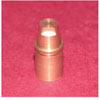
Ceramic materials investigated in this study were displayed in Table 1. A brass master die was machined to approximate dimension of a prepared molar for an all ceramic restoration with 7 mm height, 6 degree of occlusal convergence and a 90 degree shoulder of 1 mm wide finish line (Fig. 1). Preparation of master die was free of any irregularities, and was done in accordance with the current standards of full ceramic restorations.32 An antirotational design was included in the axial surface to ensure reproducible seating of the coping on the master die. Eighteen points for measurement of vertical marginal gap at 20 degree intervals were marked on a groove 3 mm below the margin by means of a high speed handpiece and a diamond needle bur. Finally, the die was embedded in an acrylic block by means of a dental surveyor (Ney Dental Surveyor, Dentsply, Balalgues, Switzerland) to ensure its long axis was perpendicular to the horizontal plane.
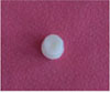
In order to eliminate the effect of impression and pouring variations, the metallic die was used as the definitive die. The die was sprayed with scan spray and scanned using a 3D-laser scanner (3ShapeD810; 3Shape, Copenhagen K, Denmark). The data were transferred to CAD software (3Shape's CAD Design software; 3Shape, Copenhagen K, Denmark) in which the copings were designed with a uniform thickness of 0.5 mm around, considering 30 µm spacer, and 1 mm short of the margin. In the occlusal surface a depression was designed to accommodate the tip of the holding device (Fig. 2). Thirty zirconia copings were milled from pre-sintered Y-TZP blanks (IPS e.max ZirCAD, Ivoclar Vivadent) in a milling machine (inLab MC XL, Sirona) in a white state. The zirconia frameworks were then sintered (Programat S1, Ivoclar Vivadent). The frameworks were examined for any imperfections and rejected if any deformation was observed.
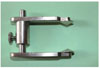
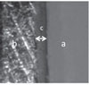
Each zirconia coping was seated on the master die and mounted on a specially holding device. Due to the cone configuration of holding device tip, the copings could seat on it only in one position (Fig. 3). A uniform load of 15 N was applied to all specimens to ensure the copings were completely seated on the die. Then the images made from the 18 previously marked points using a digital microscope (AM413FIT Dino-Lite Pro; Dino-Lite electronic corp., Taipei, Taiwan) were connected to a personal computer and photographed at ×230 magnification. These images were then analyzed with image analysis software (DinoCapture 2.0, AnMo Electronics Corp., Tainan Hsien, Taiwan). The vertical marginal gap was evaluated by measuring the perpendicular line from the most cervical external edge of the restoration to the most outer edge of the finish line of the preparation (Fig. 4).
Thirty copings were randomly divided into 3 equal groups. On each of them one of the following veneering techniques were performed (layering (L), press-over (P), and CAD-on (C) techniques). The first 10 copings were veneered using the traditional layering technique. A silicone index was used to standardize the shape and size of veneers with a homogenous veneering thickness of range between 0.7 mm at margins and 1.5 mm at occlusal surfaces.To veneer the copings with layering technique, the liner (IPS e.max, zirliner; Ivoclar Vivadent) was applied to the zirconia copings, and they fired in a compatible ceramic furnace (Programmat 700; Ivoclar Vivadent) at 960℃; then a nanofluoroapatite glass ceramic (IPS e.max Ceram; Ivoclar Vivadent) was applied in dentin and enamel layers and processed at 750℃, followed by glazing and finishing procedures to complete the restorations. The occlusal surface of the crowns was accommodated to the holding device tip to ensure the same seating of them as copings on the die (Fig. 5).
The second group was veneered by using press-over technique. In order to obtain an equivalent veneering structure as the L group, external surface of a completed crown from L group and external surface of the zirconia coping were scanned and the obtained data were used to design (3Shape's CAD Design software; 3Shape, Copenhagen K, Denmark) the veneering material with a thickness of 0.7 mm at margins and 1.5 mm at occlusal surfaces. Then, resin replicas of the veneers were milled from castable acrylate polymer blocks (IPS AcrylCAD; Ivoclar Vivadent) and attached to the zirconium oxide frameworks using a castable wax. Each framework was sprued and invested. After setting for 40 minutes, the wax and acrylate polymer were burnt out by means of heat. Then the created mold was filled with the pressable glass-ceramic ingots (IPS e.max ZirPress, Ivoclar Vivadent). Firing was performed in a proper ceramic furnace (Programat EP 5000; Ivoclar Vivadent) at a temperature of 910℃. After recovery of the restorations, they were finished according to the manufacturer's instruction and glazed at a temperature of 750℃.
Ten copings of the third group were veneered by using CAD-on technology. To obtain equivalent veneering structure as the L and P groups, the designed veneer for the P group was used to mill (inLab MC XL, Sirona) lithium-disilicate glass-ceramic blocks (IPS e.max CAD, Ivoclar Vivadent) in a pre-crystallized state. Then a fusion glass-ceramic (IPS e.max CAD Crystall./Connect, IvoclarVivadent) was applied to the inner surface of the veneer and outer surface of the coping. They were fitted to each other by applying a slight pressure. Subsequently, excess fusion ceramic was removed with a brush and fired in a ceramic furnace (Programat EP 5000; Ivoclar Vivadent) at a temperature of 840℃. This firing served as crystallization of IPS e.max CAD and the fusion process simultaneously. Finally, the crowns were completed with one glaze firing (IPS e.max Ceram Glaze & IPS e.max CAD Crystall/Glaze) at a temperature of 725℃.
The means and standard deviations were calculated in each group. Paired t-test was used to compare the amount of marginal gap of specimens before and after veneering, within the same group. One-way ANOVA and post hoc tests were used to compare the marginal gap after performing three veneering methods. The significance level of 5% was used for all of the statistical tests.
RESULTS
The means and standard deviations for the marginal gap of the specimens before and after veneering in three experimental groups are included in Table 2. Statistical analysis revealed no difference between measurements of fit values of three groups before veneering (P=.822). The vertical marginal gap of the three groups was increased after porcelain veneering (P<.001) (Table 2). The highest mean marginal gap values after veneering was found in the layering group (63.06 µm), which was higher than the other two groups (P<.001). No statistically significant difference was found between the marginal gap values of press-over and CAD-on techniques after veneering (P=.973).
DISCUSSION
The results of the current investigation revealed a significant increase in marginal gap of crowns after porcelain veneer firing. Although this event was observed by performing every three veneering methods, crowns which were veneered by using the conventional layering technique showed greater changes (P<.001). These results support the rejection of the null hypothesis.
In the present study, a single metallic die was used to standardize preparation and impede any wear of abutment during the manufacturing and measuring process. Furthermore, measurements were performed on this single die and the specimens were not cemented to prevent variability due to luting agent type, viscosity, and seating forces during cementation. Various methods have been employed to evaluate the marginal fit of restorations in the literature.515161718 Two most common nondestructive methods which permit assessment of marginal discrepancy at different fabrication stages of the restoration, are direct microscopic view and replica techniques.528 In the current investigation direct microscopic view was used to evaluate the marginal gap before and after veneering of the restorations. It is the most widely method used by the authors.5 In this technique in spite of replica technique, marginal gap could be measured in numerous points. Besides, the use of intermediate media such as impression material is not needed in the direct microscopic view which can limit the effect of material flaws used in the replica technique on the measurement of the fit.33 However, in the direct microscopic view the horizontal marginal fit could not be assessed. The exposure of cement in the margin is mostly affected by the vertical marginal discrepancy, while Horizontal marginal discrepancy is more critical for plaque control and maintainability of the restoration.34
In the present study a holding device was used to standardize the seating of the specimens on the die during measurements. The device has the essential requirements for a standard holding device firstly was proposed by Ushiwata and de Moraes.35 To standardize the seating of the restorations before and after veneering on the die, the morphology of occlusal surface was kept the same and accommodated to the holding device tip (Fig. 2, Fig. 5). The tip of the device is conical and allows orientation of the specimens only in one plane during measurements, although the rotation of the restorations is yet possible to measure around the margin (Fig. 3). An area of approximately 0.5 mm at the cervical margin was not veneered with porcelain to limit contamination of margin area with porcelain during veneering and incomplete seating of the crowns. These two events may affect the correct marginal gap evaluation.15
There are large variations regarding the amount of acceptable marginal gap of crown in the literature. Christensen et al.36 reported the range of 34-119 µm for subgingival acceptable marginal gap, and 2-15 µm for supragingival margins. However, Mclean and von Fraunhofer evaluated more than 1000 restorations within 5 years, and proposed 120 µm as the upper limit of clinically acceptable marginal opening.11 For CAD/CAM restorations, the most acceptable marginal gap range is between 50 to 100 µm.12 In the current study, the mean marginal gap was 35 µm for zirconia copings, 63 µm for crowns which were veneered by using layering technique, 50 µm for groups which veneered by using pressing technique, and 51 µm for CAD-on veneered crowns. Regarding the aforementioned studies, the amount of marginal gap for all the three groups was within the clinically acceptable range. To number the reported marginal opening for zirconia CAD/CAM restorations in former studies; Miura et al. reported the mean marginal gap of cercon zirconia CAM crowns with three different cervical margin designs to be 24-30 µm.31 Euán et al.37 found that absolute marginal gap of the Lava zirconia copings with round shoulder margin was 52.66 µm. The mean marginal gap of the Procera zirconia crown was reported to be 44.2 µm, in Kokubo et al.'s study.38 Some incompatible results of the current study and other researches maybe related to the measuring methods and possible errors in microscopic evaluation of the marginal gap, different CAD/CAM systems which are used and the criteria which is used for the marginal gap evaluation (horizontal, vertical or absolute marginal discrepancy).
In the current investigation, using each other of layering, press-over or CAD-on techniques for veneering of zirconia copings increased the marginal gap of the restorations. In comparison, Pak et al.26 demonstrated an increase in the marginal gap of Digident and lava CAD/CAM zirconia ceramics after veneering process. Also, the marginal fit of three all-ceramic crown systems (conventional In-ceram, copy milled In ceram, and copy-milled feldspathic crowns) in Balkaya et al.'s15 study changed during porcelain firing cycles. They reported that only glaze firing had no consequence on the marginal accuracy. The marginal gap of the procera crowns has been reported to increase during porcelain veneering process.39 However, Bhowmik and Parkhedlkar40 pointed out the marginal stability of glass infiltrated alumina copings during firing cycles.
Alterations of the marginal fit during veneering process could be discussed by some causes. A probable reason is the shrinkage of veneering porcelain during sintering process. This shrinkage may lead to changes in the gap, due to the ceramic lifting from the margin of the die.41 Another reason for marginal distortion during porcelain veneering process is thermal incompatibility between framework and veneering porcelain.15 Different coefficients of thermal expansion (CTE) of coping and veneer in the layered restoration causes stress formation when the restoration cools from glass transition to room temperature.42 One of the drawbacks of this event is deformation of the restoration. In metal ceramic restorations, a small positive mismatch in CTE enhances the strength of the restoration by applying compressive forces on the veneering ceramic.43 However, according to Aboushelib et al.'s44 study for all ceramic zirconia layered restorations, minimizing the thermal mismatch would be desirable. According to Isgro et al.,45 even a zero thermal mismatch does not guarantee the compatibility between ceramic core and veneering porcelain so that the fast cooling procedure, viscoelastic behavior of the porcelain, and repeated firing can lead to distortion. Among the three different veneering methods used in the current study, conventional layering method increased the marginal gap of zirconia framework more than the two other techniques (P<.001). It could be related to the less thermal mismatch of layers in the press-over and CAD-on techniques (0.9×10⁻⁶ K-1 in P group, 0.6×10⁻⁶ K-1 in C group and 1.3×10⁻⁶ K-1 in L group).
Another reason may be related to the numbers of firing cycles needed for each of these techniques. Conventional layering technique needs more firing cycles (at least four) compared to the press-over (at least three) and CAD-on (at least two) veneering techniques. Previous studies revealed that during repeated firing cycles, CTE of core, and veneer ceramic can change, producing a non-reliable thermal mismatch.4647
One of the limitations of the current study is that, the specimens were produced and tested under the ideal conditions, which may not reflect the actual clinical conditions. Besides, no attempt was made to simulate oral condition through an artificial aging process. Another limitation is that only vertical marginal gap was measured and horizontal discrepancy was not examined. Since the measurement of internal gap necessitate the cementation and sectioning of specimens, in this study, unlike the marginal gap, the internal gap was not measured.
CONCLUSION
Within the limitations of this study, following conclusions are drawn:
Three veneering methods tested in the current investigation altered the marginal fit of zirconia coping.
Conventional layering technique increased the marginal gap of zirconia framework more than the press-over and CAD-on techniques.
All ceramic crowns made through three different veneering methods, revealed clinically acceptable marginal fit.


 XML Download