

 PDF
PDF ePub
ePub Citation
 Print
Print


INTRODUCTION
Oligodontia that involves one or more teeth is a relatively common condition. In severe partial anodontia, the bilateral absence of the corresponding teeth is striking. Graber1 reported that the overall frequency of patients with congenitally missing teeth ranged from 1.6 to 9.6 per cent in various series of studies from different countries.
Although the etiology of missing teeth is unknown, there is a familial tendency for this defect in many instances. Hereditary ectodermal dysplasia might be associated with partial anodontia, and in these instances the few teeth that are present might be deformed or frequently cone-shaped.2-4 There might be a deficiency of alveolar ridges and basal bone, particularly in the maxilla.
Prosthetic treatment can play an important role in the dental management of patients whose dentition fails to develop normally. The principle and technique are essentially identical to those used in adult therapy.5-7 Without advanced surgical approaches, the application of removable dentures might be the only restorative option in oligodontia patients with insufficient bone support. Restoration of the spaces in hypodontia patients require the consideration of many factors including, the number of missing teeth, the distribution of space, the size of the teeth, and the patient's age.8
The congenital absence of teeth, along with tooth loss due to caries and traumatic injuries, is one of the most common reasons for requiring complete or removable partial dentures in young patients. This clinical report describes the multi-displinary approaches of oral rehabilitation for a young girl with severe oligodontia and maxillary hypoplasia.
CLINICAL REPORT
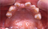
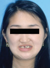
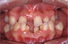
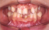
An 18 year old female with oligodontia, presenting with the chief complaint of an unesthetic appearance and masticatory difficulty due to oligodontia and inadequate occlusion was referred to the Department of Prosthodontics, Chonnam National University Hospital (Fig. 1).
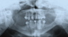
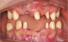
Panoramic radiographs confirmed the congenitally missing permanent teeth and the retained deciduous mandibular incisors and right mandibular cuspid. The patient showed mandibular prognathism, a concave facial profile and atrophy of the edentulous alveolus. The posterior teeth showed inadequate occlusal contact, whereas the anterior teeth exhibited a cross-bite pattern (Figs. 1, 2, 3, and 4).
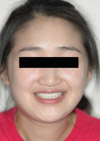
The orthodontic phase of the treatment was accomplished first. After the orthodontic treatment, a Le Fort I osteotomy was performed for the 5 mm anterior-posterior advancement of the maxilla at the department of maxillofacial surgery. After orthodontic treatment and orthognathic surgery, the facial profile was improved with proper distribution of the teeth (Fig. 5).
Among the prosthetic approaches for treating oligodontia, dental implants can be used to restore the edentulous area. In this patient, implant treatment was ruled out, because the edentulous alveolar bones showed severe atrophy (Fig. 4).
A shortened dental arch concept was the decisive treatment plan for this particular patient, because none of the posterior teeth were intact and the young female patient did not want to wear a removable partial denture. After the completion of orthodontic treatment, 4 mandibular deciduous teeth and the maxillary right central incisor from the resorptive alveolus were extracted. Using each maxillary and mandibular remaining teeth, one-piece fixed partial dentures (FPD) were planned, because the root of the residual teeth had a short and conical shape.
Face bow transfer and mounting the casts on the articulator was performed before commencing the diagnostic wax-up procedure. The vertical dimension of the patient was increased by 4 mm, considering the facial muscular harmony in the centric relation.
Based on the diagnostic wax - up, the teeth were prepared (Fig. 6) and the provisional restoration was fabricated using autopolymerizing acrylic resin (Lang, Lang Dental Co., Inc.). Unilateral balanced occlusion was applied for the provisional prosthesis. The patient was asked to visit for a regular weekly check-up. After 2 months, the definite prosthesis was set for fabrication since no specific TMJ problem or discomfort was found.
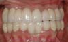
Polyvinylsiloxane impressions (Aquasil, Dentsply International, USA) were taken to fabricate one-piece metal-ceramic fixed partial dentures. On the first set-up, the metal-ceramic fixed partial dentures were luted with temporary cement (Cavitec, Kerr Corp. USA). After an evaluation of the FPDs at 2 weeks, final cementation was carried out using resin modified glassionomer cement (RelyX; 3M ESPE America, Inc. USA). After the final set-up, routine check-ups were performed for 2 years. The fixed partial dentures produced obvious improvement in the mastication and better esthetics (Figs. 7 and 8).
DISCUSSION
It is often necessary for many dental disciplines, including prosthodontics, oral and maxillofacial surgery and orthodontics, to interact in the planning and treatment of patients with complicated severe clinical situation. Multidisciplinary consultation during treatment planning and the coordination and appropriate timing of the subsequent interdisciplinary dental care enables a clinician to provide the optimum care. The scope of the orthodontic and restorative management depends on the severity of the oligodontia and alveolar bone hypoplasia.
In this particular oligodontia patient, an interdisciplinary approach was essential to evaluate, diagnose, and resolve the esthetic and functional problems using a combination of orthodontic, surgical, and prosthodontic treatments. Before the restorative treatment, the signs of facial concavity were greatly reduced by the orthognathic surgery and the remaining teeth were properly aligned by the orthodontic treatment. The final treatment outcomes in terms of the function and esthetics satisfied the expectations of both the patient and the interdisciplinary team.
The prosthodontic considerations in patient treatment include occlusal stability, establishing the correct vertical dimension, and preserving the health of the soft and hard tissues as well as that of the temporomandibular joint. The occlusal stability is determined by a number of factors, including periodontal support, the number of teeth in the dental arches, and occlusal contacts. The masticatory ability is closely related to the number of teeth.
A shortened dental arch (SDA) is defined as having an intact anterior region but a reduced number of occluding pairs of posterior teeth.9,10 The masticatory efficiency and masticatory ability are important components of the oral functionality. However, patient's adaptation to these changes in the dental arch length with the progressive loss of teeth is essential to successful treatment. If the premolar regions are intact and there is at least 1 pair of occluding molars, the SDA may not impair the masticatory efficiency. An impaired masticatory ability and associated changes are manifested only when there are less than 10 pairs of occluding teeth.9,10 The outcome of SDA therapy was found to be acceptable in approximately 82% of patients in terms of the oral function, comfort, and well-being.11
In our clinical report, there were only 4 pairs of occluding premolars and intact anterior teeth. However, the full mouth rehabilitation in this patient using the SDA concept showed long-term occlusal stability, and improved masticatory function.
CONCLUSION
This clinical report describes fixed partial dentures fabricated for the maxilla and mandible, which resulted in a significant improvement in the esthetics and function of the masticatory system. The 2 year follow up of the full mouth rehabilitation with fixed partial dentures using the SDA concept showed occlusal stability, good esthetics and improved masticatory function. The health of the periodontium and teeth as well as that of the temporomandibular joint was maintained.
A coordinated interdisciplinary evaluation and treatment will be necessary to improve the esthetics and function in severely compromised oligodontia patients.
 XML Download