

 PDF
PDF ePub
ePub Citation
 Print
Print


Abstract
Background
Pulmonary emphysema (PE) is major cause of obstructive pulmonary function impairment (OPFI), which is diagnosed by spirometry. PE by high resolution CT is known to be correlated with OPFI. Recently, low dose CT (LDCT) has been increasingly used for screening interstitial lung diseases including PE. The aim of this study was to evaluate OPFI risks of subjects with PE detected by LDCT compared with those detected by simple digital radiography (SDR).
Methods
LDCT and spirometry were administered to 266 inorganic dust exposed retired workers, from May 30, 2007 to August 31, 2008. This study was approved by our institutional review board and informed consent was obtained. OPFI risk was defined as less than 0.7 of forced expiratory volume in one second (FEV1)/forced vital capacity (FVC), and relative risk (RR) of OPFI of PE was calculated by multiple logistic regression analysis.
Results
Of the 266 subjects, PE was found in 28 subjects (10.5%) by LDCT and in 11 subjects (4.1%) by SDR; agreement was relatively low (kappa value=0.32, p<0.001). FEV1 and FEV1/FVC were significantly different between PE and no PE groups determined by either SDR or LDCT. The differences between groups were larger when the groups were divided by the findings of SDR. When PE was present in either LDCT or SDR assays, the RRs of OPFI were 2.34 and 8.65, respectively.
Figures and Tables
Figure 1
A 57-year old man (current smoker, 25-year history of dust exposure) with pneumoconiosis (profusion=1/1) and obstructive pulmonary function impairment (FEV1/ FVC=60). Pulmonary emphysema can barely be seen on simple digital radiography (A), but low dose computed tomography is seen definitely to pulmonary change (B).
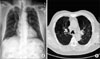
Figure 2
FEV1 and FEV1/FVC were significantly different in both simple digital radiography (SDR) and low dose computed tomography (LDCT) between groups with (■) and without pulmonary emphysema (PE) (□), and the differences were larger in SDR. *p<0.05, †p<0.01.
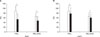
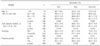
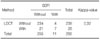
Table 3
Comparison of variables related to PE and OPFI between SDR and LDCT
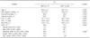
*Calculated by student t-test, †Calculated by fisher's exact test, ‡Calculated by linear by linear association, §Defined as FEV1/FVC<70.
PE: pulmonary emphysema; SDR: simple digital radiography; LDCT: low dose computed tomography; FVC: froced vital capacity; FEV1: forced expiratory volume in one second; OPFI: Obstructive pulmonary function impairment.
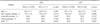
Table 5
Relative risks of obstructive pulmonary function impairment by PE detected by SDR or LDCT
Relative risks of obstructive pulmonary function impairment by PE were calculated by using multiple logistic regression after adjusting age, dust expose duration, smoking and the presence of pneumoconiosis.
PE: pulmonary emphysema; SE: standard error; SDR: simple digital radiography; LDCT: low dose computed tomography; OR: odds ratio; CI: confidence interval.
References
1. Thurlbeck WM, Müller NL. Emphysema: definition, imaging, and quantification. AJR Am J Roentgenol. 1994. 163:1017–1025.
2. Kim HK, Lee SD. Pathophysiology of chronic obstructive pulmonary disease. Tuberc Respir Dis. 2005. 59:5–13.
3. Litmanovich D, Boiselle PM, Bankier AA. CT of pulmonary emphysema--current status, challenges, and future directions. Eur Radiol. 2009. 19:537–551.
4. Bauer TT, Heyer CM, Duchna HW, Andreas K, Weber A, Schmidt EW, et al. Radiological findings, pulmonary function and dyspnea in underground coal miners. Respiration. 2007. 74:80–87.
5. Horiuchi N, Fujita J, Suemitsu I, Yamasaki Y, Higa F, Tateyama M. Low-dose multislice CT and high-resolution CT assessment of pulmonary emphysema in public school teachers. Lung. 2007. 185:25–30.
6. Lee SM, Hur J, Kim TH, Kim SJ, Kim HJ. Quantitative assessment of lung volumes using multi-detector row computed tomography (MDCT) in patients with chronic obstructive pulmonary disease (COPD). J Korean Radiol Soc. 2008. 59:91–97.
7. Kang JH, Chun JH, Gu HW, Ko KS, Yu BC, Sohn HS, et al. Diagnostic meaning of high resolution computed tomography compared with chest radiography for screening of welder's lung. Korean J Prev Med. 1996. 29:853–862.
8. Kim KI, Choi SJ, Sohn HS, Lee JW, Jung DH, Lee SH, et al. High-resolution CT findings of welders' pneumoconiosis. J Korean Radiol Soc. 1996. 34:367–371.
9. Kim KA, Kim JH, Chang HS, Ahn HS, Lim Y, Yun IG. The diagnostic role of HRCT in simple pneumoconiosis. Korean J Prev Med. 1996. 29:471–482.
10. Jung EJ, Kim YK, Lee YM, Kim KU, Uh ST, Kim YH, et al. The correlation of dyspnea and radiologic quantity in patients with COPD. Tuberc Respir Dis. 2009. 66:288–294.
11. Naidich DP, Marshall CH, Gribbin C, Arams RS, McCauley DI. Low-dose CT of the lungs: preliminary observations. Radiology. 1990. 175:729–731.
12. Diederich S, Wormanns D, Heindel W. Lung cancer screening with low-dose CT. Eur J Radiol. 2003. 45:2–7.
13. Gierada DS, Pilgram TK, Whiting BR, Hong C, Bierhals AJ, Kim JH, et al. Comparison of standard- and low-radiation-dose CT for quantification of emphysema. AJR Am J Roentgenol. 2007. 188:42–47.
14. Sim YS, Ham E, Choi KY, Lee SY, Kim SC, Kim YK, et al. Longitudinal evaluation of lung function associated with emphysema in healthy smokers. Tuberc Respir Dis. 2010. 69:177–183.
15. Omori H, Nakashima R, Otsuka N, Mishima Y, Tomiguchi S, Narimatsu A, et al. Emphysema detected by lung cancer screening with low-dose spiral CT: prevalence, and correlation with smoking habits and pulmonary function in Japanese male subjects. Respirology. 2006. 11:205–210.
16. Lopes AJ, Mogami R, Capone D, Tessarollo B, de Melo PL, Jansen JM. High-resolution computed tomography in silicosis: correlation with chest radiography and pulmonary function tests. J Bras Pneumol. 2008. 34:264–272.
17. Guideline of quality assurance for pneumoconiosis [Internet]. Occupational Safety and Health Research Institute (OSHRI). 2010. cited: Mar 10, 2010. Incheon: OSHRI;Available from: http://oshri.kosha.or.kr/board?tc=RetrieveBoardViewCmd&boardType=A&contentId=208741&pageNum=1&urlCode=T1|Y|404|371-|||||/board&tabId.
18. International Labour Office (ILO). Guidelines for the use of the ILO international classification of radiographs of pneumoconioses. 2002. Revised ed. Geneva: International Labour Office.
19. Friedman PJ. Imaging studies in emphysema. Proc Am Thorac Soc. 2008. 5:494–500.
20. Miller MR, Hankinson J, Brusasco V, Burgos F, Casaburi R, Coates A, et al. Standardisation of spirometry. Eur Respir J. 2005. 26:319–338.
21. The cause of death statistics [Internet]. Korean Statistical Information Service. cited: March 10, 2010. Daejeon: Korea National Statistical Office;Available from: http://kosis.kr/abroad/abroad_01List.jsp.
22. Kalra MK, Maher MM, Toth TL, Hamberg LM, Blake MA, Shepard JA, et al. Strategies for CT radiation dose optimization. Radiology. 2004. 230:619–628.
 XML Download